EMS MEd Blog

Article Bites #29: Is CPSS greater than or equal to 2 a reasonable tool for Large Vessel Occlusion Stroke Prediction?

Article: Crowe, R. P., Myers, J. B., Fernandez, A. R., Bourn, S., & McMullan, J. T. (2020). The cincinnati prehospital stroke scale compared to stroke severity tools for large vessel occlusion stroke predictionPrehospital Emergency Care, 1-9.


Background: Since the first trials demonstrating significant benefit from endovascular therapy for patients with stroke secondary to large vessel occlusion (LVO) were published, the EMS community has been actively refining screening and destination protocols to optimize patient outcomes through destination choice.   Indeed, given a ~ 10% decrease in good functional outcome for every 30 minutes in delay in endovascular treatment, appropriate field triage is a critical component of stroke systems of care for patients with an LVO. [1,2]

LVO Screening tools of variable complexity have been developed. Depending on the complexity, implementation of such screening tools requires significant investment in training, documentation and tracking on the part of the EMS system.  In general, the sensitivity, specificity and positive predictive value of all the LVO scales has been suboptimal. [3] In contrast, the Cincinnati Prehospital Stroke Scale (CPSS) is widely implemented and regularly performed by EMS clinicians at all scopes of practice.  The objective of this paper was to address whether newly developed LVO scales offer a clinically-meaningful advantage over the CPSS.  


Methods:  This study was a retrospective analysis of prehospital electronic care records with linked hospital outcome data for the 2018 calendar year from the ESO research database.    Inclusion criteria included 911 responses with one or more of the stroke scales of interest (CPSS, RACE, LAMS, or VAN) documented AND linked hospital diagnosis data available.  Patients were classified as having intracerebral hemorrhage, transient ischemic attack or acute ischemic stroke based the hospital ICD-10 code.  If the ICD-10 code indicated thrombosis or embolism of the middle cerebral arteries, internal carotid artery or basilar artery, the patient was categorized as having an LVO. Sensitivity and specificity for detection of LVO for each documented stroke scale was calculated.  Receiver Operating curves were used to assess the overall discrimination of each scale for LVO.


Key Results:

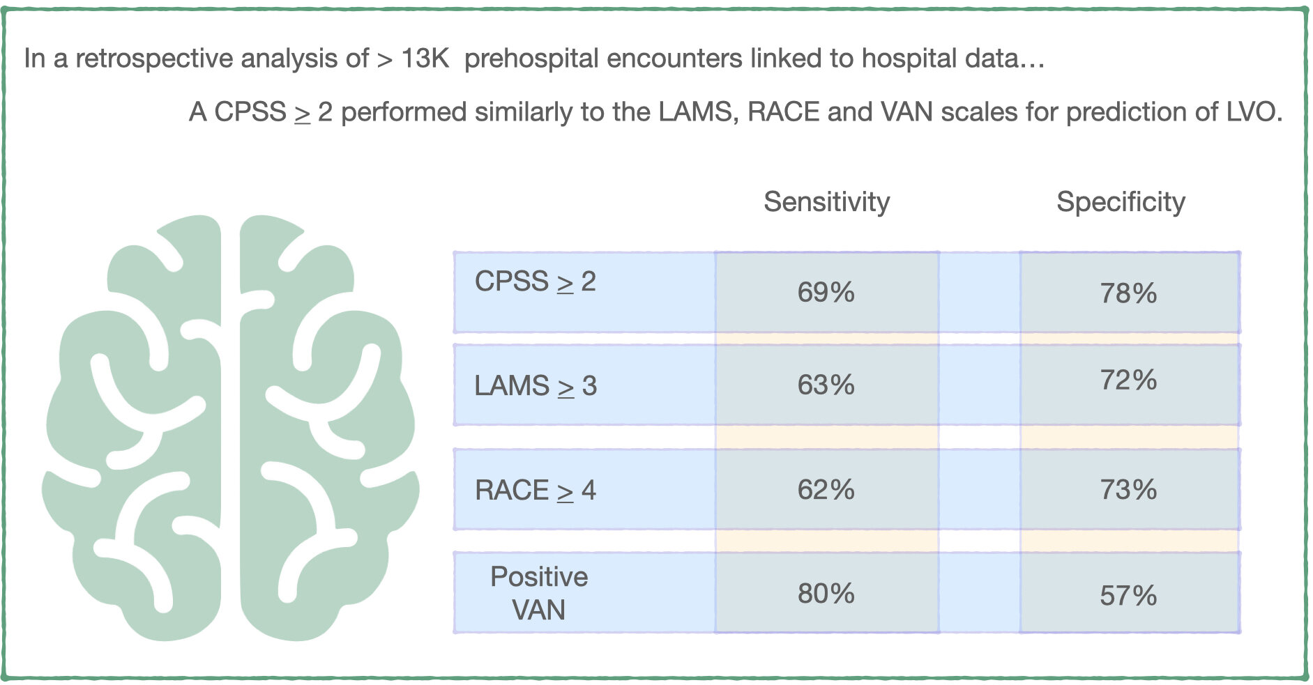
-        Among 13,596 responses from 151 EMS agencies that had one or more stroke scales documented, CPSS was the most commonly documented instrumented (83% of patients), followed by RACE (14% of patients), the LAMS (7% of patients), and VAN (4% of patients). 31% of patients with documented prehospital stroke scales had a hospital ICD-10 diagnosis indicative of stroke.

-        Breakdown of types of stroke: 57% ischemic stroke, 23% TIA, 13% with TIA, 7% with multiple types.  Of patients experiencing ischemic stroke, 26% had an ICD-10 diagnosis indicative of LVO.

-        A CPSS score > 2 (positive on 2 or more physical exam elements of facial droop or palsy; arm weakness, drift or drop; and abnormal speech) had a sensitivity of 69% and specificity of 73% for LVO.  There was no statistically significant differences between this and the performance of the RACE, LAMS or VAN scales.

-        Among patients with a CPSS score > 2, 14% were diagnosed with LVO and 9% were diagnosed with ICH.  39% did not have a stroke diagnosis.

CPSS_Crowe.001.jpeg


What this means for EMS:  Destination decision is a critical component of the prehospital management of stroke and this decision has increased in complexity with the availability of endovascular therapy for patients with large vessel occlusion.  On a system scale, optimizing patient and system outcomes requires a balance between over- and under-triage of these patients to specialty centers capable of providing endovascular therapy.  In this retrospective analysis of prehospital patients, a CPSS > 2 performed similarly to more complex LVO scales for large vessel occlusion prediction. While we await assessment of additional prehospital stroke scales, it may be more worthwhile to focus on incorporating CPSS > 2 into destination decisions and quality improvement efforts than training EMS clinicians on new stroke scales.

References:

1.     Saver, J. L., Goyal, M., Van der Lugt, A. A. D., Menon, B. K., Majoie, C. B., Dippel, D. W., ... & Cardona, P. (2016). Time to treatment with endovascular thrombectomy and outcomes from ischemic stroke: a meta-analysis. Jama316(12), 1279-1289.

2.     Jayaraman, M. V., Hemendinger, M. L., Baird, G. L., Yaghi, S., Cutting, S., Saad, A., ... & McTaggart, R. A. (2020). Field triage for endovascular stroke therapy: a population-based comparison. Journal of neurointerventional surgery12(3), 233-239.

3.     Smith, Eric E., et al. "Accuracy of prediction instruments for diagnosing large vessel occlusion in individuals with suspected stroke: a systematic review for the 2018 guidelines for the early management of patients with acute ischemic stroke." Stroke 49.3 (2018): e111-e122.


Article Summary by Maia Dorsett, MD PhD FAEMS, @maiadorsett


Article Bites #28: In most cases of OHCA, Resuscitate on Scene > Intra-arrest Transport

Article: Grunau B, Kime N, Leroux B, et al. Association of Intra-arrest Transport vs Continued On-Scene Resuscitation With Survival to Hospital Discharge Among Patients With Out-of-Hospital Cardiac Arrest. JAMA. 2020;324(11):1058–1067. doi:10.1001/jama.2020.14185

Background

Treatment for a non-traumatic Out-Of-Hospital Cardiac Arrest (OHCA) can occur on scene or during transport. It continues to be the standard in some systems that OHCA patients are transported early, while in other systems patients are treated on scene until return of spontaneous circulation (ROSC) is attained. There is a perception among many in the lay public, hospitals, and even EMS that optimal care is given in the Emergency Department, and all OHCA victims should be transported for best outcomes. However, treatment on scene utilizing similar protocols to the ED until ROSC is felt amongst many to provide a higher quality care than can be attained during transport.

Methods

Data was obtained from the Resuscitation Outcomes Consortium (ROC) registry and collection prospectively from consecutive nontraumatic adult EMS-Treated OHCA. There were 10 sites in North America, data was obtained between 2011 and 2015, and was at the ALS and BLS level. Exclusion criteria included age < 18, a DNR order, cardiac arrest after initiation of transport, or those with missing data. Primary outcome was survival to hospital discharge. Secondary outcome was neurologically-intact survival.

Results

A total of 43,969 consecutive OHCAs were included with 11,625 underwent intra-arrest transport and 32,344 were treated on-scene until ROSC or termination of resuscitation. Survival to hospital discharge was 3.8% for patients who received intra-arrest transport and 12.6% for those who received on-scene resuscitation. Of those transported prior to ROSC, 16% achieved ROSC prior to hospital arrival.

To account for differences between the on-scene resuscitation vs. intra-arrest transport groups, the authors performed a propensity-matched analysis.  Survival of intra-arrest transport patients was 4.0%, while survival of transport after ROSC was 8.5%. This result was statistically significant, with an adjusted risk ratio of 0.48 (95% CI, 0.43-0.54).   Neurologically-intact survival, defined as a mRS < 3, was also higher in the group with continued on-scene resuscitation: 7.1% for on-scene vs. 2.9% for intra-arrest transport with a statistically-significant adjusted risk ratio of 0.60 (95% CI, 0.47-0.76). This trend held for both shockable and non-shockable rhythms.

image.001.jpeg

What does this mean for EMS?

While not a randomized-controlled trial, this study uses a large prospectively gathered dataset with cohort matching. Therefore the data gathered is likely to represent real-world conditions. In addition this data continues to add to a growing trend of evidence and standard of practice that most patients suffering from cardiac arrest have a higher chance of survival if treated on-scene until ROSC. There are likely subgroups of patients, such as in pregnancy or those who may benefit from eCPR after arrival at a hospital, where transport early or after the 15 minute time mark may lead to some benefit.

Article Summary by Joshua Stilley, MD FAEMS, @JoshuaStilley

Article Bites #27: Is eCPR the future of refractory Vfib Management?

Yannopoulos D, Bartos J, Raveendran G, et al. Advanced reperfusion strategies for patients with out-of-hospital cardiac arrest and refractory ventricular fibrillation (ARREST): a phase 2, single centre, open-label, randomized controlled trial. Lancet 2020; Nov 13

Background – ECMO and CPR (eCPR)

Refractory ventricular fibrillation (rVF), often called electrical storm, has dismal outcomes which is exceedingly frustrating because we know the cause is likely acute coronary occlusion (~75%).  Recent therapies for rVF include double-sequential defibrillation, esmolol, and even ultrasound-guided stellate ganglion nerve blocks.  Others have suggested early ECMO in this patient population.  The logic behind this approach makes intuitive sense; CPR/ACLS are unlikely to work if you have a 100% left main lesion causing OHCA.  One potential solution for this dilemma is to use ECMO as a bridge to cardiac catheterization and revascularization (eCPR). Dr. Demetris Yannopoulos at The University of Minnesota has been an early champion of eCPR with multiple retrospective studies suggesting improved survival when using ECMO for rVF and OHCA.  However, the ARREST study is the first randomized trial of standard ACLS vs. eCPR.

Methods

This study occurred from August 2019 through June 2020 at The University of Minnesota and included three transporting EMS systems. Patients  were included in the study if they were 18-75yo, presented with shockable rhythm with no ROSC after three shocks, had a body habitus compatible with use of the LUCAS mechanical CPR device, and had a transfer time of < 30 minutes.  There were multiple exclusionary criteria, as well, that were all applied in an attempt to select for patients who would have the best opportunity for good functional outcomes AND be most likely to have acute coronary occlusion as the cause of their cardiac arrest: 

Exclusion Criteria

·       Trauma

·       Overdose

·       Pregnancy

·       Prisoners

·       DNR/Nursing home patients

·       Terminal cancer

·       Opt-out bracelet

·       Contrast allergy

·       Active bleeding

Patients were randomized on arrival to the emergency department into one of two arms: immediate ECMO with subsequent catheterization/revascularization or standard ACLS in the ED.   This is important, as the current standard in many systems is continue to manage the arrest in place.  

The eCPR group went directly to the cath lab for immediate cannulation and catheterization.  The standard ACLS group got at least 15min of resuscitation or until 60min from arrest.  If ROSC was obtained, the standard ACLS group received immediate angiography.  If the patient survived to admission, those in both groups got standard cardiac ICU care with targeted temperature management and no neuro prognostication for at least 72hr.

What outcomes were measured?

The primary outcome was survival to hospital discharge with secondary outcomes of safety and functionality (measured by mRS and CPC score) at hospital discharge, three months, and six months.


Results:

Were the two groups similar?

Although many historical specifics were unknown (not surprising for a study of OHCA), the two groups were similar at baseline although there were more women and ESRD patients in the standard ACLS arm.  All patients presented with an initial rhythm of rVF.  Rates of witnessed arrest and bystander CPR, time to EMS arrival, and prehospital ACLS treatments were similar.  No patients had ROSC on arrival to the ED.  These patients were SICK with hospital arrival lactates of 10-11, pH values of 6.9-7, and measured ETCO2 levels of 28-33.  Six patients were excluded:  two with initial PEA, one with a transport time >30min, and three with ROSC after the 2nd shock. 

What were the key results?

Survival to discharge in the eCPR group was 43% (6/14) vs. 7% (1/15) in those treated with standard ACLS.

But what was the functional status of the survivors?  At the 6-month mark, all 6 ECMO survivors had an mRS of 3 or lower.  Only 2 of the 15 standard ACLS patients even made it to the cardiac catheterization lab.  One died in hospital from cerebral edema and the other after hospital discharge from anoxic injury.  Two patients in the eCPR/ECMO group were declared dead prior to cannulation (2 or more of ETCO2<10/PaO2<50/lactate>18 = dead).  Six ECMO patients died before hospital discharge due to anoxia/cerebral edema.  ECMO complications included one tubing break, two access site bleeds, and one retroperitoneal bleed.

One criticism of this study will obviously be the small patient numbers, but the data safety monitoring board actually stopped the study early due to lack of equipoise.


What would a program like this require?

From a prehospital standpoint, our portion of care will be the beginning in a long process that will require coordination of care across EMS/ED/cath lab/ICU and even rehabilitation.  That said, the authors specifically stated that the time from 911 call to ECMO cannulation is the prime predictor of survival.  Implementation of an eCPR program will not be something that EMS can tackle alone.  A streamlined system of care with a dedicated resuscitation center will be critical and must include ECMO availability with ED, cardiology, an intensive care buy-in.  Yes, an eCPR approach is expensive and labor-intensive, but improving survival to almost 50% with good functional outcomes in a condition with an otherwise bleak prognosis seems promising at the very least.

Article Summary by Casey Patrick, MD

Figure by Maia Dorsett, MD PhD

Do push dose pressors have a role in prehospital care?

In November, we posted these cases and discussion stem to the blog discussion forum and got a number of insightful comments regarding the use of push dose vasopressors in the prehospital environment.  This commentary is summarized here.

The term “Push dose pressors” (also known as Bolus-Dose Vasopressors) refers to the intravenous administration of small, discrete doses of vasopressors, such as epinephrine or phenylephrine, to rapidly treat hypotension.[1-2]  The routine use of push dose vasopressors (PDPs) originated in the operating room setting to offset hypotension as a side effect of anesthetic medications, such as hypotension induced by spinal anesthesia. [3-4]  Subsequently their use gained traction in the emergency department and out-of-hospital setting as a result of popularization by the Free Open Access Medical Education (FOAMed) movement. [1, 5, 6]  

 While PDP use has become increasingly common, evidence regarding the benefits and/or harms of their use outside of the operating room remains limited. [1]  As such we asked our readers the following question regarding their EMS system:

-       Are you using push dose vasopressors?  If not, why not? 

-       If so, what are the indications and dosing/frequency? 

-       What is the protocol for their use and what safeguards against medication error are in place?  Are you tracking any quality measures for their use?


Yes… BUT

 The Austin-Travis County EMS system uses PDPs as a temporizing measuring pending initiation of vasopressor infusion or to prevent peri-intubation cardiac arrest in a hemodynamically unstable patient in need of invasive airway management… an approach shared by Jeff Jarvis of Williamson County EMS.  

The reason cited for using only as a bridge?  A high risk of medication error. The risk of medication errors in a high stakes, low frequency case is certainly a concern.  What does the current literature say regarding risk of medication errors with PDPs?

Rotando et al. (2018) performed a retrospective analysis of PDP use practice patterns, efficacy and safety in critically ill patients ICU patients at a single institution. [7]  146 patients met their inclusion criteria.  As expected, PDP use improved blood pressure.  However, 17/146 (11.6%) patients had an adverse event associated with push-dose vasopressor use, including dosing errors, administration when patient was not documented to be hypotensive, or administration when patient was already on a continuous infusion vasopressor.

A subsequent retrospective study of Emergency Department PDP use by Cole et al. (2019) also demonstrated a high rate of dosing errors during administration of PDPs. [8] One of the most interesting aspects of this study was that instead of only using standardized chart review, the authors reviewed video of the resuscitations to obtain the majority of their data.  They retrospectively identified 249 patients who received PDPs in the ED between 2010-2017 and then reviewed videos of the resuscitation when available (68% of all cases) for dosing errors, hemodynamic consequences of administration and documentation errors.  They found that adverse hemodynamic events (defined as HR > 140, new bradycardia (HR < 60), SBP > 180, or development of ventricular tachycardia) occurred in 39% of all cases (27% in the phenylephrine group and 50% of the epinephrine group).  Human errors occurred in the care of 19% of patients, including administering a dose different than the intended dose (3%).  Dosing errors included anywhere from 10-fold overdoses of phenylephrine or epinephrine to a 100-fold dosing error of phenylephrine.  The risk of medication error was significantly increased when an ED pharmacist was not present. Of note, the average time for patients to be started on an epinephrine drip was 8 min, whether or not an ED pharmacist was present, leading the authors to making a conclusion very similar to that of @ATCEMS/Jason Pickett:

In select patients with shock whose physiology cannot tolerate the several minutes required to prepare a vasopressor infusion, the improvement of blood pressure and vital organ perfusion imparted by push dose pressors may outweigh potential adverse effects. However, push dose pressors could be deleterious to patients whose outcomes are not likely to be improved with this stop-gap therapy (e.g., those with hypotension who will rapidly correct with no intervention or fluid resuscitation, or those with hypotension but not shock). An important area of future research will be determining the patient population most likely to benefit from this therapy.”

What about risk of error in EMS-based studies of PDP use?

Nawrocki et al. (2019) characterized hemodynamic effects and adverse events associated with push dose epinephrine use during critical care aeromedical transport.[9]  They retrospectively studied patients who received at least one dose of push dose epinephrine to correct documented hypotension (SBP < 90 mmHg or MAP < 65) following the introduction of a protocol directing administration of 10-20 mcg of push dose epinephrine every 2 minutes until vitals normalize or the vasopressor infusion is ready to start. In this study, they found that of the 100 doses of push dose epinephrine administered over a two year period, 94% were deemed “appropriate” in terms of dosing and indication.

Patrick et al. (2020) evaluated the use of push dose epinephrine in ground-based EMS transport. [10]   They performed a one year retrospective chart review of all patient care records where a patient was treated for hypotension (defined as SBP < 90 mmHg) with push dose epinephrine following a protocol very similar to that of Nawrocki et al. They specifically excluded patients whose initial presentation was cardiac arrest.  There was one documented medication error amongst 43 cases (2.3%), which was related to a change in the epinephrine stock concentration from 1:10,000 concentration to 1:1,000 concentration secondary to drug shortages.

Of note, in neither of the above EMS studies was the primary objective to evaluate the incidence of medication error.

Is a Bridge Really Necessary?

The most common cited indication for the PDPs is as a bridge to vasopressor initiation.  In the Cole et al. study, the average time was 8 min and neither of the prehospital studies were set up to measure this interval.  It is thus worth considering whether there is, in general, a different time to initiation a vasopressor initiation in the field versus in the ED.   

Some of the commentary addressed this very issue:

 The benefit for PDP is iatrogenic hypotension from a procedure (RSI) or you walk in and the patient is peri arrest… Appropriate check lists and references after training can help avoid medication errors if pre made medication syringes aren't available. Depending on crew configuration you may not be saving significant time mixing up a stick of pressor vs a bag.

-Shane O'Donnell

We do not currently utilize push dose pressors in the University of Missouri system. We have a pretty aggressive protocol for initiating pressor infusions, including getting one setup for all post-ROSC patients. I have not seen patients decompensate to cardiac arrest without PDP, and to me a couple minutes of hypotension while getting a pressor infusion set up is reasonable and much safer. We also have a strong push for delaying intubation in unstable patients, and my EMS Clinicians have done a good job of embracing that. – Joshua Stilley

Interestingly, in the prehospital study of PDP use by ground EMS [10], almost one half of the administrations were treated with PDPs pre-delayed sequence intubation, most only required 1-2 doses as they received adjunctive treatments such as fluid administration, and only 48% required continuous vasopressor infusion.


Countin’ Drips

When considering the risk of dosing error in infusions versus PDPs, there remains one key distinction between hospital and most prehospital systems: the availability of IV pumps.  This complexity was captured in the following comments by Joshua Borkosky, Jeff Jarvis and Ryan Jacobsen:

We do not use Push-Dose pressors in my EMS System currently. We do use (and I personally use) in the academic ED I practice in however. I've seen no convincing evidence in the literature other than the popular gurus (ex. Weingart) espousing their amazingness. In theory I like it, but several caveats. We use norepinephrine drips currently if pressor needed. We don't have pumps however and use a metronome (checklist) to calibrate a rate. It is very imperfect and not awesome. Balancing the risk/benefit of norepinephrine drip without a pump versus mixing a syringe of epinephrine to 10mcg/ml in high risk/low frequency environment is challenging. I have recently found we can get pre-mixed push dose epinephrine (2 month shelf life) so mixing would not be a concern, but likelihood of use versus constantly rotating stock every 2 months in large service has logistics/cost issues. I like the concept in theory. If you have a phenylephrine AND epinephrine premade syringe for push dose you could avoid all need for "drips/pumps" in the 911 urban/suburban environment. I have mixed feelings. - Ryan Jacobsen MD, Medical Director, Johnson County EMS System, Johnson County, KS

Before we jump to suggesting a cheaper “dial-a-drip” or “dial-a-flow” set up, a 2018 study by Loner et al. tested these devices and found them to be inaccurate and associated with both underdosing and overdosing errors. [11]

Given the concern over accuracy of off-pump IV infusions, some EMS systems, particularly urban/suburban transport systems with shorter transport times, opt for PDPs in place of vasopressor infusions given lack of availability of IV pumps.  Given the short prehospital transport times, the prehospital PDP may act as a bridge to the in-hospital infusion in these scenarios:

We use PDPs in LA County. We don’t have pumps, and maybe we should, but most of our transports are less than 15 minutes and PDPs are far simpler to do. We use cardiac Epi diluted 10X (10mcg/mL), 1mL Q 3-5 mins. I just can’t reconcile counting drops/min in the 21st century with all of the tech that we bring to the patient these days. – Clayton Kazan

My system is doing both PDPs and IV drip. I personally stick to PDPs though since we do not have IV pumps. We only have dial a flow devices which are inaccurate at best. I personally feel PDPs are safer because you know exactly how much you are giving because you are in control. I mix 1mg Epi in 500ml NS and give 10-20mcg pushes. - Matt King


So, what about that most important quality measure – patient outcome?

So far, all the research we have discussed has evaluated the effect of push dose pressors on hemodynamic parameters, which theoretically serve as surrogate markers for improved outcome because of the known association between hypotension and adverse outcomes, especially for cardiac arrest and traumatic brain injury.

But what if we examined actual patient outcomes?

So far, data is limited to retrospective studies alone.

Guyette et al. (2019) examined the relationship between push dose epinephrine and mortality in patients cared for by aeromedical critical care transport service.  They performed a retrospective case-cohort study that included patients > 14 years of age with SBP < 70 mmHg.  In 2015, the service introduced a protocol for administration of 100 mcg of IV epinephrine (a dose ~ 5-10 times higher than typical PDP dose) to patients with preserved pulses and SBP < 70 mmHg.  This was to be accompanied by volume resuscitation followed by norepinephrine infusion. Each patient who received push dose epinephrine was matched to two historical controls using demographic and prognostic data including age, gender, shock type, weight, index vital signs at first hypotensive episode, and pretreatment characteristics including CPR, defibrillation, transcutaneous pacing, needle thoracostomy, vasopressor administration, intubation or arrhythmia.  Interestingly, 30-day survival was lower in patients who received push dose epinephrine (36% vs. 56%) and was significantly different in an adjusted analysis (p=0.01).  However, given the retrospective nature of the study, it remains possible that the patients receiving PDPs were still sicker despite attempts to control for likely confounders.

Summary & Take Home

There is variability in prehospital PDP use in EMS practice.  While PDPs may have benefit to rapidly address iatrogenic hypotension from a procedure or as a bridge to vasopressor infusion (initiated prehospital or at the hospital destination in the case of short transport times), there are concerns in terms of medication safety and lack of demonstrated benefit in terms of patient outcomes.  This remains an area in need of high-quality prehospital research to inform best practice.

Summary post by Maia Dorsett, MD PhD (@maiadorsett)

Edited by Brian Miller, MD (@BrianMillerMD)

References:

[1] Holden, D., Ramich, J., Timm, E., Pauze, D., & Lesar, T. (2018). Safety considerations and guideline-based safe use recommendations for “bolus-dose” vasopressors in the emergency department. Annals of Emergency Medicine71(1), 83-92.

[2] Cole, J. B. (2017). Bolus-Dose vasopressors in the emergency department: first, do no harm; second, more evidence is needed. Ann Emerg Med1, 3.

[3] Siddik-Sayyid SM, Taha SK, Kanazi GE, et al. (2014).  A randomized controlled trial of variable rate phenylephrine infusion with rescue phenylephrine boluses versus rescue boluses alone on physician interventions during spinal anesthesia for elective cesarean delivery. Anesth Analg. 118:611-618.

[ 4] Doherty A, Ohashi Y, Downey K, et al. (2012). Phenylephrine infusion versus bolus regimens during cesarean delivery under spinal anesthesia: a double-blind randomized clinical trial to assess hemodynamic changes. Anesth Analg. 115:1343-13501

[5] Weingart S. https://emcrit.org/emcrit/bolus-dose-pressors/

[6] Weingart, S. (2015). Push-dose pressors for immediate blood pressure control. Clinical and experimental emergency medicine2(2), 131.

[7] Rotando, A., Picard, L., Delibert, S., Chase, K., Jones, C. M., & Acquisto, N. M. (2019). Push dose pressors: Experience in critically ill patients outside of the operating room. The American journal of emergency medicine37(3), 494-498.

[8] Cole, J. B., Knack, S. K., Karl, E. R., Horton, G. B., Satpathy, R., & Driver, B. E. (2019). Human Errors and Adverse Hemodynamic Events Related to “Push Dose Pressors” in the Emergency Department. Journal of Medical Toxicology15(4), 276-286.

[9] Nawrocki, P. S., Poremba, M., & Lawner, B. J. (2019). Push dose epinephrine use in the management of hypotension during critical care transport. Prehospital Emergency Care.

[10] Patrick, C., Ward, B., Anderson, J., Fioretti, J., Keene, K. R., Oubre, C., ... & Dickson, R. (2020). Prehospital Efficacy and Adverse Events Associated with Bolus Dose Epinephrine in Hypotensive Patients During Ground-Based EMS Transport. Prehospital and Disaster Medicine35(5), 495-500.

[11] Loner, C., Acquisto, N. M., Lenhardt, H., Sensenbach, B., Purick, J., Jones, C. M., & Cushman, J. T. (2018). Accuracy of intravenous infusion flow regulators in the prehospital environment. Prehospital Emergency Care22(5), 645-649.

[12] Guyette, F. X., Martin-Gill, C., Galli, G., McQuaid, N., & Elmer, J. (2019). Bolus dose epinephrine improves blood pressure but is associated with increased mortality in critical care transport. Prehospital Emergency Care23(6), 764-771.

Article Bites #26: Time for a new location or Technique in infants? Success rates of proximal tibia IO placement in pediatric patients as determined by post-mortem CT

Article: Harcke, H. T., Curtin, R. N., Harty, M. P., Gould, S. W., Vershvovsky, J., Collins, G. L., & Murphy, S. (2020). Tibial Intraosseous Insertion in Pediatric Emergency Care: A Review Based upon Postmortem Computed Tomography. Prehospital Emergency Care, 1-7.

 Background:  The most commonly used (and recommended) site for intraosseous (IO) access in pediatric patients is the proximal tibia. The primary objective of this study was to determine the accuracy of emergency IO placement in pediatric patients by both prehospital providers and emergency department providers.

Methods: The authors determined accuracy of tibial IO placement using post-mortem CT.  They reviewed 92 cases referred by the state medical examiner for post-mortem CT and found 31 where a tibial IO had been placed. Successful IO placement was defined by needle placement between the proximal 5% to 30% of the tibia with the needle tip in the medullary cavity.  Needle length was determined by measuring via CT or appearance of the needle hub color.

 Key Results: Among 31 cases, there were 42 total tibial IO insertions.  The authors found that:

·      Infants < 6 months of age accounted for 30/42 IO placements.  Overall success in this age group was 47%.  There was variability in success by IO needle size (56% for 15 mm needle and 0% success for 25 mm needle).

·      Success rate amongst patients 6 months to 2 years of age was 83% (n=6)

·      Success rate among patients > 2 years of age was 100% (n=6)

·      The most common reason for failure varied by needle size.  For 15 mm needle it was that the needle was outside the bone (45%, n=11) or embedded in the cortex  (45%, n=11), while for the 25 mm it was perforation of the tibia (83%, n=6).

·      Rates of failure were not significantly different between EMS and ED personnel (30% failure rate for ED personnel, 46% failure rate for EMS).

Conclusions: While there is some risk of bias as this study only examined non-survivors, the failure rate of proximal tibial IO placement in pediatric patients, in particular those < 6 months of age, was alarming.  Size mattered: 15 mm needles were much more likely to be successful that 25 mm needle (which was unsuccessful in 6/7 patients under 2 years of age).  These results are in line with a prior cadaver study by Maxien et. al. demonstrating a high rate of malposition (64%) of IO in infants < 1 year.

These findings raise the question of whether how can we improve success rates: manual insertion over drill? Mandatory needle sizes? Increased training? Alternative site such as the distal femur?

 What this means for EMS: Pediatric IO placement has a high failure rate, especially in infants < 6 months of age.  At minimum, to improve success rate a 15 mm needle should be chosen in this age group.   Further research is needed to address whether alternative sites or methods may be preferable.

Article Summary by Maia Dorsett, MD PhD FAEMS FACEP, @maiadorsett

Interested in a more in depth discussion of this article and pediatric IO? - see the PEC podcast Deep Dive Episode.

Discussion Forum: A pressor in a push…

Consider Three Clinical scenarios:

Scenario A:

cartoon.jpeg

EMS responds to the scene of a cardiac arrest.  Patient is found in PEA and ROSC is achieved after 5 cycles of CPR.  Immediately Post-ROSC and 3 minutes since the last dose of epinephrine, blood pressure is 110/80, HR 125, EtCO2 45 mmHg, SpO2 of 95%.  Post-ROSC EKG shows an inferior MI.  As the patient is being packaged for transport, a repeat blood pressure of 70/30 is obtained, HR is now 90, and EtCO2 has declined to 30 mmHg.  The paramedic considers starting an epinephrine or norepinephrine drip, but thinks that utilization of push dose pressors may suffice and make it easier to rapidly extricate patient and begin transport towards the local STEMI receiving center… 


Scenario B:

EMS responds to an assisted living facility for altered mental status in a 78 year-old male.  The patient has been becoming progressively more altered over the course of the day and the nursing home reports a fever.  The patient is obtunded and responds only to painful stimuli.  An indwelling Foley catheter is noted to be draining cloudy urine.  VS obtained show a BP 68/30, HR 110, SpO2 89% on room air, RR 24, EtCO2 22 mmHg.  IV access x 2 is obtained on scene, and fluids and oxygen administration are initiated.  The paramedic considers whether push dose pressors may be indicated for this patient… 

Scenario C:

EMS responds to a motor vehicle collision.  The patient has suffered major blunt trauma to the head and pelvis.  Assisted ventilations are initiated, the patient is placed in spinal motion restriction, and a pelvic binder is applied.  Transport is initiated to the nearest trauma center which is 15 minutes away.  VS are BP 80/60, HR 120, RR 12 (assisted), SpO2 96%, EtCO2 30 mmHg.  IV access is initiated and the paramedic considers push dose pressors to support the blood pressure in anticipation of need for airway management en route or in the ED…

Push dose pressor use has become increasingly common in emergency departments and has been introduced to several EMS systems, however the evidence regarding the benefits and/or harms of their use outside of the operating room remains limited.  

We are interested in what your current EMS system is doing and why?  Are you using push dose vasopressors?  If not, why not?  If so, what are the indications and dosing/frequency?  What is the protocol for their use and what safeguards against medication error are in place?  Are you tracking any quality measures for their use?

Please share your comments below by NOVEMBER 20th to be included in the subsequent summary post.

Article Bites #25. Compassion First: Mortality risk of Patients with Psychogenic Nonepileptic seizures

Nightscales, R et al. Mortality in Patients with Psychogenic Nonepileptic Seizures. Neurology 95.6 (2020): e643–e652. Neurology

Background

Caring for psychogenic nonepileptic seizure (PNES) patients (the condition formerly known as “pseudoseizures”) in the emergency setting can be exceedingly difficult and frustrating.  This is understandable because discerning between true seizure activity and a psychogenic event leads to drastically different treatment pathways.   Additionally, PNES patients have high rates of underlying substance use disorder and psychiatric illness which can further cloud the clinical picture.  This is a difficult diagnosis for even the hospital neurologists demonstrated by the fact that PNES patient account for 25% of EEG unit admissions and a final diagnosis of PNES takes an average of eight years!  If that’s not enough, between 5-20% of PNES patients also have true epilepsy.  The objective of this study was to look at the disease progression and mortality characteristics of PNES patients.  

Methods

This was a retrospective cohort study that included all admissions to three video EEG (the gold standard for epilepsy diagnosis) monitoring units in Victoria, Australia.  Data from 1995 through 2015 was reviewed. 

For diagnostic purposes, patients admitted for video EEG monitoring (VEM) were separated into three groups: PNES, epilepsy or the combination of the two.  Then, chart review was linked to the Australian national death index (NDI) to correlate mortality rates and causes. 

Key Results

Article_Bites_PNES.001.jpeg

There were 5,508 admissions to the VEM units during the study period.  (674 with PNES, 3064 with Epilepsy, 75 with both and 1595 diagnosed as other). Brain tumor patients were excluded.  The death rates over the 20-year study period for the three groups were as follows 8.2% PNES (55/674) 9.4% Epilepsy (288/3,064) and 8% Both (14/175).

Compared to the standard population, the relative risk of death in PNES patients increased 8.8-fold in patients less than 30 years old.  The authors saw NO significant difference in mortality between the epilepsy and PNES groups.  Surprisingly, 24% of the PNES group had a cause of death listed as epilepsy. The authors then specifically reviewed those patient’s VEM results and found no hospital EEG findings to suggest epilepsy.

Another concerning finding was that ~20% PNES deaths were due to suicide or poisoning as compared to a 6.6% rate in the general population and an 11% rate in the epilepsy group. Also, PNES patients were more likely to live in the lowest socioeconomic areas.

What Else Do We Know About PNES?

There have been two prior PNES mortality studies.  One from Scotland (n=260) that demonstrated an increased rate of premature death in PNES patients and another from Denmark (n=472) that showed a 3-fold increase in mortality in patients with PNES.  

 What Should We Do Now?

Be kind and empathetic, period - PNES is a “REAL” disease.  Emergency providers must eliminate the idea that these patients are “fakers.” Patients with PNES are struggling both medically and socially with high rates of suicide, substance-use disorder and low socioeconomic status.

Also, beware of being too certain. 3% of VEM admits had diagnoses of both PNES and epilepsy and ~25% PNES deaths were due to “epilepsy.”  Distinguishing between epilepsy and PNES can be impossible even for neurologists and the coroners.

The BOTTOM LINE…

PNES mortality is EQUAL to that of treatment resistant epilepsy.  There is nothing “fake” or “pseudo” about that.  We must treat these patients with kindness and empathy without passing judgment.  This topic warrants much more discussion with additional research and work toward better treatment options in both the prehospital and hospital settings.

Article Bites Summary by Casey Patrick, MD (twitter @cpatrick_89)

Pre-Hospital Pediatric Cardiac Arrest: Should Children Be Treated Like Small Adults?

By Louis Fornage, MD

Over the last 10 years, advances in out of hospital cardiac arrest (OHCA) have resulted in significant improvements in patient outcome. While this has definitely been the case for adult cardiac arrest, this unfortunately has not translated to improved outcomes in pediatric cardiac arrest, which continues lag behind. Even in cases with improved bystander CPR – a known positive prognostic factor in adults – pediatric outcomes are still not meeting their adult counterparts [1]. It is uncertain why this phenomenon occurs. Do we, as pre-hospital providers, need to significantly change how we care for children in cardiac arrest? 

On-Scene Resuscitation

In 2019, Banerjee, et al studied the effect of prolonged scene times in pediatric cardiac arrest. They hypothesized that rapid transport was a disservice to patient care because critical interventions would have to be performed en route. By prolonging on-scene time, they would give the medics a better chance of success with chest compressions, securing an airway, and obtaining vascular access. They studied this by implementing a new protocol that mandated IO access, iGel placement, and administration of epinephrine before initiating transport. Education was provided to the medics before implementation of this new protocol, along with education on preventing hyperventilation during airway management. The results were impressive. Over this 4 year prospective period, rates of return of spontaneous circulation (ROSC) increased from 5.3% to 30.4% and neurologically intact outcomes increased from 0% to 23.2% (both of which were statistically significant). [1] There was no difference in the number of patients who received bystander CPR, incidence of shockable rhythm, and time to arrival of ALS between the pre-and post-study groups. Interestingly, the additional interventions did not significantly increase the amount of on-scene time. [1] They also noted that absence of adult field termination criteria (non-shockable rhythm and unwitnessed arrest) were not predictive of better outcomes. This further supports the need for further study before we can implement pediatric field termination protocols.

Taken from Banerjee et al., 2019

Taken from Banerjee et al., 2019

Medications

For the most part, medications used in cardiac arrest are the same for pediatric and for adult patients – one of which is epinephrine. A 2019 study by Matsuyama, et al. uses the Japanese nationwide OHCA registry to examines the use of epinephrine in pediatric patients to look for any correlation between epinephrine use and patient outcome. Unlike prior studies, which showed improvement in both rates of ROSC and neurological outcome [3,6], this study had a larger patient population and used propensity matching in order to control for confounders such as resuscitation time and other interventions. They were able to conclude that epinephrine administration in pediatric cardiac arrest improves return of spontaneous circulation (ROSC), but does not lead to improvement in other patient-centric outcomes such as neurological outcome. However, this study is limited in some ways. First, it is retrospective. Second, there are major differences between the Japanese system and domestic EMS agencies including the absence of prehospital IOs in Japan and the more restrictive age range for prehospital IV epinephrine administration in cardiac arrest – the patients had to be at least 8 years old. Unfortunately, the exclusion of the younger half of pediatric patients due to local standards of care limits the external validity of this study’s results. Similarly, other studies on medications used during pediatric cardiac arrest, such as sodium bicarbonate, have demonstrated similar findings.[7] In summation, current evidence supports the statement that pediatric cardiac arrest patients respond to pharmacologic treatment as the adult population.

Therapeutic Hypothermia

In 2015, Moler et al. studied the use of therapeutic hypothermia, following the work that had been done in adults a decade earlier. The equivocal results in the adult population, which showed no improvement in neurological outcomes, concerned pediatric providers, who until then only had observational studies to guide their practice. Furthermore, it has been documented to show harm in pediatric TBI patients. [9] As such, it was imperative to conduct a randomized clinical trial to better elucidate potential benefits.  

Moler et al. evaluated the effects of therapeutic hypothermia in 260 unconscious pediatric patients with OHCA (all-comers, not pre-selected based on presenting rhythm) who had complete data recorded. This study took place at 38 hospitals in the US and Canada. Exclusion criteria included inability to consent, severe concurrent trauma, existing DNR, high dose epinephrine infusion prior to randomization, certain blood disorders and pregnancy to name a few. Patients were either randomized to 120 hours of normothermia at 36.8 degrees C or 48 hours of cooling to 33 degrees C, followed by 16 hours of rewarming and the rest at normothermia, until they reached the 120th hour mark. Unfortunately, despite all their efforts, there was no difference in functional scores between the two groups at 12 months. Their 1 year survival was also the same. There was no significant difference in life-threatening adverse effect between both groups (such as bleeding, arrhythmia at 7 days or 28 day mortality). [10]

Discussion

All in all, there is a strong case to be made that it is safe to approach pediatric cardiac arrest in the same way we treat adult cardiac arrest. This means several things. First, it will allow for better translation of paramedic skill and experience from adult OHCA care to pediatric OHCA. Second, this can lead to increased paramedic confidence in treating this high risk/low incidence condition, which is particularly important because, as has been seen in athletes, a confident provider will perform better than one that doubts their ability to perform. [4] The hope is that education on these findings will lead to similar improvements in neurological outcome as seen in the Banerjee study.

As a final note, it is important to not only use evidence to build similar protocols and to monitor results, but also to continue to look at ways to improve pediatric cardiac arrest outcomes. Here are some possible areas for additional research:

  • If non traumatic pediatric OHCA victims are more likely to survive if taken to a pediatric ED versus a general ED [8]

  • Review of the ideal timing of epinephrine administration (some evidence suggests longer intervals between doses lead to better outcomes) [2,5,11]

In conclusion, this may be one of the few situations where thinking of pediatric patients as small adults might be best, especially if it allows us to bring out the best in our paramedics and allow them to excel in this high stakes situation.

Editor: Alison Leung, MD (@alisonkyleung)

References

  1. Banerjee, Paul R., et al. “Early On-Scene Management of Pediatric Out-of-Hospital Cardiac Arrest Can Result in Improved Likelihood for Neurologically-Intact Survival.” Resuscitation, vol. 135, 2019, pp. 162–167., doi:10.1016/j.resuscitation.2018.11.002.

  2. Faria, João Carlos Pina, et al. “Epinephrine in Pediatric Cardiorespiratory Arrest: When and How Much?” Einstein (São Paulo), vol. 18, 2020, doi:10.31744/einstein_journal/2020rw5055.

  3. Fukuda, Tatsuma, et al. “Time to Epinephrine and Survival after Paediatric out-of-Hospital Cardiac Arrest.” European Heart Journal - Cardiovascular Pharmacotherapy, vol. 4, no. 3, 2017, pp. 144–151., doi:10.1093/ehjcvp/pvx023.

  4. Hays, Kate, et al. “The Role of Confidence in World-Class Sport Performance.” Journal of Sports Sciences, vol. 27, no. 11, 2009, pp. 1185–1199., doi:10.1080/02640410903089798.

  5. Hoyme, Derek B., et al. “Epinephrine Dosing Interval and Survival Outcomes during Pediatric in-Hospital Cardiac Arrest.” Resuscitation, vol. 117, 2017, pp. 18–23., doi:10.1016/j.resuscitation.2017.05.023.

  6. Loomba, Rohit S., et al. “Use of Sodium Bicarbonate During Pediatric Cardiac Admissions with Cardiac Arrest: Who Gets It and What Does It Do?” Children, vol. 6, no. 12, 2019, p. 136., doi:10.3390/children6120136.

  7. Matsuyama, Tasuku, et al. “Pre-Hospital Administration of Epinephrine in Pediatric Patients With Out-of-Hospital Cardiac Arrest.” Journal of the American College of Cardiology, vol. 75, no. 2, 2020, pp. 194–204., doi:10.1016/j.jacc.2019.10.052.

  8. Michelson, Kenneth A., et al. “Cardiac Arrest Survival in Pediatric and General Emergency Departments.” Pediatrics, vol. 141, no. 2, 2018, doi:10.1542/peds.2017-2741.

  9. Moler, Frank W., et al. “Rationale, Timeline, Study Design, and Protocol Overview of the Therapeutic Hypothermia After Pediatric Cardiac Arrest Trials.” Pediatric Critical Care Medicine, vol. 14, no. 7, 2013, doi:10.1097/pcc.0b013e31828a863a.

  10. Moler, Frank W., et al. “Therapeutic Hypothermia after Out-of-Hospital Cardiac Arrest in Children.” New England Journal of Medicine, vol. 372, no. 20, 2015, pp. 1898–1908., doi:10.1056/nejmoa1411480.

  11. Warren, Sam A., et al. “Adrenaline (Epinephrine) Dosing Period and Survival after in-Hospital Cardiac Arrest: A Retrospective Review of Prospectively Collected Data.” Resuscitation, vol. 85, no. 3, 2014, pp. 350–358., doi:10.1016/j.resuscitation.2013.10.004.

The 2020 EMS LLSA Article Summaries

LLSA_Cover.jpg

The 2020 EMS LLSA Articles and test have been released by ABEM.

The EMS MEd team & NAEMSP Education committee are happy to bring to you concise summaries of all 14 articles via the Article Bites section of the blog. Click on the article link to be connected to the article summary.


1. Jarvis JL, Gonzales J, Johns D, Sager L. Implementation of a clinical bundle to reduce out-of-hospital peri-intubation hypoxia. Ann Emerg Med 2018 Sep;72(3):272-9.

2. Patterson PD, Higgins JS, Van Dongen HPA, Buysse DJ, Thackery RW, Kupas DF, et al.Evidence-based guidelines for fatigue risk management in emergency medical services. Prehosp Emerg Care 2018 Feb;22(sup1):89-101.

3. Klebacher R, Harris MI, Ariyaprakai N, Tagore A, Robbins V, Dudley LS, et al.Incidence of naloxone redosing in the age of the new opioid epidemic.Prehosp Emerg Care Nov-Dec 2017;21(6):682-7.

4. Benger JR, Kirby K, Black S, Brett SJ, Clout M, Lazaroo MJ, et al.Effect of a strategy of a supraglottic airway device vs tracheal intubation during out-of-hospital cardiac arrest on functional outcome: The AIRWAYS-2 randomized clinical trial. JAMA 2018 Aug;320(8):779-91.

5. PARAMEDIC2 Collaborators. A randomized trial of epinephrine in out-of-hospital cardiac arrest. N Engl J Med 2018 Aug;379(8):711-21.

6. Wang HE, Schmicker RH, Daya MR, Stephens SW, Idris AH, Carlson JN, et al. Effect of a strategy of initial laryngeal tube insertion vs endotracheal intubation on 72-hour survival in adults with out-of-hospital cardiac arrest: A randomized clinical trial. JAMA 2018 Aug;320(8):769-78.

7. Bosson N, Sanko S, Stickney RE, Niemann J, French WJ, Jollis JG, et al. Causes of prehospital misinterpretations of ST elevation myocardial infarction. Prehosp Emerg Care 2017 May-Jun;21(3):283-90.

8. Kupas DF, Melnychuk EM, Young AJ. Glasgow coma scale motor component (“patient does not follow commands”) performs similarly to total glasgow coma scale in predicting severe injury in trauma patients. Ann Emerg Med Dec 2016:68(6):744-50.

9. Sacramento County Prehospital Research Consortium.Out-of-hospital triage of older adults with head injury: a retrospective study of the effect of adding “anticoagulation or antiplatelet medication use” as a criterion.Ann Emerg Med 2017 Aug;70(2):127-38.

10. Spaite DW, Hu C, Bobrow BJ, Chikani V, Barnhart B, Gaither JB, et al.The effect of combined out-of-hospital hypotension and hypoxia on mortality in major traumatic brain injury. Ann Emerg Med 2017 Jan;69(1):62-72.

11. Remick K, Redgate C, Ostermayer D, Kaji AH, Gausche-Hill M. Prehospital glucose testing for children with seizures: A proposed change in management. Prehosp Emerg Care 2017 Mar-Apr;21(2):216-21.

12. Fischer PE, Perina DG, Delbridge TR, Fallat ME, Salomone JP, Dodd J, et al.Spinal motion restriction in the trauma patient - A joint position statement. Prehosp Emerg Care 2018 Nov-Dec;22(6):659-61.

13. PAMPer Study Group. Prehospital plasma during air medical transport in trauma patients at risk for hemorrhagic shock. N Engl J Med 2018 Jul;379(4):315-26.

14. Smith ER, Shapiro G, Sarani B. The profile of wounding in civilian public mass shooting fatalities. J Trauma Acute Care Surg 2016 Jul;81(1):86-92.

A big thank you to the Article Bites summary authors & the content review group.

Article Bites #24: More than a Moment in Time: The Association between Out-of-Hospital Hypoxia and Hypotension on Traumatic Brain Injury

ArticleSpaite DW, Hu C, Bobrow BJ, Chikani V, Barnhart B, Gaither JB, et al.The effect of combined out-of-hospital hypotension and hypoxia on mortality in major traumatic brain injury. Ann Emerg Med 2017 Jan;69(1):62-72.

Background:  Hypoxia and hypotension are known to independently increase mortality in patients with severe traumatic brain injury, but their combined effects are less clear.  The objective of this study was to evaluate the association between survival with out-of-hospital hypoxia (SpO2 < 90%), hypotension (SBP< 90 mmHg) or both following traumatic brain injury.

Methods: This was a retrospective study utilizing EPIC (Excellence in Prehospital Injury Care) database which links the Arizona State Trauma Registry data with detailed out-of-hospital data for patients with major traumatic brain injury.  The EPIC trial (subsequently published) evaluated the effect of implementing out-of-hospital traumatic brain injury guidelines on outcome following major traumatic brain injury.  This study analyzed the EPIC pre-intervention cohort, including patients age > 10 years of age and treated between January, 2007 and March 2014.

Blood pressure and oxygen saturation data were evaluated by reviewing every documented measurement for each patient during the out-of-hospital phase of care.  A patient was categorized as having hypotension if there was single documented SBP < 90 mmHg and categorized as having hypoxia if there was a single documented SpO2 < 90%.  The primary outcome of interest was survival to hospital discharge.

The association between mortality, hypotension and hypoxia was examined by logistic regression with adjustment for potential confounding variables including age, sex, race, ethnicity, payment source, trauma type and head region injury score.

Key Results:  

·      13,151 patients met study inclusion criteria.  11,545 (87.8%) had neither hypotension nor hypoxia, 604 (4.6%) had hypotension only, 790 (6.0%) had hypoxia only, and 212 (1.6%) had both hypotension and hypoxia.

·      Hypotension and hypoxia had additive effects on mortality.  Adjusted Odds Ratio (aOR) for death in each group was

  • hypotension only: 2.49 (95% CI, 1.87-3.32)

  • hypoxia only:  3.00 (95% CI, 4.20 – 8.86)

  • both hypoxia and hypotension: 6.10 (4.20-8.86)

Conclusions: The combination of out-of-hospital hypotension and hypoxia is associated with more than doubling of the risk of death compared either alone following major traumatic brain injury.

What this Means for EMS: The prehospital phase of care, even though it may be brief, can have dramatic impact on patient survival following traumatic brain injury.  This suggests that anticipating and minimizing hypoxia and hypotension as part of a clinical bundle of care may have a dramatic impact on patient mortality, as was confirmed by the EPIC trial results. Incidence of hypoxia and hypotension should be tracked as quality measures for traumatic brain injury.

Article Summary by Maia Dorsett, MD PhD FAEMS, @maiadorsett

Article Bites #23: Applying the K.I.S.S. Principle to Trauma Triage.

Article: Kupas, D. F., Melnychuk, E. M., & Young, A. J. (2016). Glasgow coma scale motor component (“patient does not follow commands”) performs similarly to total glasgow coma scale in predicting severe injury in trauma patientsAnnals of emergency medicine68(6), 744-750.

 Background: Trauma patients are often triaged to trauma centers based on their GCS.  Indeed, a GCS < 13 is one of the physiologic criteria within the CDC Field Trauma Triage Guidelines for transport to a trauma center.  However, when GCS is calculated by providers, there is a great deal of variability and inaccuracy. One proposed substitution is a simpler binary assessment of a GCS-motor (GCS-m) score < 6 (“patient does not follow commands”).  The objective of this study was to compare the total GCS score < 13 with a GCS-m<6 in predicting trauma-related outcomes.

Methods:  This was a retrospective study of prospectively-collected data over a four-year period (1999-2013) from the Pennsylvania Trauma System Foundation registry, which captures all patients with a trauma diagnosis who are admitted to state-accredited Level I, II, II or IV trauma centers.  Only patients age > 18 yo were included. 370,392 complete patient care encounters were included in the study. 

The out-of-hospital GCS score, out-of-hospital GCS-m score, and Injury Severity Score (ISS) were obtained for each patient. In addition, they collected CDC Field Trauma Triage Box 1 physiologic data including systolic blood pressure and respiratory rate.

 The primary outcome by which GCS was compared to GCS-m was an ISS>15, which is the established definition of a major trauma or polytrauma.  They also evaluated secondary outcomes including ISS > 24, death, ICU admission, need for craniotomy, any surgery, intubation, and a composite variable of “Trauma center need” which was defined as a ISS > 15, ICU admission 24 hrs or greater, need for surgery or death before discharge.   

Figure.001.jpeg

The sensitivity and specificity of the two scores for both the primary outcome of major trauma and the secondary outcomes were compared using receiver operating curves. This was done both for all patients who had a GCS and GCS-m recorded, and also a secondary analysis which excluded patients who met CDC Field Trauma Triage criteria for trauma center need based on other physiologic parameters or documented injuries.   They defined a difference of < 5% in the area under the receiver operating characteristic curve as clinically unimportant.

 Key Results:  The differences between the GCS < 13 and GCS-m < 6 fell below the pre-specified 5% threshold for clinical importance for both analyses.

 Conclusions: The simpler decision point of “does not follow commands” is just as predictive of outcomes and trauma center need as the full GCS.

What this Means for EMS:  The physiologic trauma triage criteria should be simplified to incorporate the binary assessment of “not following commands” in place of calculating a full GCS.  Strongly consider transport of trauma patients who are not following commands directly to a trauma center.


Article Summary by Maia Dorsett, MD, PhD, FAEMS

Article Bites #22:  Moving Beyond First Pass Success: A Clinical Bundle to Reduce the Incidence of Out-of-Hospital Peri-Intubation Hypoxia.

Article:  Jarvis, J. L., Gonzales, J., Johns, D., & Sager, L. (2018). Implementation of a clinical bundle to reduce out-of-hospital peri-intubation hypoxia. Annals of Emergency Medicine72(3), 272-279.

 Background:  Peri-intubation hypoxia is an important quality measure for EMS agencies performing rapid sequence intubation, as it is associated with poor patient outcomes including peri-intubation cardiac arrest and death and poor neurologic outcome following traumatic brain injury.  The aim of this study was to determine if implementation of a “clinical bundle encompassing positioning,  apneic oxygenation, delayed sequence intubation and goal-directed pre-oxygenation is associated with decreased peri-intubation hypoxia compared with standard out-of-hospital rapid sequence intubation.”

 Methods:  This was a retrospective before-and-after study utilizing data from Williamson County EMS in Texas.  Williamson County EMS is a suburban EMS system that staffs Paramedic-Paramedic, utilizes the King Vision video laryngoscope for intubation, has regular training in airway management and a baseline first pass intubation success rate of 85%. The study included all adult non-arrest patients requiring intubation.  The primary outcome of interest was the proportion of patients with peri-intubation hypoxia, defined as an oxygen saturation < 90% occurring at any time during intubation attempts.  

In the “before period”, the agency used standard rapid sequence intubation approach for non-cardiac arrest patients consisting of NRB, ketamine + paralytic, apneic oxygenation and intubation.  Following a training and implementation period, the agency implemented a clinical bundle consisting of the following components: ketamine administration (without paralytic), BVM+PEEP and NC oxygenation, patient positioning (head up/ear to sternal notch), minimum of 3 minutes of an SpO2 of > 93%, followed by paralytic administration and intubation.  If they were unable to achieve an acceptable pre-intubation SpO2, a supraglottic airway was inserted.  The clinical bundle required the second paramedic to act as an observer and timer.

Physiologic events were measured by manual review of monitor data, including continuous waveform capnography and pulse oximetry.  Patients were excluded from the analysis if they had incomplete pulse oximetry data or if in the after group, the clinical bundle was initiated but intubation was not attempted secondary to inability to meet pre-oxygenation goals (18/105 patients, 17%).

v2.001.jpeg

Key Results: The study included 104 patients in the before group and 87 patients in the after group. The before and after groups were not significantly different in terms of initial hypoxia (40.4% vs. 41.4%), first pass success (84.6% vs. 88.5%) or overall success (95.2% vs. 93.1%).  However, the rate of peri-intubation hypoxia dropped significantly (44.2% vs. 3.5%).  The median scene time increased by 9 minutes (95% CI 5.0 to 13.0 minutes) following bundle implementation. Even if you include the 17% of patients who were excluded for the “After” group for failure to meet pre-oxygenation goals, this represents a significant improvement.  There was no difference in incidence of peri-intubation cardiac arrest, but the study was not powered to detect a difference in this low frequency event.

 Conclusions: Implementation of a “clinical bundle emphasizing patient head positioning, apneic oxygenation, delayed sequence intubation, and goal-directed pre-oxygenation” has the ability to decrease the rate of peri-intubation hypoxia in non-cardiac arrest patients undergoing rapid sequence intubation.

What This Means for EMS: This study is an excellent potentially practice-changing study that blurs the lines between improvement science and research.  EMS providers, agency leadership, and medical oversight should be monitoring incidence of patient-oriented quality measures such as peri-intubation hypoxia to ensure quality of care of delivery.  Implementation of clinical care bundles and processes geared at improving performance on such measures have potential to greatly improve patient care.

Article Summary by Maia Dorsett, MD PhD FAEMS

Article Bites #21: Spinal Motion Restriction in Trauma: A Joint Position Statement

Article: Peter E. Fischer, Debra G. Perina, Theodore R. Delbridge, Mary E. Fallat, Jeffrey P. Salomone, Jimm Dodd, Eileen M. Bulger & Mark L. Gestring (2018) Spinal Motion Restriction in the Trauma Patient – A Joint Position Statement, Prehospital Emergency Care, 22:6, 659-661, DOI: 10.1080/10903127.2018.1481476

Podcast on this position statement: https://pecpodcast.libsyn.com/prehospital-emergency-care-podcast-24

Background:  Immobilization of a patient’s spine with a spinal backboard has been viewed as an essential procedure in EMS for decades.  Recent research has questioned the effectiveness of backboards ability to immobilize the spine and the potential harms associated with this intervention.  A joint position statement issued by the American College of Surgeons on Trauma (ACS COT), American College of Emergency Physicians (ACEP), and the National Association of EMS Physicians (NAEMSP) attempts to clarify this controversy to in this joint position statement.

Methods:  This is not a comprehensive review of all the spinal immobilization literature.  This document, however, is a consensus statement between organizations based on published evidence and available peer review.   This is an expert opinion by three prominent organizations.  

Key Results: The highlights of this position statement are as follows:

  • The term “Spinal Motion Restriction (SMR)” is the preferred terminology and its goal is to minimize unwanted movement of a potentially injured spine. 

  • SMR can be achieved by the use of scoop stretcher, vacuum splint, ambulance cot, or similar devices in which the patient is secured. 

  • There are 5 reasons to use SMR:

    • Acutely altered level of consciousness (e.g., GCS<15, evidence of intoxication)

    • Midline neck or back pain and/or tenderness

    • Focal neurologic signs and/or symptoms (e.g., numbness or motor weakness)

    • Anatomic deformity of the spine

    • Distracting circumstances or injury (e.g., long bone fracture, degloving, or crush injuries, large burns, emotional distress, communication barrier, etc.) or any similar injury that impairs the patient’s ability to contribute to a reliable examination

  • When performing SMR, restricting movement of the entire spine decreases the risk of noncontiguous injuries.  An appropriate fitting C-collar is essential for SMR of the cervical spine while keeping the head, neck, and torso in alignment.  

    • While SMR cannot be performed while the patient is sitting up, the elevation of the head can still be achieved. This can be done by elevating the head utilizing the SMR device while maintaining the alignment of the neck and torso.  

  • Patient transfer to the stretcher is one of the riskiest areas for secondary injury to the spine.  Careful attention should be paid to the patient transfer.  Utilize a long spine backboard, scoop stretcher, or vacuum mattress to assist in patient transfer. 

  • Once safely positioned on the ambulance cot, consideration must be made to remove the extrication device.  The team must weigh the risks of patient transfer versus the benefits of device removal.

  • There is no role for SMR in penetrating trauma. 

  • When considering SMR in the pediatric population:

    • Age alone is not the ONLY factor in determining use of SMR

    • Based on the best available evidence from the Pediatric Emergency Care Applied Research Network (PECARN) cervical collars should be applied if there is: 

      1. The complaint of neck pain

      2. Torticollis

      3. Altered Mental Status that includes GCS < 15, intoxication, and other signs like agitation, somnolence, etc. 

      4. Mechanism of injury including high-risk motor injury, high impact driving injury, and substantial torso injury 

    • Consider minimizing time on backboards

    • Consider additional padding under the shoulders to avoid excessive cervical spine flexion with SMR given the anatomic size difference of head to body in pediatrics.

Takeaways: There are now more options for EMS Medical directors and EMS clinicians to use when considering SMR.  At the same time remember the considerations and contraindications of utilizing SMR.


What this means for EMS:  You now have the support of three prominent organizations when practicing progressive prehospital medicine when it comes to SMR.

Article Summary by H. Phil Moy, MD FAEMS, @pecpodcast

Article Bites #20: Computers as adjuncts to Humans: Causes of Prehospital misinterpretation of STEMI

ArticleBosson, N., Sanko, S., Stickney, R. E., Niemann, J., French, W. J., Jollis, J. G., ... & Koenig, W. (2017). Causes of prehospital misinterpretations of ST elevation myocardial infarction. Prehospital Emergency Care21(3), 283-290.

 Background:   Prehospital STEMI identification plays a critical role in ensuring appropriate destination decision and shortening times to reperfusion for patients with acute myocardial infarction.  Computer-based ECG interpretation is used in many systems as an adjunct to paramedic interpretation to facilitate prehospital STEMI identification, but like clinician interpretation, remains imperfect.   The purpose of this study was to evaluate cases in which the computer algorithm disagreed with the clinical diagnosis of STEMI in order to identify reasons for the discordance and opportunities for improving prehospital STEMI identification. 

 Methods:  The study reviewed 44, 611 consecutive ECGs obtained by Los Angeles County Fire Department using LifePak 15 monitors  on patients > 18 years of age with a chief complaint of chest pain, discomfort, or other symptoms in whom paramedics suspect a cardiac etiology, as well as patients at high risk for an acute cardiac event based on medical history, patients with new dysrhythmia, and patients resuscitated from cardiac arrest.  In this system, patients are triaged as a STEMI if the LifePak 15 interprets “ACUTE ST ELEVATION MI CRITERIA” and cardiac catheterization lab activation is determined by review of the transmitted ECG by an Emergency Physician at the receiving hospital, sometimes in consultation with Interventional Cardiology. 

Only one ECG was reviewed per patient.  This preferred ECG was the first ECG of sufficient quality (interpretation and no quality statement).   Cases were classified as emergent angiography indicated if the Los Angeles County EMS STEMI database (which tracks all cases of STEMI brought in by EMS), either indicated any of the following outcomes: PCI was performed, PCI was not performed due to need for emergent CABG, intra-aortic balloon placement, difficult catheterization, multivessel coronary artery disease, coronary artery vasospasm or patient death, or the Cardiac catherization lab was canceled due to patient factors such as dye allergy, refusal of treatment, lack of availability, presence of DNR or significant comorbidity.   Cases were classified as emergent angiography not indicated if patient underwent catheterization with no lesion or vasospasm, CCL was cancelled or not activated secondary to physician interpretation, or ECG with prehospital diagnosis of not STEMI was not found in the STEMI registry which includes all cases of STEMI diagnosed in the field or in the ED.   In cases where the prehospital ECG was interpreted as STEMI, but the patient was not included in the registry, ECGs were reviewed by Cardiology in blinded fashion.

All False Negative and False positive ECGs were classified according to the reason for discordance.

Key Results: 44,611 ECGs were included in the study.  There were 482 true positives (1.1%), 711 False positives (1.6%), 43,371 True negatives (97.2%) and 47 (0.11%) false negatives.   Of the 126 of the ECGs classified as false positives were subsequently classified as appropriate for emergent coronary angiography when causes of FP STEMI were later assessed and ECGs demonstrated STEMI equivalent or ST elevation in a vascular distribution.   With reclassification of these ECGs as false positives to true positives, the sensitivity of computer interpretation for diagnosis of STEMI was 92.8% [95% CI 90.6, 94.7%], Specificity of 98.7% [98.6%, 98.8%], positive predictive value 51.0% [48.1%, 53.8%], and negative predictive value of 99.9% [99.9%, 99.9%].  The high negative predictive value is inflated by the very low prevalence of STEMI in this population.

 Leading causes of false positives were ECG artifact (20%), early repolarization (16%), probable pericarditis/myocarditis (13%), indeterminate (12%), left ventricular hypertrophy (8%) and right bundle branch block (5%).   Leading causes of false negatives were borderline ST segment elevation (40%), and tall T waves that reduced the ST/T ration below the algorithm threshold (15%).

Bosson_STEMI.001.jpeg

 Conclusions:  Primary opportunities for improving prehospital 12 lead interpretation include minimizing ECG artifact, including paramedic and/or physician interpretation into decision making and improving software performance.

What this means for EMS:  While automated ECG interpretation has reasonable sensitivity for prehospital STEMI identification, it should not be used in isolation.  Paramedic and physician judgement should be used as well.  In addition, ECG quality matters and is the most common reason for false positive cath lab activation, representing a significant improvement opportunity.

Article Summary by Maia Dorsett, MD PhD FAEMS, @maiadorsett

Article Bites #19: Evidence Based Guidelines for Fatigue Risk Management

Article: Patterson PD, Higgins JS, Van Dongen HPA, Buysse DJ, Thackery RW, Kupas DF, et al. Evidence-based guidelines for fatigue risk management in emergency medical services. Prehosp Emerg Care 2018 Feb;22(sup1):89-101.

Background:

Fatigue_Guidelines.001.jpeg

There is not a large amount of evidence in the EMS literature that guides how to mitigate workplace fatigue, which affects over half of all EMS personnel.  Fatigue has been linked to injury of personnel and patients, errors in patient care, and adverse events.  The shift work of EMS has also been shown (in other groups) to disrupt sleep patterns and contribute to fatigue.  There are no guidelines for fatigue risk management in EMS, unlike in other high risk industries (rail, aviation, nuclear power); however, they are not based on evidence-based-guidelines.  The goal of this investigation was to create and evidence-based guideline for fatigue management tailored to EMS operations.

Methods:

A systemic search was performed based on seven research questions from a Population, Intervention, Comparison, and Outcome (PICO) framework.  Investigators included literature that was specific for EMS personnel or similar shift workers.  The panel that was brought together reviewed summaries based on the Grading of Recommendations, Assessment, Development, and Evaluation (GRADE) methodology.  Recommendations were then made based on the evidence and the Content Validity Index (CVI) was used to quantify the panels' agreement on each recommendation (for clarity and relevance).  The panel consisted of experts in sleep medicine, fatigue science, emergency medicine, EMS, risk management, administration, and consumerism.

Key Results:

Question 1: Are there reliable and valid instruments for measuring fatigue among EMS personnel?

Recommendation 1: Recommend using fatigue/sleepiness survey instruments to measure and monitor fatigue in EMS personnel.

Strength of Recommendation: Strong

Quality of Evidence: Low

Remarks: Evidence was supportive for survey instruments, but none has undergone comprehensive reliability and validity measurements.  There is no gold standard for assessment, so there were questions raised about the accuracy of surveys.  Answers from surveys may be able to be used to leverage policies that limit work and invoke mandatory rest periods or personnel may answer questions to avoid lost opportunities for work/overtime.


Question 2: In EMS personnel, do shift-scheduling interventions mitigate fatigue, mitigate fatigue-related risks, and/or improve sleep?

Recommendation 1: Recommend that EMS personnel work shifts shorter than 24 hours in duration.

Strength of Recommendation: Weak

Quality of Evidence: Very Low

Remarks: Shifts less than 24 hours were found to be more favorable towards critical or important outcomes, but shorter shifts did not show a difference in comparison of 8 vs 12 hours (or other combinations).  Shorter shifts were associated with reduced fatigue, improved alertness, better sleep and sleep quality, and increased safety but could cause higher costs to the system, reduced access to care for patients, and increased risks to personnel.  This was found to be more important in rural/remote areas where personnel have to travel greater distances to work and the longer shifts may allow for additional employment due to less time spent traveling and transitioning to and from work.


Question 3: In EMS personnel, does the worker's use of fatigue countermeasures mitigate fatigue, mitigate fatigue-related risk, and/or improve sleep?

Recommendation 3: Recommend that EMS personnel have access to caffeine as a fatigue countermeasure.

Strength of Recommendation: Weak

Quality of Evidence: Low

Remarks: Evidence shows that there are positive effects of caffeine on psychomotor vigilance, but health and safety Issues such as anxiety and cardiac arrhythmias may cause negative symptoms.  Also, long-term effects of caffeine use for fatigue mitigation is not well known.


Question 4: In EMS personnel, does the use of sleep or rest strategies and/or interventions mitigate fatigue, fatigue-related risk, and/or improve sleep?

Recommendation 4: Recommend that EMS personnel have the opportunity to nap while on duty to mitigate fatigue.

Strength of Recommendation: Weak

Quality of Evidence: Very Low

Remarks: The use of naps on shift is an effective strategy to positively impact fatigue-related outcomes and improve alertness and performance.  However, sleep inertia (reduced alertness or impaired cognition) may occur immediately after wakening and the evidence did not address any optimal nap time.  Naps <10 minutes decrease sleep inertia and naps 15-120 minutes area associated with better performance.  No evidence was provided to demonstrate an impact on personnel safety.


Question 5: In EMS personnel, does fatigue training and education mitigate fatigue, fatigue-related risks, and/or improve sleep?

Recommendation 5: Recommend that EMS personnel receive education and training to mitigate fatigue and fatigue-related risks.

Strength of Recommendation: Weak

Quality of Evidence: Low

Remarks: A favorable relationship exists between education and training in fatigue and sleep health and important outcomes of patient and personnel safety, with improvements shown in 4-8 weeks after education and training.  Some undiagnosed sleep disorders may be identified from this but costs of programs vary.  However, they should be repeated with new orientation and every 2 years to prevent decay in knowledge and skills of fatigue mitigation.


Question 6: In EMS personnel, does implementation of model-based fatigue risk management mitigate fatigue, mitigate fatigue-related risks, and/or improve sleep?

Recommendation 6: No recommendation because of insufficient confidence in effect estimates.

Strength of Recommendation: Not applicable

Quality of Evidence: Very Low

Remarks: Findings from one study were favorable, but the body of evidence was insufficient to make a recommendation.


Question 7: In EMS personnel, do task load interventions mitigate fatigue, fatigue-related risk, and/or improve sleep?

Recommendation 7: No recommendation because of insufficient confidence in effect estimates.

Strength of Recommendation: Not applicable

Quality of Evidence: Very Low

Remarks: Definitions of task load and work load vary and none of the evidence reviewed investigated the relationship between work/task load and safety and cost.


Takeaways:

The panel does recommend using survey instruments for assessing and monitoring fatigue, scheduling shifts < 24 hours, providing caffeine during shifts, incorporating naps into on-duty time, and providing education in fatigue risk management.  

What This Means for EMS:

There is no gold standard for mitigation of fatigue and fatigue-related risks in EMS, but with evidence-based guidelines, an initial set of recommendations can be made.  However, these are going to have to take into account the type and location of EMS service and the ability to implement these recommendations before we can make broad ranging recommendations for all services. 

Article Summary by Mark Levine, MD FAEMS

Here comes the baby! Out of hospital principles in Neonatal resuscitation

 by David Rayburn MD, MPH

Scenario:

A crew is dispatched for OB/Childbirth. The baby, delivered as the EMS crew arrives, is  pale, limp, and apneic.

What are the next steps of care for this patient?  How do we prepare to manage this scenario?

Review:

Delivery of a baby by a prehospital provider is not an infrequent event with approximately 62,000 out-of-hospital births happening each year in the United States. [3] The vast majority of deliveries require only warming and drying of the newborn, but approximately 10% of newborns require some assistance to establish breathing at birth. Of the small overall percentage of pediatric transports in an EMS system, infants less than 1 year make of the majority, which makes this a low frequency, high-stakes scenario for prehospital providers. A pediatric education needs assessment by Hansen et al found neonatal resuscitation to be one of the top three technical or procedural skills needs along with advanced airway and IV/IO access for prehospital providers. Knowing all of these facts, there is limited data from the prehospital setting on the delivery of care for this unique patient population.


A review of Priorities of Care for neonatal resuscitation

In any circumstance, resuscitation is geared towards addressing the inciting event.  In adults, this is most often cardiac.  In most pediatric patients, respiratory.  But neonates are unique in that the inciting event is a failure to transition from fetal circulation to the post-natal circulation, triggered by the decrease in pulmonary vascular resistance brought on by the baby taking its first breath.  As such, the focus of neonatal resuscitation, as outlined by the NRP algorithm (See Figure 1 for algorithm adapted to prehospital scope of practice), is rapid assessment of the neonate and initiation of measures to stimulate or assist respirations as soon as possible.  The initial measure to resuscitate a blue, limp neonate is drying and stimulation for 30 seconds.  If this fails to get the heart rate above 100, ventilations are assisted at a rate of 40-60 breaths per minute.  There needs to be thirty seconds of effective ventilations before compressions are initiated for a heart rate < 60. The compression to ventilation ratio is 3:1, in order to enable at least 30 breaths/min to continue during the resuscitation.   From a prehospital perspective, providers should consider that there is a “golden 90 seconds” of drying, stimulation and initiation of effective positive pressure ventilation where a tragic situation may be reversed with prioritized action. Importantly, these 90 seconds encompass BLS measures only.

Figure 1: The NRP algorithm adapted to prehospital care (predominantly BLS with exception of epinephrine administration and LMA). As the most common reason for a need to initiate resuscitation in a neonate is a failure to transition from the fetal to the post-natal circulation, initial resuscitation focuses entirely on initiation of ventilation to decrease pulmonary vascular resistance and initiate oxygenation via the lungs. (Image credit: Maia Dorsett)

Because the priorities of care to be fundamentally different for neonates when compared with any other patient population, maintenance of competency is challenging. For most of us who care predominantly for adults with a focus on compressions and minimizing over-ventilation, this algorithm feels foreign and contains no muscle memory.  But doing the right thing in the first minute can make the difference between life and death or hypoxic injury, making it paramount that prehospital providers train for this rare, but high stakes scenario.

Unique Challenges

The prehospital environment provides several unique challenges to EMS providers when taking care of neonates including frequency of calls, age-appropriate pediatric equipment availability and the unpredictable nature of the prehospital environment. There is also different anatomy and physiology for neonates compared to older pediatric patients with a higher incidence of hypothermia and hypoglycemia in this age group.

Many of our prehospital providers have limited training regarding pediatrics as well as limited experience with taking care of this patient population. One survey study reported 66% of providers had not received NRP training or that training was >2 years ago. [3] Often NRP is not required for prehospital providers, and often not provided as part of ongoing education within agencies. [1]

Huynh et. al. also found in their simulated patients drying only occurred in 20% of the time and warming only 2% within the first 30 seconds as recommended by NRP. Another finding was that providers bagged at a rate too slow (<40/min) in over 80% of cases, as well as provided too much volume when bagging.

While our providers often have appropriately sized pediatric equipment, this is not always the case for neonatal equipment, and they are often forced to adapt and used the equipment they do have to provide care. This can lead to inadequate or inappropriate care in some circumstances.

Timing provides another challenge in the prehospital environment. If the provider is on scene and delivers the infant, they are able to provide initial NRP care including warming, drying, and providing stimulation as well as providing positive pressure ventilation as necessary. Neonatal Resuscitation Program (NRP) guidelines recommend drying and warming to maintain normothermia as well as initiating bag valve mask within the first minute after delivery. In times where the provider is not on scene when the infant in initially born this puts the providers against the clock before they have even established contact with the patient.

Educational and operational implications

There is a lack of education regarding pediatrics and more specifically neonatal resuscitation among prehospital providers. This is an area that providers note as a deficiency and are interested in receiving more education about. Previous studies have identified skills decay after 6 months, which means this is also a skill that not only needs to be added to prehospital education at baseline but needs to be reassessed at minimum bi-annually. [4] There are multiple avenues of education that are already employed for providers including learning modules, case-based reading, skills stations and even simulation. Enhancing neonatal education or in some cases adding it to the education curriculum would likely be beneficial and improve neonatal patient care.

At the very least, prehospital providers should be providing the basic interventions immediately after delivery of a newborn include warming and drying, which in a small study were shown to be performed infrequently. We know that hypothermia can have severe consequences in this patient population, and delivery in the prehospital environment increases the risk of this. Prehospital providers need to remain vigilant in preventing hypothermia after delivering an infant or responding to a call involving a newborn baby. These concepts should be a focus of education for our prehospital providers. This is likely an area where dispatchers could also have an effect by providing information to individuals over the phone before providers get on scene.

Appropriately sized equipment is important for taking care of pediatric patients and this is even more important when caring for our neonatal population. When drying and stimulating are not sufficient, focusing on providing effective positive pressure ventilation before compressions is essential. For the 10% of neonates born in the out of hospital environment, having an appropriately sized BVM can have dramatic effect on the resuscitation being provided. While prehospital providers are experts at adapting to the situation at hand, they should be provided with all of the necessary tools to take care of all of the patient populations they encounter.

Take Home Points

Pediatrics is a source of great anxiety for prehospital providers and this is only enhanced when their pediatric patient is a newborn. Prehospital providers would likely benefit from more formalized EMS-specific neonatal training as well as EMS equipment that is specific to the neonatal population.  To account for the low frequency but high stakes of these resuscitation scenarios as well as changes in clinical practice, planning for spaced repetition of this education is critical.  


Case Resolution

Infant was lowered to the level of the placenta and patient was vigorously stimulated for thirty seconds.  As the heart rate remained under 100, the EMS provider initiated assisted respirations at a rate of 40-60 seconds.  After thirty seconds of assisted respirations, the infant began to have a weak cry and starts to breathe spontaneously. Pt was wrapped in warm blankets including head and was secured for transport. Received blow by oxygen during transport. Color and oxygen saturation improved during transport. Taken to NICU on arrival to Children’s Hospital.

About the author: Dr. Rayburn is currently an EMS Fellow at Indiana University and will complete his training in June 2020. He previously completed Emergency Medicine-Pediatrics residency prior to fellowship training. He currently serves as the deputy medical director for Speedway Fire Department and Wayne Township Fire Department. 

EMS MEd Editor, Maia Dorsett, MD, PhD, FAEMS @maiadorsett


References  

1.     Duby R., Hansen M., Meckler G., Skarica B., Lambert W., Guise JM. Safety Events in high Risk Prehospital Neonatal Calls. Prehospital Emergency Care. 2018; 22(1): 34-40.

2.     Hansen M., Meckler G.,  Dickinson C., Dickenson K., Jui J., Lambert W., Guise JM. Children’s Safety Initiative. A National Assessment of Pediatric Educations Needs among Emergency Medical Services providers. Prehospital Emergency Care. 2014. 19:2, 287-291.

3.     Huynh T.  When seconds matter: Neonatal Resuscitation in the Prehospital Setting

4.     Lammers R., Byrwa M., Fales W., Hale R. Simulation-based Assessment of Paramedic Pediatric Resuscitation Skills. Prehospital Emergency Care. 2009. 13:3, 345-356.

5.     Neonatal Resuscitation Program. https://www.aap.org/en-us/continuing-medical-education/life-support/NRP/Pages/NRP.aspx Accessed on January 3, 2020.

6.     Robinson S. Pre-Hospital Newborn Resuscitation: The Ten-Minute Dilemma. Journal of Emergency Medical Services. 2019. https://www.jems.com/2019/08/05/the-ten-minute-dilemma/ Accessed on 12/28/19.

7.     Su E., Schmidt T., Mann C., Zechnich A. A Randomized Controlled Trial to Assess Decay in Acquired Knowledge among Paramedics Completing a Pediatric Resuscitation Course. Academic Emergency Medicine. 2000. 7(7): 779-786.


COVID-19: The Basics, Prevention, and EMS Transport

by Nick Wleklinski, MD and Hashim Zaidi, MD

Introduction:

An EMS crew is called to residence of a 39-year old, otherwise healthy male with diarrhea. The patient reports 2 days of diffuse abdominal pain and diarrhea. On evaluation, the patient’s abdomen is non-tender, but he is surprisingly tachycardic, mildly tachypneic, and saturating at 88% on room air with clear lungs on exam. Under normal circumstances, a primary viral respiratory illness would not be the highest on the differential, but in the setting of a pandemic, this is not the case. Precautions need to be taken to ensure safety of the patient and of the crew.

SARS-CoV-2 (named for “severe acute respiratory syndrome-coronavirus-2”), commonly referred to as COVID-19 or “The Coronavirus”, is an RNA virus in the same family as the viruses responsible for the SARS and MERS epidemics in 2003 and 2012, respectively. This virus causes a viral pneumonia, which can lead to respiratory failure and poor oxygenation. As the illness progresses, some patients experience a hyperinflammatory response, leading to further clinical deterioration. [1] Those who are older and with multiple co-morbidities are at highest risk for deterioration, but young, healthy, patients have also shown a need for critical care support.

COVID-19 has become a strain for healthcare systems around the world, highlighting the lack of personal protective equipment (PPE) necessary to prevent transmission.  Social distancing measures put in place by many communities have helped slow transmission or “flatten the curve”, but many people will still become ill with COVID, making it imperative that we continue to take precautions and prepare. Healthcare professionals are a limited resource and are unable to contribute to patient care if sick or on home quarantine, so protecting EMS personnel is key! EMS providers, administrators, and medical directors need to be vigilant about how to identify potential COVID patients, to know how to properly protect EMS crew members while conserving precious PPE supplies, and to be ready to safely transport these patients.


Clinical Considerations:

Symptomatic patients will most commonly have fever and a dry cough, but this is not always the case. Providers should consider COVID infection in anyone with: [2-6]

  • Fever

  • Chills

  • Any respiratory symptoms

  • Hypoxia (even if the patient does not have dyspnea!)  

  • Sore throat

  • Muscle aches/myalgias

  • GI symptoms (abdominal pain, diarrhea), which can precede respiratory complaints

  • New loss of taste/smell

  • Recent exposure to anyone with the above symptoms

Those who are more at risk for contracting COVID include healthcare workers, nursing home or long-term residential facility residents, those who are unable to socially distance or isolate, and those who continue to travel.

The progression of the illness varies, but there is a tendency for patients to deteriorate very quickly after an otherwise stable course. Therefore, it may be helpful to keep a general timeline (Figure 1) for those at higher risk for severe illness. [7-9]

Patients at a higher risk for severe illness include people 65 years or older, people who live in a nursing home or long-term care facility, and people of all ages with underlying medical conditions, particularly if not well controlled, including chronic lung disease, serious cardiac conditions, immunocompromised states, severe obesity, diabetes, chronic kidney disease, or liver disease. [10]

Figure 1: Median days of symptom onset following infection. ICU bed availability was limited in these studies, resulting in patients developing ARDS prior to transfer to ICU.

Figure 1: Median days of symptom onset following infection. ICU bed availability was limited in these studies, resulting in patients developing ARDS prior to transfer to ICU.

PPE Considerations:

Transmission: Table 1 outlines the various modes of transmission of the COVID-19 virus. The main means of transmission is attributed to droplets created by coughing/sneezing, but there is data that suggests the virus survives in an aerosolized form for a prolonged period. The virus can survive on surfaces. It survives for the longest time on stainless steel and plastic and for the shortest time on cardboard and copper. [11]

Table 1: Transmission modes of COVID19 and how to prevent said transmission

Table 1: Transmission modes of COVID19 and how to prevent said transmission

When compared to past epidemics, COVID-19 is proving to be more infectious. R0 (reproductive number) refers to number of secondary cases that result from one person. The higher the number, the more people one person can infect. The R0 for COVID-19 is estimated to be 4.7-6.6 compared to H1N1 and SARS, which have an R0 of 1.2-1.6 and 2.2-3.6, respectively. However, with social distancing and stay at home orders, that number drops to 2.3-3.0. [12, 13] This illustrates the importance of limiting contact to prevent further transmission of the virus.

Preventing Transmission: Curbing the transmission of COVID-19 starts by preventing unnecessary exposure. Therefore, keeping a 6-foot distance from the patient, when able, is important. Most of the assessment can be done from this distance and frank respiratory distress can be observed. Limit the number of providers who have direct contact with the patient. Have the patient put on a mask him or herself or provide one on arrival. Since there is evidence the virus spreads via asymptomatic carriers, masks should be worn on all patient encounters regardless of symptoms. [14-16] Furthermore, universal masking policies have shown to be effective at preventing transmission in Hong Kong during this pandemic. [17] If the patient is able, have them walk to the ambulance themselves so that providers do not have to enter the residence. While N95 masks are the most effective protection against aerosolized viral particles, there has been a nationwide shortage of masks. For this reason, surgical masks are a reasonable alternative to N95 masks provided that no aerosolizing procedures are being performed (Table 2). For patients that present in extremis, don full PPE early to allow for uninterrupted and safe care. [18]

Table 2: PPE to use when treating suspected or confirmed COVID patients based on supply availability

Table 2: PPE to use when treating suspected or confirmed COVID patients based on supply availability

Reusing PPE: The lifespan of N95 masks or respirators can be extended by wearing the same mask throughout a shift (extended use) or by donning and doffing the same mask during the shift (reuse) [19]. A surgical mask may also be used over the N95 to protect it from foreign material.

If electing to reuse a mask, hand hygiene is crucial when adjusting or removing the respirator.  Touching a contaminated mask can facilitate transmission of the virus via contaminated hands.

Extended use: Keeping the same mask on throughout the entire day or shift (maximum of 8-12 hours)

  • Discard after :

    • Contamination

    • Aerosolizing procedures

    • Contact with patients requiring contact precautions (e.g. C. diff)

Reuse: Taking the mask on and off. This is riskier as it requires touching the mask more frequently, which increases risk of self-contamination.

  • Discard after contamination as with extended use

  • Keep in breathable (i.e. paper) bag when not in use

  • DO NOT touch the inside of the respirator; discard if this occurs  

  • Use gloves when donning the mask and performing a seal check, take them off after, and put on a fresh set

  • Limit to approximately 5 uses or check manufacturer recommendations


Management of Respiratory distress:

Oxygenation tends to be the principal issue before patients deteriorate. However, many interventions that improve oxygenation increases risk of transmission as they also aerosolize viral particles. Aerosolizing procedures include:

  • CPAP/BiPAP

  • Suctioning

  • Nebulized medications

  • BVM

  • Intubation (ETT or supraglottic)

How to treat:

  • Keep patients upright if possible; this decreases the amount of de-recruitment and improves oxygenation

  • Keep the patient’s SpO2 >90%

  • If using a nasal cannula, place a mask over the nasal cannula

  • Use metered dose inhalers (MDIs) instead of nebulizers

    • Consider using the patient’s medication to conserve supply

  • Use epinephrine for severe respiratory distress

    • Adult: 0.3 mg of 1:1000 IM

    • Peds: 0.01 mg/kg 1:1000 IM

  • Use a supraglottic airway if more definitive airway management is required: 

    • Precautions: Stop compressions while placing a supraglottic airway and consider placing the device before entering the ambulance to decrease potential spread to providers. A proper seal is required to limit unintended aerosolization of viral particles (see figure 2). [20, 21]

    • Consider placing a plastic sheet over the patient

    • Use a HEPA filter with BVM (figure 3)

Figure 2: Use of supraglottic airway for COVID patients. Capnography waveforms indicates proper placement with good seal (checkmark) as opposed to a waveform indicating improper placement (X).

Figure 2: Use of supraglottic airway for COVID patients. Capnography waveforms indicates proper placement with good seal (checkmark) as opposed to a waveform indicating improper placement (X).

Figure 3. Always use HEPA filters when using a BVM

Figure 3. Always use HEPA filters when using a BVM

Who to transport: Some services have adopted protocols to limit unnecessary transport to the emergency room. Figure 4 is an example of a protocol from Alabama used to identify who truly requires transport to prevent overcrowding of hospitals. Details are available on NAEMSP’s COVID-19 resource page. The figure also highlights important differential diagnosis to consider when approaching these patients as well as PPE considerations to keep in mind.

Figure 4: Sample protocol for non-transport of patients [22]

Figure 4: Sample protocol for non-transport of patients [22]

Transportation to care facility:

  • Keep the door or window between driver and patient compartment closed

    • If unable to do so, the driver should also be wearing the appropriate PPE

  • Turn on ventilation (non-circulating mode) with rear exhaust at max.  

  • Notify the receiving hospital so they can prepare and minimize exposure to others

  • Leave rear doors open while transporting patient into hospital

  • Cleaning:

    • If no aerosolizing procedure performed: clean all surfaces that the patient had contact with

    • If aerosolizing procedure performed: clean all surfaces regardless of patient contact


Summary:

COVID-19 is a tremendous stressor on the healthcare system, requiring rapid development of protocols that need to remain flexible as PPE supply becomes limited and new information regarding this illness emerges. EMS providers are some of the first providers to come in contact with these patients and the risk of transmission is high. PPE is crucial to prevent spread, but judicious use is imperative in order to prevent unnecessary depletion of an already limited supply. Respiratory support should be provided using only nasal cannula, making sure to avoid aerosolizing procedures such as CPAP, BIPAP, and intubation. Consider preferentially using supraglottic devices if prehospital advanced airway management is required. Encourage non-transport of low-risk patients if your local protocols permit to prevent unnecessary exposure and always notify receiving hospital that a potential COVID-19 positive patient is in route.

References:

  1. Gattinoni, L., et al., COVID-19 pneumonia: different respiratory treatment for different phenotypes? . Intensive Care Medicine, 2020.

  2. Guan, W.J., et al., Clinical Characteristics of Coronavirus Disease 2019 in China. N Engl J Med, 2020.

  3. Del Rio, C. and P.N. Malani, COVID-19-New Insights on a Rapidly Changing Epidemic. Jama, 2020.

  4. Xie, J., et al., Critical care crisis and some recommendations during the COVID-19 epidemic in China. Intensive Care Med, 2020.

  5. Symptoms of Coronavirus. 2020 3/20/2020 [cited 2020 April 30]; Available from: https://www.cdc.gov/coronavirus/2019-ncov/symptoms-testing/symptoms.html.

  6. Wang, D., et al., Clinical Characteristics of 138 Hospitalized Patients With 2019 Novel Coronavirus-Infected Pneumonia in Wuhan, China. Jama, 2020.

  7. Arnold, F., Louisville Lectures: Internal Medicine Lecture series, in COVID-19 (SARS-CoV-2) Epidemic with Dr. Forest Arnold, D.F. Arnold, Editor. 2020: YouTube.

  8. Huang, C., et al., Clinical features of patients infected with 2019 novel coronavirus in Wuhan, China. Lancet, 2020. 395(10223): p. 497-506.

  9. Thomas-Ruddel, D., et al., Coronavirus disease 2019 (COVID-19): update for anesthesiologists and intensivists March 2020. Anaesthesist, 2020.

  10. People Who Are at Higher Risk for Severe Illness. 2020 4/15/2020 [cited 2020 May 1st]; Available from: https://www.cdc.gov/coronavirus/2019-ncov/need-extra-precautions/people-at-higher-risk.html.

  11. Van Doremalen, N., et al., Aerosol and Surface Stability of SARS-CoV-2 as Compared with SARS-CoV-1. N Engl J Med, 2020.

  12. Sanche, S., et al., The Novel Coronavirus, 2019-nCoV, is Highly Contagious and More Infectious Than Initially Estimated. medRxiv, 2020: p. 2020.02.07.20021154.

  13. Fraser, C., et al., Pandemic potential of a strain of influenza A (H1N1): early findings. Science, 2009. 324(5934): p. 1557-61.

  14. Kimball, A., et al., Asymptomatic and Presymptomatic SARS-CoV-2 Infections in Residents of a Long-Term Care Skilled Nursing Facility - King County, Washington, March 2020. MMWR Morb Mortal Wkly Rep, 2020. 69(13): p. 377-381.

  15. Hoehl, S., et al., Evidence of SARS-CoV-2 Infection in Returning Travelers from Wuhan, China. N Engl J Med, 2020. 382(13): p. 1278-1280.

  16. Moriarty, L.F., et al., Public Health Responses to COVID-19 Outbreaks on Cruise Ships - Worldwide, February-March 2020. MMWR Morb Mortal Wkly Rep, 2020. 69(12): p. 347-352.

  17. Cheng, V.C.C., et al., The role of community-wide wearing of face mask for control of coronavirus disease 2019 (COVID-19) epidemic due to SARS-CoV-2. J Infect, 2020.

  18. Prevention, C.f.D.C.a. Interim Infection Prevention and Control Recommendations for Patients with Suspected or Confirmed Coronavirus Disease 2019 (COVID-19) in Healthcare Settings. 2020 4/13/2020 [cited 2020 4/13]; Available from: https://www.cdc.gov/coronavirus/2019-ncov/hcp/infection-control-recommendations.html.

  19. PANDEMIC PLANNING. 2020 3/37/20 [cited 2020 April 13th]; Available from: https://www.cdc.gov/niosh/topics/hcwcontrols/recommendedguidanceextuse.html.

  20. I-gel Supraglotic Airway from Intersurgical: An introduction. 2016  [cited 2020 April 30th]; Available from: https://www.quadmed.com/product/i-gel-supraglottic-airway.

  21. Duckworth, R.L. How to Read And Interpret End-Tidal Capnography Waveforms. 2017 08/01/2017 [cited 2020 April 13th]; Available from: https://www.jems.com/2017/08/01/how-to-read-and-interpret-end-tidal-capnography-waveforms/.

  22. Emerging Infectious Disease COVID-19 Transport. 2020 3/2020 [cited 2020 April 30th]; Available from: https://naemsp.org/resources/covid-19-resources/clinical-protocols-and-ppe-guidance/.


Edited by Alison Leung, MD (@alisonkyleung)

COVID-19 Pandemic: Expect the Unexpected

by Aaron Farney, MD

Case Presentation

EMS is dispatched priority 1 for an 86-year-old female unconscious/fainting call.  Despite 911 center implementation of the Medical Priority Dispatch System (MPDS) 36 pandemic card, this incident is coded via the 31 card – unconscious/fainting.

On arrival, EMS discovers family in an upstairs bathroom surrounding an elderly female who is weak and has fallen off the toilet after defecating.  Family reports they were nearby, saw her start to faint and helped her down to the floor.  She reportedly did not strike her head or completely lose consciousness.  The patient has been experiencing diarrhea for approximately five days, has had decreased fluid intake, and worsening generalized weakness culminating in a fall.  She denies chest pain.  Family confirms the patient is full code.

Her medical history is most pertinent for diabetes mellitus, high blood pressure, and coronary artery disease with a prior myocardial infarction.  Medications are most notable for aspirin, insulin, torsemide and supplemental potassium.  She does not take anticoagulants.  She has continued to take her medications throughout her present illness.

On exam, the patient is alert but confused, able to follow commands, with a small contusion over her forehead.  She is ill-appearing and partially covered in stool.  Heart rate is 120 and irregular, blood pressure is 128/67, she is breathing 30 respirations/minutes, and room air pulse oximetry is 87%.  She appears to be in respiratory distress with poor perfusion.

Initial rhythm strip is below:


EMS 12 lead EKG is below:


EMS applies supplemental oxygen via a non-rebreather mask, grossly decontaminates her, and rapidly moves her to the stretcher via a patient mover tarp and stairchair.  Transport is commenced to the nearest STEMI center.  Enroute, vascular access is established, an IV fluid bolus is initiated, and the receiving hospital is notified of an inbound potential STEMI candidate.  Blood glucose is 300.

While enroute, the patient suddenly loses consciousness and the paramedic notes a change in rhythm.  A synchronized shock is delivered at 200 J, resulting in successful cardioversion as seen below.

On arrival at the hospital, EMS is met by the Emergency Medicine and cath lab team.  Initial hospital EKG is below. 

The STEMI alert is called off and intravenous calcium is administered while laboratories are pending.  A repeat EKG following calcium administration is below:

 The patient is further stabilized with intravenous insulin/dextrose, sodium bicarbonate, IV fluids, furosemide, and antibiotics.  A foley catheter is inserted and no urine output is observed.  Nephrology is emergently consulted while labs are pending.  Potassium returns at 8.3.  The patient is admitted to the ICU.  A COVID-19 swab is sent off and subsequently results positive.

Case Summary

This is a case of a patient presenting with a substantially abnormal EKG mimicking a STEMI, associated with electrical instability & multiple runs of wide complex tachycardia, ultimately found to have hyperkalemia.  The hyperkalemia is likely secondary to acute renal failure, in turn caused by severe dehydration secondary to diarrhea from COVID-19 infection, complicated by continued compliance with diuretic medication and potassium supplementation.

The history and EKG are highly suggestive of hyperkalemia and warranted emergent empiric treatment with IV calcium.  Rapid administration of intravenous calcium is a life-saving intervention that immediately shields the cardiac myocytes from the effects of potassium, mitigating electrical disturbances seen in severe hyperkalemia.  IV calcium can be administered prehospital, either by standing order when applicable, or by consultation with online medical control.  The impact is realized very quickly, as evidenced by the repeat EKG demonstrated above following calcium administration.

EMS took all appropriate PPE precautions throughout this incident.


Take Home Points

·      Hyperkalemia can be seen in patients with no prior history of renal disease

·      Diarrhea is a known cause of dehydration and acute renal failure than can cause electrolyte disturbances

·      Severe hyperkalemia causes a variety of EKG changes, including peaked T waves and widened QRS complexes that may mimic a STEMI

·      Patients with EKG changes from hyperkalemia warrant IV calcium administration

·      COVID-19 can present in a variety of chief complaints and symptoms, and may manifest in odd ways due to downstream impact on end organ systems

·      It is imperative that EMS remains vigilant and exercises a high degree of suspicion for COVID-19

farney_bio.001.jpeg

The Post Naloxone Patient: Optimizing Opioid Overdose Refusals

banner.001.jpeg

by Brent Olson, NRP & Hashim Zaidi, MD

Clinical Scenario

You answer a call for on-line medical control for a 28-year-old male patient refusing transport to the hospital. The paramedics report that he was initially cyanotic with pinpoint pupils and snoring, shallow respirations. The paramedics administered a single dose of 2 mg intranasal naloxone. The patient subsequently became alert, oriented, conversant, and admitted to opioid use approximately half an hour prior.  His physical exam is unremarkable without signs of trauma, a normal set of vitals including pulse oximetry, and a normal blood glucose level. They have been on scene for 15 minutes and are requesting input as the patient is adamant that they do not want to be transported to the hospital. Can you safely allow the patient to refuse care on scene? Are there other factors that may be assessed to help mitigate risk or prompt transportation for further care?

Many EMS Systems Are Evaluating Ways to Mitigate the Risks of Refusal in Post Naloxone Patients

Many EMS Systems Are Evaluating Ways to Mitigate the Risks of Refusal in Post Naloxone Patients

Literature Review

Numerous studies have evaluated the safety of patient refusal after naloxone resuscitation and have found extremely low mortality rates, ranging from 0-0.48% in the 24-28 hours after refusal.[1-7] In these studies, patients who passed the EMS system’s refusal criteria were allowed to decline transport to the hospital. Although the studies used different criteria to determine whether a patient is eligible to refuse, they all similarly cross-checked patient records with medical examiner records in the area during the designated time frame. Many of these studies chose to narrow their focus by reviewing only the records of deaths deemed solely secondary to heroin or morphine metabolites.[1-3]Another study that compared the patients’ GCS upon arrival to the ED against their mortality outcome found zero deaths and low rates of repeat naloxone dosing in patients with a GCS  14 in comparison to those with a GCS < 14.[8] While this study does not comment on the initial field GCS that EMS crews evaluated immediately post resuscitation, we can cautiously extrapolate the data to presume that higher GCS scores in the field will also be predictive of better patient outcomes.

Another critical aspect examined in the literature is the post naloxone resuscitation time frame in which adverse events have been shown to occur. Early studies have reported adverse effects, such as delayed respiratory depression, over a wide range of up to 120 minutes. [9] More recent studies have shown this window can be narrowed to within 1 hour for evaluating for signification complications.[3,10]Unsurprisingly, longer acting opioids such as methadone often led to delayed respiratory depression.[9] Another literature reported complication feared to be due to naloxone is noncardiogenic pulmonary edema. Sporer and colleagues found this to be an extremely rare finding (0.9%) and one that, after a field overdose reversal, was evident with hypoxia upon arrival to the ED. In applying this to the prehospital world, signs of respiratory depression and pulmonary edema will likely present while EMS crews are still on scene and any patients that begin to show signs of sustained or rebound hypoxia after a resuscitation should be transported to the Emergency Department. Refusing patients and their caretakers should be educated on the risk of adverse respiratory effects within the next hour, and advised to call 911 if they begin to experience shortness of breath or respiratory depression after EMS crews leave the scene.


Methadone and Other Long Acting Opioid Agonists Are High Risk for Delayed Respiratory Depression

Methadone and Other Long Acting Opioid Agonists Are High Risk for Delayed Respiratory Depression

More recent studies have attempted to create a validated set of objective criteria that allows ED physicians to combine physical exam findings with clinical gestalt to make a decision about patient discharge at the 1-hour mark. The components of the criteria include 1) patient can mobilize as usual 2) SpO2 on room air : 92% or above, 3) respiratory rate >10 breaths/min and <20 breaths/min, 4) temperature >35°C and <37.5°C, 5) heart rate >50bpm and <100bpm, and 6) GCS of 15.[11-13] Together, these criteria makes up the St. Paul’s Early Discharge Rule. A validation study in 2017 done by Clemency and colleagues found that combining these rules with clinical gestalt resulted in missing adverse effects in only 1.8% of resuscitated overdoses.[13] But what about EMS? With these recent studies, there is adequate research demonstrating that allowing certain naloxone resuscitated opioid overdoses to refuse care by EMS on scene results in few adverse events. These adverse events are rare, and often evident within 1-2 hours for respiratory depression or immediately, as in the case of pulmonary edema. Patients have best outcomes when presenting as alert and oriented with a GCS of 15, with normal vital signs including blood sugar, and without oral or long acting opioid agents on board.

Hospital Observation Upon Reversal (HOUR) Criteria13

Hospital Observation Upon Reversal (HOUR) Criteria13

Evidence suggests that patients who have overdosed on short-acting opiates and have been reversed with naloxone have a very low rate of adverse outcomes.  While this suggests that refusal of transport is therefore reasonable, there are a number of additional measures that should be considered to ensure a safe refusal. While there is a low level of documented evidence to support some of these factors, the combination of these in the low risk patient may help to reduce adverse events in this patient population. The first is to ensure that the patient is going to be released to a caregiver. Ideally, this would occur in a setting when both the patient and their caregiver will be awake for the next few hours. In one study evaluating rates of rebound toxicity deaths, all were due to patients going to sleep after being released by EMS and subsequently being found deceased in the morning.[7] The magnitude of the dose required for a naloxone response has also been a factor considered in the safety of a refusal. Although with limited data, doses larger than 0.4 mg could point to more potent synthetic agents having caused the overdose and may need extended observation.[14] In addition to dose, route of administration must also be considered with the IN route being most commonly utilized in the validation study of the St. Paul’s Early Discharge Rule.[13] It has also been shown to maintain higher blood concentration than IV over a 120-minute time frame.[15] McDonald and colleagues showed that the “2 mg IN dose produced speed of onset and early exposure comparable to 0.4 mg IM, while maintaining plasma levels for the next 2 hours at twice the level of the IM reference.”[16] Depending on the dosing, this helps to categorize IV and IM dosing as potentially being more at risk for adverse events as higher analogous concentrations required for resuscitation may be underlying more potent opioid toxicities. Ideally many of these opioid toxicities would have predictable pharmacokinetics. Therefore, patients that are resuscitated with low doses of IN naloxone are potentially at lower risk of rebound toxicity.

Pure IV Heroin Overdose Reversal Has the Most Predictable Pharmacokinetics; More Potent and Mixed Opioid Overdose Reversals Display More Unpredictable Pharmacokinetics

Pure IV Heroin Overdose Reversal Has the Most Predictable Pharmacokinetics; More Potent and Mixed Opioid Overdose Reversals Display More Unpredictable Pharmacokinetics

In ideal circumstances, post resuscitation field refusals would apply strictly to IV heroin overdoses, as this is what much of the data that we have available from the late 90s and early 2000s evaluated. When we venture outside of this realm into other drugs, co-ingestions, and alternate routes of ingestion, the pharmacokinetics get more difficult to predict. Watson and colleagues’ study showed that higher rates of recurrent toxicity occurred with long-acting opioids such as methadone, sustained-release morphine, and propoxyphene.[9] Similarly, almost half of the opioid toxicity recurrence “misses” that occurred after applying the St. Paul’s Early Discharge Rule and clinical judgment in the HOUR study were due to oral ingestions, such as methadone.[13] Since many of the earliest studies excluded co-ingestions and the pharmacokinetic alterations they could cause, it seems to be in the provider’s best interest to consider these patients for further evaluation. Obviously, it is often extremely difficult to determine the source of the overdose or intoxication while on scene with the patient. The decision will have to be made by asking the patient the substance and route they have used, evaluation of the scene, known history of the patient, and clinical evaluation of the patient. In support of this, recent studies have found the largest predictors of death in the months after an overdose-related ED visit to be patients using opioid agonist therapy or benzodiazepines in the past 12 months, opioid abuse plus another substance abuse disorder, a previous nonfatal heroin overdose, and  3 previous nonfatal overdoses.[17,18] Therefore, we encourage providers to attempt to transport patients with drug abuse histories that meet these criteria to the ED for possible early intervention. If patients are deemed appropriate to allow for on-scene refusal, adverse effects can be minimized when the overdose was due to IV heroin use without co-ingestions, when the resuscitation did not require more than 0.4 mg of IV/IM naloxone or preferably its 2 mg IN equivalent, and when the patient and their caretaker are advised to remain awake for the next few hours.

Although we have strong evidence to suggest that it is safe to allow for on-scene refusals post naloxone administration, much of the current research is still with limitations. A large barrier that EMS providers and ED physicians currently face is the recent surge in synthetic agents. Much of the research that was done on this topic came from the late 90s or early 2000s, before synthetics had hit the market yet. These studies that were able to look purely at heroin seem to have reliably reproducible results. The synthetic agents that are sometimes found in today’s drug pool make pharmacokinetics much more difficult to predict. Most of the data we have covered also is looking specifically at mortality and not morbidity. This means we are excluding any sort of health complications that do not result in death, i.e. anoxic brain injuries or pulmonary complications. The research method of cross-checking patient information from EMS refusals with death records from the medical examiner’s office that we have previously described is also not a perfect system. Patients could easily cross geographical borders and their death would not be recorded at that coroner’s office, for example. Additionally, we are missing opportunities for addiction interventions. Getting the patient to the ED could be the first step in a chain of events that could lead to recovery, consisting of social work consultations, medication assisted treatment e.g. buprenorphine, rehabilitation centers, or providing resources and patient education. There are some EMS systems who have initiated linking patients to addiction/recovery programs in the field to address this very issue. As discussed, much of the recent data reported originates from the ED setting, looking at when a patient can be safely discharged. Unfortunately, EMS time and personal constraints make it impractical to observe the patient on scene for an hour. These clinical decision rules still require validation in the prehospital world.

Case Conclusion

Let’s return to our paramedic crew on scene with the 28-year-old male patient. He was resuscitated with one dose of 2 mg IN naloxone. He is now alert and oriented, all of his vital signs are within normal limits, and he is not showing any signs of recurrent hypoxia. His sober partner is also on scene and agrees to watch over him for the rest of the afternoon, making sure he stays awake. He admitted to injecting IV heroin, but states he is not under the influence of any other drugs or alcohol. He denies having any medical history, taking any medications including benzodiazepines, and has not had any previous overdoses requiring resuscitation or hospitalization. You ask your EMS crew to explain all risks of refusing transport up to and including death, to advise the patient and caregiver to call 911 if he begins to experience any recurrence of respiratory depression or adverse symptoms such as shortness of breath, and you accept the refusal as medical control.  The EMS crew completes the refusal of transport paperwork, but leaves behind a flyer outlining substance-abuse/addiction resources that are available in the local area.


AUthor_bios_Olsen_zaidi.001.jpeg

References

1.         Vilke GM: Assessment for Deaths in Out-of-hospital Heroin Overdose Patients Treated with Naloxone Who Refuse Transport. Academic Emergency Medicine. 2003;August 1;10(8):893–6.

2.         Vilke GM, Buchanan J, Dunford JV, et al.: Are heroin overdose deaths related to patient release after prehospital treatment with naloxone? Prehospital Emergency Care. 1999;January;3(3):183–6.

3.         Boyd JJ, Kuisma MJ, Alaspää AO, et al.: Recurrent opioid toxicity after pre-hospital care of presumed heroin overdose patients. Acta Anaesthesiologica Scandinavica. 2006;November;50(10):1266–70.

4.         Heyerdahl F, Hovda KE, Bjornaas MA, et al.: Pre-hospital treatment of acute poisonings in Oslo. BMC Emerg Med. 2008;December;8(1):15.

5.         Wampler DA, Molina DK, McManus J, et al.: No Deaths Associated with Patient Refusal of Transport After Naloxone-Reversed Opioid Overdose. Prehospital Emergency Care. 2011;June 8;15(3):320–4.

6.         Levine M, Sanko S, Eckstein M: Assessing the Risk of Prehospital Administration of Naloxone with Subsequent Refusal of Care. Prehospital Emergency Care. 2016;September 2;20(5):566–9.

7.         Rudolph SS, Jehu G, Nielsen SL, et al.: Prehospital treatment of opioid overdose in Copenhagen—Is it safe to discharge on-scene? Resuscitation. 2011;November;82(11):1414–8.

8.         Fidacaro GA, Patel P, Carroll G, et al.: Do Patients Require Emergency Department Interventions After Prehospital Naloxone?: Journal of Addiction Medicine. doi: 10.1097/ADM.0000000000000563 (Epub ahead of print).

9.         Watson WA, Steele MT, Muelleman RL, et al.: Opioid Toxicity Recurrence After an Initial Response to Naloxone. Journal of Toxicology: Clinical Toxicology. 1998;January;36(1–2):11–7.

10.       Smith DA, Leake L, Loflin JR, et al.: Is admission after intravenous heroin overdose necessary? Annals of Emergency Medicine. 1992;November;21(11):1326–30.

11.       Willman MW, Liss DB, Schwarz ES, et al.: Do heroin overdose patients require observation after receiving naloxone? Clinical Toxicology (15563650). 2017;February;55(2):81–7.

12.       Christenson J, Etherington J, Grafstein E, et al.: Early Discharge of Patients with Presumed Opioid Overdose: Development of a Clinical Prediction Rule. Academic Emergency Medicine. 2000;7(10):1110–8.

13.       Clemency BM, Eggleston W, Shaw EW, et al.: Hospital Observation Upon Reversal (HOUR) With Naloxone: A Prospective Clinical Prediction Rule Validation Study. Academic Emergency Medicine. 2019;26(1):7–15.

14.       Cole JB, Nelson LS: Controversies and carfentanil: We have much to learn about the present state of opioid poisoning. The American Journal of Emergency Medicine. 2017;November 1;35(11):1743–5.

15.       Clemency BM, Eggleston W, Lindstrom HA: Pharmacokinetics and Pharmacodynamics of Naloxone. Academic Emergency Medicine. 2019;26(10):1203–4.

16.       McDonald R, Lorch U, Woodward J, et al.: Pharmacokinetics of concentrated naloxone nasal spray for opioid overdose reversal: Phase I healthy volunteer study. Addiction. 2018;March;113(3):484–93.

17.       Leece P, Chen C, Manson H, et al.: One-Year Mortality After Emergency Department Visit for Nonfatal Opioid Poisoning: A Population-Based Analysis. Annals of Emergency Medicine. 2020;January;75(1):20–8.

18.       Krawczyk N, Eisenberg M, Schneider KE, et al.: Predictors of Overdose Death Among High-Risk Emergency Department Patients With Substance-Related Encounters: A Data Linkage Cohort Study. Annals of Emergency Medicine. 2020;January;75(1):1–12.

Edited by Maia Dorsett, MD PhD, @maiadorsett

All photographs are unlicensed and copyright free from pixabay.


Article Bites #18: Trauma triage of older adults: Anticoagulants Matter.

Article Bites Summary by Clare Wallner MD

Infographic by Jeffrey Stirling MSc(c), PCP, @jeffrey_stirlin

Article: Sacramento County Prehospital Research Consortium. Out-of-hospital triage of older adults with head injury: a retrospective study of the effect of adding “anticoagulation or antiplatelet medication use” as a criterion. Ann Emerg Med 2017 Aug;70(2):127-38.

Background and objectives: The CDC Field trauma triage guidelines exist to help with appropriate hospital destination decision making, directing those who need it most to a designated Trauma center.  It is known that older adults tend to be undertriaged despite a higher risk of clinically significant injury. Anticoagulant and antiplatelet use increase this risk and “Step 4” of the trauma triage guidelines, special considerations, incorporated language focused on elder adults as well as head injury of any patient using these medications in 2011. This study examines the sensitivity and specificity for intracranial hemorrhage on initial CT as well as death or neurosurgical intervention for patients meeting steps 1-3 of the guidelines with and without meeting the special considerations within step 4.

Methods:  This was a retrospective chart review of a period of 12 months of 2012, including all EMS agencies that utilize the Sacramento County Trauma Triage Tool within the Sacramento County area.  All patients 55 years and older, with isolated head injury, and transported to hospital were included. Those that were interfacility transfers, penetrating head trauma, prisoners, or had no matching hospital data available were excluded.  Patients were identified by review of EMS billing data and ICD9 codes.  Data from EMS patient care records was reviewed and matched to Emergency Department and Hospital records.  Isolated head injury was determined by those patients with abbreviated injury score of less than 3 in body areas other than the head, per review of hospital records.  Descriptive statistics were used to characterize the study population.  Test characteristics were evaluated for four criteria groups, based on the triage tool: 1) Steps 1-3, 2) Steps 1-3+ anticoagulant or antiplatelet use, 3) actual transport, and 4) actual transport + anticoagulant or antiplatelet use. 

Key Results: 2110 patients were included. Median age was 73 and 28% had pre-injury anticoagulant or antiplatelet use.  The most common mechanism was fall from standing or lower (68%) and most had GCS 15 (80%).  Of these patients, 6.5% (131/2110) were diagnosed with intracranial hemorrhage and 1.9% (31/2110) had death or neurosurgical intervention.  Of the 2110 patients, 162 met steps 1-3 of the field triage criteria, 566 did not meet steps 1-3 but had anticoagulant or antiplatelet use, with 1382 patients remaining.  Of the patients that did not meet steps 1-3, those with anticoagulant or antiplatelet use had a higher rate of intracranial hemorrhage compared to those who did not (9.2% CI 6.9-11% vs 3.8% CI 2.9-5%). Of the 52 patients who were diagnosed with traumatic intracranial hemorrhage, 69% were on aspirin, 25% on warfarin, 19% were on clopidogrel and 13% were on more than one antiplatelet or anticoagulant medication.

INFO Out-of-hospital triage of older adults with head injury_ a retrospective study of the effect of adding “anticoagulation o.jpg

Take Home: Only a small percentage (8%) of older adults with isolated head injury met steps 1 to 3 of the field triage criteria. Of those remaining, a third were on anticoagulant or antiplatelet medications.  Among this older population, the sensitivity of steps 1-3 of the field triage criteria only is quite low.  Including anticoagulant or antiplatelet use in addition to those criteria increased the sensitivity for intracranial hemorrhage and death or neurosurgical intervention and remains an important consideration is determining transport destination.