EMS MEd Blog

Can Situational Awareness Be Taught?

Article by Karlee De Monnin

CASE

During my first ambulance ride-along as a fourth-year medical student on an EMS elective, my unit responded to a call for abdominal pain. On arrival to the apartment building, we stepped inside to find a tearful, but otherwise well-appearing young woman lying on the floor of her living room.

She was an otherwise healthy 23-year-old who called us for abdominal pain and vomiting. She noted a positive home pregnancy test several days prior, for which she hadn’t yet sought prenatal care. Her abdominal pain, localized to the left lower quadrant, had been present for several days, but she had become severely nauseated and vomited several times over the past hour since awakening that morning. The paramedics assisted the patient into the ambulance. Her vitals were stable, and the team placed an IV. We began transport to the hospital.

With the truck in motion, the paramedic focused his attention on his computer screen, asking the patient several questions and inputting her information. All the while, sweat began to bead up along the patient’s hairline. Glancing up, the paramedic commented on her increasing clamminess and turned on the air conditioning before turning back to his computer.

I was focused on exploring the truck as it was my first time transporting a patient in an ambulance. As seconds passed, the patient began squirming in her seat and moaning. This quickly progressed to dry heaving. At this point, the paramedic jumped up from his seat, lunged over the patient to slide open a cabinet above my head and obtained an emesis bag. The bag made it just in time. “We’re out of Zofran, unfortunately,” he murmured to me.

REFLECTION

At first glance, this might appear to have been a relatively smooth encounter. We picked up a patient, evaluated her, and transported her safely to the hospital. And yet, there’s a tiny detail in this case that highlights a potential area for improvement: this patient had established a pattern of vomiting prior to our arrival on scene, of which we were fully aware. In the rig, she started showing clear signs that she was about to have another episode of emesis, and yet, we failed to get her an emesis receptacle until the very last second, just before it was too late. We lacked appropriate situational awareness, and narrowly avoided the consequences.

Situational awareness, of which anticipation is a major component, relies to a large degree on experience. This is a skill that is built upon time and mistakes – the team in the back of the truck that day was comprised of a relatively newer paramedic, and me, a medical student riding along in an ambulance for the first time. I was in a completely new environment and experienced the consequences of an elevated cognitive burden. Neither of us anticipated emesis until it was almost too late.

To a team of more experienced prehospital clinicians, it may have been second nature to reach for an emesis bag upon entry to the vehicle. There is no substitute for experience. Yet, regardless of experience level, it is important to consider how EMS education can teach situational awareness. This may be critically important in complicated clinical scenarios, such as being able to predict the trajectory of an unstable patient and intervene appropriately, or being able to recognize a lack of scene safety and take appropriate precautions before an accident happens. The skill of anticipation is broadly applicable across prehospital care, and is likely to be amplified in stressful situations, during which a person’s cognitive load is increased.

Since it is impossible to staff all response vehicles with only the most experienced of providers, how can we best prepare newer prehospital clinicians?

LITERATURE REVIEW

Does research show situational awareness can be taught or can it only be gained through experience? In the hospital, situational awareness is an important factor in human error analysis, a critical aspect of healthcare, given that medical errors are a remarkably common cause of death. There is some literature on situational awareness across clinical settings, and a small number of studies in the prehospital setting, which can be loosely extrapolated to offer insight into effective EMS training.

From a research perspective, the concept of situational awareness was initially developed in the aviation sector, but has since been applied broadly to other systems, including healthcare (Williams 2013). Justin Hunter and colleagues (2019) describe situational awareness as “being aware of what is happening around you, understanding what that information means now, and predicting what it will mean in the future.” In the example above, situational awareness might then be explained as recognizing the initial chief complaint of vomiting and abdominal pain, along with the patient’s increasing diaphoresis and discomfort en route.

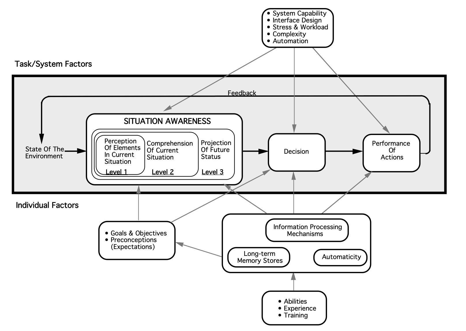
Expanding on their definition above, Hunter and colleagues (2019) offer several theoretical frameworks by which situational awareness can be understood, of which they argue that Mica Endsley’s three-level framework of situational awareness (comprised of identification, interpretation, and prediction) is most applicable to paramedicine. Because situational awareness is a form of cognitive processing, it is unsurprisingly difficult to quantify and study. Hunter and colleagues have attempted to put forth additional studies on the topic of situational awareness, using Endsley’s framework as a guide. For example, they conducted a pilot study (2021) which considered the situational awareness of paramedic students during a simulation.  Situational awareness was low and a common theme identified was the lack of an organized approach to the situation. Inability to achieve “prediction” level situational awareness was limited also by stress and failure of recognition/perception.

Image from Endsley, M. R. (1995). Toward a theory of situation awareness in dynamic systems. Human factors37(1), 32-64.

A small number of validated techniques have been employed for measuring situational awareness in healthcare. For example, Endsley developed the Situation Awareness Global Assessment Technique (SAGAT), which has been employed to evaluate trauma skills among medical trainees (Hogan 2006) and recognition of patient deterioration among student nurses (Kinsman 2009, Cooper 2019). This test requires halting a simulated scenario to assess participant insight into situational awareness, utilizing standardized reflective questions. An alternative metric, the Situation Present Assessment Method (SPAM), has also been employed (Williams 2013). Additionally, some interest has been expressed in examining eye tracking as a means of measuring situational awareness, though the efficacy of this, as well as its utility in clinical education, remains to be seen (Williams 2013). Little of this research has touched the prehospital arena.

Within the prehospital setting, Hunter’s group has recently started to examine educational strategies to modify situational awareness, with a statistically significant increase in situational awareness with an educational intervention in a quasi-experimental before-after study in 2022. The educational tool employed by Hunter and colleagues focused on elements of Endsley’s situational awareness model and some adapted principles from aviation, Crew Resource Management, designed to improve situational awareness.

Though the study is small, there is something to be said for acknowledging the potential value of education on situational awareness in paramedic training. The limited research on this topic is illustrative of how difficult it is to quantify and study. In the absence of clear, compelling evidence, an important question arises as to how medical directors can identify the need for targeted teaching and feedback of nontechnical skills like this, as they will not necessarily be reflected on chart review, and whether there is even value in broadly incorporating education on this skill.

CONCLUSION

Situation awareness education and training for prehospital clinicians and scenarios needs additional investigation. As research continues, we may discover evidence for interventions to improve situational awareness. Although there are time and financial costs to education and training, it important to remember that situational awareness itself is critical to prehospital patient care.

REFERENCES

  • Cooper S, Kinsman L, Buykx P, McConnell-Henry T, Endacott R, Scholes J. Managing the deteriorating patient in a simulated environment: nursing students' knowledge, skill and situation awareness. J Clin Nurs. 2010;19(15-16):2309-2318. doi:10.1111/j.1365-2702.2009.03164.x

  • Hogan MP, Pace DE, Hapgood J, Boone DC. Use of human patient simulation and the situation awareness global assessment technique in practical trauma skills assessment. J Trauma-Injury Infect Crit Care. 2006;61(5):1047–1052.

  • Hunter, J., Porter, M., & Williams, B. (2019). What Is Known About Situational Awareness in Paramedicine?: A Scoping Review. Journal of allied health48(1), e27–e34.

  • Hunter, J., Porter, M., Phillips, A., Evans-Brave, M., & Williams, B. (2021). Do paramedic students have situational awareness during high-fidelity simulation? A mixed-methods pilot study. International emergency nursing56, 100983. https://doi.org/10.1016/j.ienj.2021.100983

  • Hunter, J., Porter, M., Cody, P., & Williams, B. (2022). Can a targeted educational approach improve situational awareness in paramedicine during 911 emergency calls?. International emergency nursing63, 101174. https://doi.org/10.1016/j.ienj.2022.101174

  • Kinsman, L., Endacott, R., Cooper, S. J. R., Scholes, J., Buykx, P., & McConnell-Henry, T. E. (2009). Situational awareness of patient deterioration in a simulated environment.

  • Williams, B., Quested, A., & Cooper, S. (2013). Can eye-tracking technology improve situational awareness in paramedic clinical education?. Open access emergency medicine : OAEM5, 23–28. https://doi.org/10.2147/OAEM.S53021

About the Author:

Karlee is an emergency medicine-bound fourth-year medical student at Washington University in St. Louis. She has a strong clinical interest in medical education. Outside the hospital, she can be found running with her dog, baking, or getting lost in a book.

Editing by James Li, MD
Commercial Tourniquet Use in Pediatrics

Article by Veronica “Vee” Smith, MD

Case

It’s 11 o’clock in the morning on a sunny autumn day. Your radio alerts you about a mass casualty event and you are then dispatched to what turns out to be a school shooting. The estimated casualty count is over 20 with an unknown number of injured victims on the scene, their ages are estimated to range from 5-58 years of age. As your partner begins to drive light and sirens to the scene, several questions quickly race through your mind “Do I have enough tourniquets”, “Can I use a commercial tourniquet on a pediatric patient? And if so, is there an age cutoff?”. Before you know it you and your partner have arrived on the scene.

Introduction

Traumatic injuries are the leading cause of death among children and adolescents aged 1-17 years of age in the United States. Many of those deaths have been attributed to motor vehicle accidents, homicide, suicide and non-accidental trauma [1]. In 2020, penetrating trauma from firearm injuries became the leading cause of traumatic deaths in the pediatric population, surpassing deaths caused by motor vehicle accidents [1]. Traumatic brain injuries are the most common injury complex associated with death in children with traumatic injuries, followed by anoxia and hemorrhage [2]. Most deaths occur within the first 24 hours of the primary injury emphasizing the importance of interventions performed in the prehospital setting and the immediate hospital care in the emergency department [2]. Death due to hemorrhagic shock is more commonly associated with gunshot wounds with death occurring earlier in the first 24 hours in comparison to deaths from TBIs and anoxia [2]. As school shootings have continued to be on the rise in the US, there has been increased focus on hemorrhage control in the prehospital setting due to its significant morbidity and mortality. Data on tourniquet use in the pediatric population is currently sparse, much of the information that has been used to support the use of tourniquets in this population has been provided from research that has focused on the adult population or children injured in military war zones.

Literature Review

A study conducted at a combat support hospital in Iraq compared the outcomes of patients with hemorrhaging extremity injuries who had tourniquets applied in the prehospital setting vs tourniquet placement in the Emergency Department. Survival rates were higher in patients who received pre-hospital tourniquet placement [3]. The survival rate was also higher if the tourniquet was placed prior to the onset of shock, 90% of those patients survived their injuries in comparison to only 18% of those with tourniquets placed after the onset of shock [3]. Likewise, domestic studies involving tourniquet use in traumatic extremity injuries have shown similar results. A retrospective study at a level 1 trauma center in New Orleans examined commercial tourniquet use for penetrating extremity trauma over an 8-year period. Patients with a tourniquet were compared to those without. In comparison to the cohort without tourniquets, those who received tourniquets: required fewer blood transfusions, had higher systolic blood pressures (prehospital and on arrival to the ED), lower incidence of shock on arrival to the ED and shorter length of hospital stay [4]. In addition, the non-tourniquet cohort a higher incidence of fasciotomy and secondary amputation [4].

Many of the current commercial tourniquet styles have been used and tested in the military, with most of the use occurring in patients over the age of 18. For a tourniquet to function properly, it must be tightened to apply adequate circumferential pressure to halt blood flow distally. Some commercial tourniquets have a rigid mechanical system (i.e. windlass or ratchet) used to tighten the tourniquet onto an extremity; this can cause difficulty in fitting the tourniquet onto extremity circumferences that are smaller than the mechanism. Pediatric limb circumferences are smaller than that of an adult, which raises the question: Can enough circumferential pressure be applied to stop bleeding in small children? 

A 2019 study examined the use of the Combat Application Tourniquet (CAT) in sixty children (aged 6-16 years) with a goal in determining whether the CAT would be able to apply enough pressure to occlude extremity arterial blood flow. The CAT was applied to an upper arm and thigh while peripheral pulses were monitored by Doppler. The number of windlass turns to occlude arterial blood flow were also recorded (maximum allowed was 3 turns (1080 degrees)). The CAT was found to have a 100% success rate in occluding arterial blood flow in the upper extremities tested [5]. In comparison the success rate in the lower extremity was 93% [5]. There was an increase in the number of windlass turns required to occlude blood flow as patients age and BMI increased [5]. Though this study provided demonstration that CATs could successfully be used in pediatric human subjects, the question of whether there is an age limit for successful use in commercial tourniquets remains. A 2020 study also examined the application of CATs to pediatric upper and lower extremities, this time examining children aged 2-7 years [6]. Although the sample size was smaller (24 extremities tested), the study found a 100% success rate in occluding arterial blood flow in both upper and lower extremities [6]. The smallest arm circumference and leg circumference included in the study were 13cm and 24.5cm respectively [6]. It may be possible that commercial tourniquets may be successful in children under the age of 2, but currently there has not been human subject research performed on this age group to provide that evidence.

Conclusion

High profile mass casualty events such as the Boston Marathon bombing have increased public interest in tourniquet use. With educational campaigns such as Stop the Bleed, commercial tourniquet use in the prehospital setting will likely increase and provide more information on tourniquet use and outcomes in the pediatric population. Although commercial tourniquets such as the CAT were designed for adult use particularly that of servicemen and servicewomen, at least two studies have shown that they can be successfully applied to pediatric patients aged 2-16 years [5,6]. The patients in these studies were volunteers with no extremity trauma, prehospital research could establish more efficacy of commercial tourniquet use in pediatric patients experiencing extremity trauma with exsanguinating hemorrhage. Despite limited research data the Pediatric Trauma Society supports (PTS) the usage of tourniquets in the prehospital setting during the resuscitation of pediatric patients with severe extremity trauma and hemorrhage [7]. In addition, PTS also supports the Stop The Bleed campaign, placing emphasis on prehospital hemorrhage control by use of direct pressure, wound packing with hemostatic gauze and tourniquet use.

References

  1. Centers for Disease Control and Prevention. Web-based Injury Statistics Query and Reporting System. https://www.cdc.gov/injury/wisqars/index.html

  2. Theodorou CM, Galganski LA, Jurkovich GJ, Farmer DL, Hirose S, Stephenson JT, Trappey AF. Causes of early mortality in pediatric trauma patients. J Trauma Acute Care Surg. 2021 Mar 1;90(3):574-581. doi: 10.1097/TA.0000000000003045. PMID: 33492107; PMCID: PMC8008945.

  3. Kragh JF Jr, Littrel ML, Jones JA, Walters TJ, Baer DG, Wade CE, Holcomb JB. Battle casualty survival with emergency tourniquet use to stop limb bleeding. J Emerg Med. 2011 Dec;41(6):590-7. doi: 10.1016/j.jemermed.2009.07.022. Epub 2009 Aug 31. PMID: 19717268.

  4. Smith AA, Ochoa JE, Wong S, Beatty S, Elder J, Guidry C, McGrew P, McGinness C, Duchesne J, Schroll R. Prehospital tourniquet use in penetrating extremity trauma: Decreased blood transfusions and limb complications. J Trauma Acute Care Surg. 2019 Jan;86(1):43-51. doi: 10.1097/TA.0000000000002095. PMID: 30358768.

  5. Harcke HT, Lawrence LL, Gripp EW, Kecskemethy HH, Kruse RW, Murphy SG. Adult Tourniquet for Use in School-Age Emergencies. Pediatrics. 2019 Jun;143(6):e20183447. doi: 10.1542/peds.2018-3447. Epub 2019 May 7. PMID: 31064797.

  6. Kelly JR, Levy MJ, Reyes J, Anders J. Effectiveness of the combat application tourniquet for arterial occlusion in young children. J Trauma Acute Care Surg. 2020 May;88(5):644-647. doi: 10.1097/TA.0000000000002594. PMID: 31977996.

  7. Cunningham A, Auerbach M, Cicero M, Jafri M. Tourniquet usage in prehospital care and resuscitation of pediatric trauma patients-Pediatric Trauma Society position statement. J Trauma Acute Care Surg. 2018 Oct;85(4):665-667. doi: 10.1097/TA.0000000000001839. PMID: 29462083.

Author Bio: Dr. Veronica "Vee" Smith is a second year Pediatric Emergency Medicine Fellow at St. Louis Children's Hospital/Washington University in St. Louis. She enjoys cooking, writing, playing & watching basketball when she is not working. 

Website Editing and Layout by James Li, MD

Calcium in Out-of-Hospital Cardiac Arrest

Article by Erin Lincoln, MD

Case Scenario:

You are dispatched to a 68-year-old male in cardiac arrest.  His family has been performing bystander, and report that he suddenly collapsed just a few minutes ago. CPR is taken over by responding crews, and he is placed on a cardiac monitor/defibrillator. He is found to be in ventricular fibrillation (VF).  After several cycles of defibrillation, epinephrine, and amiodarone, the patient remains in cardiac arrest.  The medic on scene calls on-line medical control to ask for advice, and specifically asks if calcium can be given, as she has “seen it work before” to get pulses back as a “last ditch effort.”

Background:

Calcium chloride or gluconate was originally utilized in cardiac arrest resuscitation in the 1950’s after a single study was published in 1951 (Kay & Blalock, 1951). However, evidence emerged in the 1980’s demonstrating that calcium chloride had no effect on return of spontaneous circulation (ROSC) rates, and in fact could be detrimental (Landry, Foran, & Koyfman, 2014). Current AHA guidelines do not recommend routine use of calcium in cardiac arrest (Panchal, et al., 2020). Calcium acts as a vasopressor and inotropic agent (Lindqwister, et al., 2020) thus lending itself to a potential drug for cardiac arrest. Calcium is also frequently used in the treatment of hyperkalemia, calcium channel blocker overdose, hypermagnesemia, and hypocalcemia and may be more likely to be used when one of these diagnoses is the suspected cause of cardiac arrest. Additionally, low ionized calcium levels have been correlated with increasing mortality in sepsis and other critical illnesses in adults and children (Bora, Ramazan, Oznur, Emre, & Basar, 2021), (Sanchez, et al., 1989). Thus, calcium may be a drug considered in these and similar etiologies as appropriate due to known association with low calcium and mortality.

However, as calcium is still used for both presumed benefit in special cases, as well as a “last ditch effort” new literature continues to be published addressing the use of calcium in cardiac arrest, including a significant recent RCT. 

What does the literature say?

Since the 1980’s, literature has been routinely published regarding the use of calcium in cardiac arrest.  Several recent papers have come out, including a double blind, randomized controlled trial of calcium in cardiac arrest, and these papers are nicely summarized in a 2014 Annals of Emergency Medicine article by Landry, Foran, and Koyfman. The take home message: “Irrespective of presenting rhythm, in patients with cardiac arrest, there is no conclusive evidence that administration of calcium during cardiopulmonary resuscitation (CPR) improves survival.” This paper also notes that many of the studies were retrospective, had varied results, and that to truly answer this question, more randomized trials were needed.

Since the publication of this review, several new studies have been published including several randomized controlled trials. 

General Adult Medical Cardiac Arrest

The Calcium for Out-of-hospital Cardiac Arrest (COCA) Trial

This trial was conducted in Denmark and demonstrated through a double blind, randomized, placebo-controlled trial that calcium likely causes harm and was stopped early at a planned interim analysis due to concern for harm in the calcium arm. 397 patients were randomized- 193 received calcium, 198 received saline; of these 37 (19%) of the calcium group achieved ROSC, 53 (27%) of patients in the saline group received ROSC, risk ratio 0.72 [95% CI 0.27-1.18]. This CI does include 1; and further and further analysis of the data showed that the likelihood that calcium has a beneficial effect (e.g. risk ratio >1) was 4% for ROSC, 6% for 30 day survival, and 4% for survival with a favorable neurologic outcome at 30 days (Vallentin, et al., 2021). Visual abstract from this study:

Calcium use during cardiac arrest: A systematic review

This systematic review published in 2022 reviewed prior literature and identified that a meta-analysis was not possible due to only three available RCTs, and only one of those was considered low risk of bias. The overall conclusion was that as less than 1% of cardiac arrest etiologies fall into a group that would potentially benefit from calcium, that routine use should be avoided (Padrao, et. al., 2022).

Association between calcium administration and outcomes during adult cardiopulmonary resuscitation at the emergency department

A small retrospective study from Thailand showed again that there is no benefit to calcium given during Emergency Department resuscitation. This study also reported decreased chances of ROSC in hypocalcemic cardiac arrest patients who received calcium, and potential harm with calcium administration during traumatic arrest. This study did not account for time of administration, so survival bias may have influenced results (Wongtanasarasin, et al., 2022).

Special Cause Cardiac Arrest: Hyperkalemia and Calcium Channel Blocker Overdose

Calcium is regarded as a mainstay treatment for patients with hyperkalemia and EKG changes, and is one of two indications listed in the European Resuscitation Council guidelines on Cardiac Arrest(Lott, et al., 2021), and AHA guidelines also maintain this use (Panchal, et al., 2020). Other AHA/European guideline indications include calcium channel blocker overdose, hypermagnesemia, or hypocalcemia

The effects of calcium and sodium bicarbonate on severe hyperkalaemia during cardiopulmonary resuscitation: A retrospective cohort study of adult in-hospital cardiac arrest

This study out of Taiwan looked at known hyperkalemic cardiac arrests who received both Sodium Bicarbonate (bicarb) and calcium during in-hospital cardiac arrest. The study included 109 hyperkalemic cardiac arrest patients from 2006 through 2012. Of these, 40 (36.7%) patients achieved sustained ROSC, but only four (3.7%) patients survived to hospital discharge. Patients were grouped based on if they received: a) neither calcium nor bicarb; b) bicarb only; c) calcium only; d) calcium AND bicarb. After analysis, bicarb was positively associated with sustained ROSC when serum potassium level was <7.9 mEq/L (odds ratio [OR]: 10.51; 95% confidence interval [CI]: 1.50−112.89; p=0.03); administration of both calcium and bicarb was positively associated with sustained ROSC when serum potassium level was <9.4 mEq/L (OR: 51.11; 95% CI: 3.12−1639.16; p=0.01). This study was limited by small sample size and does NOT look at the effect of calcium alone on known hyperkalemic arrest due to the small available numbers. As no study patients survived with favorable neurologic outcome, no outcome data is available (Wang, et al., 2016).

Secondary analysis of COCA Trial

This assessed the effect of calcium in patients with PEA/ECG characteristics that could potentially have been associated with hyperkalemia and ischemia. 104 patients from the trial were found to have PEA as their last known rhythm prior to receiving the trial drug (calcium or placebo). The rhythm obtained by the defibrillator pads was analyzed for signs of hyperkalemia including loss of P waves, wide QRS complexes and large T wave amplitude.  Of these patients, 45 received calcium, 59 received placebo; 9 patients (20%) in the calcium group achieved ROSC as compared with 23 patients (39%) in the placebo group (risk ratio 0.51, 95% CI 0.26-1). While this again does not demonstrate statistical significance to imply harm, it certainly suggests that calcium may not be as helpful as previously expected when findings of hyperkalemia are present on EKG. (Vallentin, Povlsen, Granfeldt, Terkelsen, & Andersen, 2022). The forest plot for ROSC summarizes some of this data:

Pediatrics

AHA PALS guidelines recommend against routine administration of IV calcium during pediatric cardiac arrest (Topjian, et al., 2020), but IV Calcium is still used routinely in some cases in the critical care setting, such as congenital heart disease. While literature discussing prehospital administration of calcium in pediatric cardiac arrest is sparse, in-hospital literature suggests not only that calcium doesn’t demonstrate benefit, but also is associated with worse outcomes. 

Get With The Guidelines-Resuscitation (GWTG-R) Registry

This study demonstrated worse outcomes in pediatric patients with heart disease who received calcium in cardiac arrest- survival to hospital discharge was 39% in calcium recipients vs. 46% in non-recipients (P=0.02)  (Dhillon, Kleinman, Staffa, Teele, & Thiagarajan, 2022).

An editorial responding to this study does suggest that while the above paper is effective in many ways, it fails to fully account for the fact that pediatric patients who receive calcium are most likely sicker at baseline than those who do not receive calcium, and are more likely to have worse outcomes irrespective of calcium administration (Savorgnan & Acosta, 2022).

ICE-RESUScitation Project Secondary Analysis

This in-hospital analysis initially included 1,100 patients and was designed to evaluate a CPR quality improvement bundle vs usual care, researchers also found worse outcomes in patients who received calcium, INCLUDING some subgroups that had previously been hypothesized to have potential to benefit from receiving calcium during CPR including sepsis or renal insufficiency. This study attempted to mitigate bias in pre-arrest characteristics between groups by data weighting and included a PRISM (Pediatric Risk of Mortality) score when available from 2-6 hours prior to the arrest. While this study can still only prove correlation, the weighting of variables reduces bias and further supports the association of the calcium alone and the decline in outcomes (Cashen, et al., 2023). 

Take Home Points:

Calcium (chloride or gluconate) is not recommended in routine or unknown etiology cardiac arrest for both adult and pediatric patients, and this is consistent with both the AHA and European resuscitation guidelines. This continues to be supported by new literature. Special causes of cardiac arrest to include hyperkalemia and calcium channel blocker overdose, have limited data regarding efficacy but do still carry the recommendation for calcium administration.

References:

1.     Bora, C., Ramazan, K., Oznur, A. N., Emre, A. S., & Basar, C. (2021). Ionized calcium level predicts in-hospital mortality of severe sepsis patients: A retrospective cross-sectional study. Journal of Acute Disease, 10(6), 247-251.

2.     Cashen, K., Sutton, R., Reeder, R., Ahmend, T., Bell, M., Berg, R., . . . Meert, K. (2023). Calcium use during paediatric in-hospital cardiac arrest is associated with worse outcomes. In Press.

3.     Dhillon, G. S., Kleinman, M. E., Staffa, S., Teele, S., & Thiagarajan, R. (2022, November). Calcium administration during Cardiopulmonary Resuscitation for In Hospital Cardiac Arrest in Children With Heart Disease Is Associated With Worse Survival-- A Report From the American Heart Association's Get With The Guidelines- Resuscitation (GWTG-R) Re. Pediatric Critical Care Medicine, 23(11), 860-871.

4.     Kay, J., & Blalock, A. (1951). The use of calcium chloride in the treatment of cardiac arrest in patients. Surg Gynecol Obstet, 93, 97-102.

5.     Landry, A., Foran, M., & Kyofman, A. (2014, August). Does Calcium Administration During Cardiopulmonary Resuscitation Improve Survival for Patients in Cardiac Arrest? Annals of Emergency Medicine, 64(2), 187-189.

6.     Lindqwister, A. L., Lampe, J. W., Gould, J. R., Kaufman, C., Moodie, K. L., & Paradis, N. A. (2020, Sep 4). Intravenous calcium as a pressor in a swine model of hypoxic pseudo-pulseless electrical mechanical activity-a preliminary repo. Intensive Care Med Exp, 8(1), 50.

7.     Lott, C., Truhlar, A., Alfonzo, A., Barelli, A., Gonzales-Salvado, V., Hinkelbein, J., . . . Soar, J. (2021). European Resuscitation Council Guidelines 2021: Cardiac arrest in special circumstances. Resuscitation, 161, 152-219.

8.     Padrao, E., Bustos, B., Mahesh, A., Castro, M., Randhawa, R., Dipollina, C., . . . Besen, B. (2022). Calcium use during cardiac arrest: A systematic review. Resuscitation Plus, 12, 1-9.

9.     Panchal, A., Bartos, J., Cabanas, J., Donnino, M., Drennan, I., Hirsch, K., . . . Berg, K. (2020). Part 3: Adult Basic and Advanced Life Support: 2020 American Heart Association Guidelines for Cardiopulmonary Resuscitation and Emergency Cardiovascular Care. Circulation, 142, S366-S468.

10.  Sanchez, G., Venkataraman, P., Pryor, R., Parker, M., Fry, H., & Blick, K. (1989). Journal of Pediatrics, 114(6), 952.

11.  Savorgnan, F. M., & Acosta, S. P. (2022). Calcium Chloride Is Given to Sicker Patients During Cardiopulmonary Resuscitation Events. Pediatric Critical Care Medicine, 23(11), 939-940.

12.  Topjian, A., Raymond, T., Atkins, D., CHan, M., Duff, J., Jr., B. J., . . . Schexnayder, S. (2020). Part 4: Pediatric Basic and Advanced Life Support: 2020 American Heart Association Guidelines for Cardiopulmonary Resuscitation and Emergency Cardiovascular Care. Circulation, 142, S469-S523.

13.  Vallentin, M., Granfeldt, A., Meilandt, C., Povlsen, A., Sindberg, B., & Andersen, L. (2022). Effect of Calcium vs. placebo on long term outcomes in patients with out of hospital cardiac arrest. Resuscitation, 179, 21-24.

14.  Vallentin, M., Granfelt, A., C Meilandt, P. A., Sindberg, B., Holmberg, M., Iversen, B., . . . Mortensen, L. (2021, Dec 14). Effect of Intravenous or Intraosseous Calcium vs. Saline on Return of Spontaneous Circulation in Adults With Out-of-Hospital Cardiac Arrest. JAMA, 326(22), 2268-2276.

15.  Vallentin, M., Povlsen, A., Granfeldt, A., Terkelsen, C., & Andersen, L. (2022). Effect of calcium in patients with pulseless electrical activity and electrocardiographic characteristics potentially associated with hyperkalemia and ischemia- sub-study of the Calcium for Out-of-hospital Cardiac Arrest (COCA) trial. Resuscitation, 181, 150-157.

16.  Wang, C.-H., Huang, C.-H., Chang, W.-T., Tsai, M.-S., Yu, P.-H., Wu, Y.-W., . . . Chen, W.-J. (2016). The effects of calcium and sodium bicarbonate on severe hyperkalemia during cardiopulmonary resuscitation: A retrospective cohort study of adult in-hospital cardiac arrest. Resuscitation, 98, 105-111.

17.  Wongtanasarasin, W., Ungrungseesopon, N., Namsongwong, N., Chotipongkul, P., Visavakul, O., Banping, N., . . . Phinyo, P. (2022). Association between calcium administration and outcomes during adult cardiopulmonary resuscitation at the emergency department. Turkish Journal of Emergency Medicine, 22, 67-74.

Editing by Brian Miller, MD

Website Editing and Layout by EMS MEd Editor James Li, MD

Why should quality improvement concepts be integrated into EMS education?

By Tiffany Pleasent, MD

Case Review

A 64-year-old female calls 911 for severe shortness of breath and chest pain. An ALS crew arrives on scene to find a woman who is tachypneic, hypoxic, tachycardic, and hypotensive, with diffuse rales and rhonchi throughout her lung fields. She becomes altered and severely hypoxic. The attending paramedics begin bag-valve-mask (BVM) ventilation, correct her hypoxia and hypotension, and subsequently intubate her on scene with ketamine and rocuronium. This is followed by appropriate confirmation with waveform capnography and all other appropriate adjuncts. Upon arrival to the ambulance, the EMT notices that the patient’s heart rate is 22 bpm. Shortly after they discover that she is pulseless and apneic. They begin CPR en route to the emergency department where the patient’s rhythm deteriorates into asystole. Further resuscitation is futile, and she dies.

The ER physician in the destination emergency room determines that the endotracheal tube was misplaced in the esophagus. This EMS system promotes a “just culture” that influences the crew to self-report their difficult cases to medical direction for evaluation and feedback.

The EMS medical director reviews the case and its associated monitor file which includes the timing and quality of all vital signs, including chest compression depth and rate. The monitor file shows an initial 4-phase waveform capnography of 25 mmHg for 8 breaths immediately following intubation. It further shows a subsequent loss of end-tidal waveform. This is followed by an abrupt bradycardia occurring simultaneously with rapidly dropping SpO2. The period between loss of initial waveform capnography and initiation of chest compressions was over 7 minutes. The endotracheal tube was never evaluated, removed, nor exchanged in the field.

The EMS medical director wonders if this is a “one-off” case or an indicator of more systemic issues. She decides to evaluate other similar cases and discovers that 5% of all intubations in her system lose waveform capnography and progress to cardiac arrest. She decides to implement system-level changes to manage these cases.

Keeping patient safety as our priority

Application of a systematic approach to quality improvement allows us to do the greatest good for the greatest number of people on a consistent basis. The case above describes a system with multiple unrecognized failed airways (UFAs), some likely resulting in patient harm. Fortunately, the medical director and the service leadership have a committed desire to implement change.

Several considerations arise:

  • How does the medical director develop ideas and choose what she wants to change in order to decrease UFAs?

  • How should she know what to monitor?

  • Should she use qualitative or quantitative data to drive system change?

  • How frequently should she be analyzing the data?

  • How does she appropriately analyze the data in order to obtain the right information? When should she abort a change idea?

  • How does she know that an improvement truly is an improvement?

Both EMS physician and EMS clinician involvement are required in this process, and the system is best served when both understand key quality management principles.

What are some key quality management principles that are applicable to EMS?

The Model for Improvement

The Institute of Medicine’s (now known as the National Academy of Medicine) Model for Improvement was initially adopted from the Associates in Process Improvement as a commonly used framework for healthcare providers to improve their systems [5,7,8]. It requires clinicians a) to create a measurable aim, b) to establish quantifiable measures that can be used to evaluate improvement over time, and c) to develop change ideas to implement. This is followed by small-scale tests of change using PDSA (plan-do-study-act) cycles that are scalable if the cycle is successful.

The difference between quality improvement and quality assurance

These two terms are frequently used interchangeably, albeit incorrectly. Quality assurance describes the detection of an error after the error has occurred [8]. Quality improvement describes evaluation of the system to ensure it appropriately produces the desired results and prioritizes error avoidance. Quality improvement is proactive whereas quality assurance is reactive, and both are important.

Case Review Application

The medical director decides to apply small tests of change to improve her system. She creates an aim statement, establishes quantifiable outcome measures, and develops several change ideas. Following implementation of each change idea, she says “show me the data!”

PDSA Cycles

#1 – An e-mail memo is sent to the EMS system reminding clinicians to establish and monitor continuous waveform capnography of all airway devices while en-route to the hospital and during patient movement and handoffs. After one month, there is no change in the system’s UFA outcome measures.

Model for Improvement: PDSA Cycle (Institute for Healthcare Improvement)

#2 – A podcast is developed explaining waveform capnography and the importance of continuous monitoring of advanced airways. Additionally, a protocol revision is made outlining advanced airway confirmation and management requirements.  Unfortunately, there is minimal change in the UFA outcome measures.

#3 – To identify further change theories, the medical director facilitates a case review with the EMS clinicians involved in each of the UFA cases that resulted in cardiac arrest. Nearly all the crews recall difficulty seeing the cardiac monitor in their ambulances due to the monitor mount being poorly positioned. Furthermore, they report their monitor never alarmed that capnography was lost. Clinicians provide recommendations for better placement of the mount in the ambulance and the change is made to the entire fleet. Crews report in the following weeks a significant improvement in their ability to monitor their patient.

#4 – The cardiac monitor settings are reviewed and nearly all alarms have been turned off by crews because of frequent inappropriate alarms resulting in alarm fatigue. The team involves EMS clinicians to identify the most important alarms to maintain, including the capnography apnea alarm, and an admin password is established to prevent these settings from being changed. These monitor changes are trialed by a group of crews for a few shifts and additional small refinements are made based on their feedback prior to deployment on all the system’s monitors.

There is subsequently a dramatic and sustained system-wide improvement and no further UFAs occur over the next year.

The EMS physician and the EMS clinician are key players in quality improvement

While this case example presents an example of a single quality improvement project, there are innumerable opportunities for improvement among the diversity of EMS systems worldwide. The process of improving healthcare delivery is most efficient when there is valuable input and advocacy from both EMS physicians and EMS clinicians. Understanding of quality management fundamentals begins with initial education.

In the above case, imagine if the EMS clinicians’ initial education taught the importance of and the process by which system improvement is achieved. Several considerations arise:

  • Would they have been proactive in telling her their difficulty in seeing the monitor?

  • Would someone have told her how the unnecessary alarms caused them to turn all alarms off on all of their monitors?

  • Would there be increased compliance and accuracy of documentation within electronic health records that are used for quality improvement & research?

  • Could this swing the pendulum from seeking buy-in to achieving advocacy among key stakeholders?

The most efficient shift from quality assurance to quality improvement requires input from the “boots on the ground” who offer critical perspective and input regarding their needs and challenges in delivering healthcare within the vision and oversight of the EMS physician. If the fundamentals of quality management were provided in initial education, the impact would be profound. 

Should we integrate quality improvement concepts within EMS education?

Yes! The Future of EMS begins NOW!

As new EMS medical directors and EMS clinicians enter the workforce, it is incumbent upon their educators - including educators for the medical directors - to provide the skills that are required to be successful in this changing healthcare environment. Educators should be trained in key quality improvement concepts, which include tools that may be infrequently taught in traditional healthcare-related settings. Examples include process control charts and other data analysis skills. We must maintain a systematic, data-driven, and evidence-based approach to system quality management and performance improvement [1,2,3,5]. Within our evolving healthcare climate, this effort requires creativity and innovation.

The EMS Agenda 2050 describes six guiding principles to its “people-centered vision for the future of emergency medical services.” These include: inherently safe and effective, integrated and seamless, reliable and prepared, socially equitable, sustainable and efficient, and adaptable and innovative [3]. As outlined in the NAEMSP position statement “Defining Quality in EMS”, we must provide quality and value-based assessments of care in an evidence-based manner which prioritizes patient outcomes [2]. We must be uniform in our terminology and presentation of data, and we must utilize evidence-based methods through open and transparent communication. In order to meet these goals, we must ensure these core principles are provided within our foundation – our EMS education.

Edited by: Jeffrey Jarvis, MD, EMT-P; Ray Fowler, MD, FACEP, FAEMS; Brian Miller, MD; and Al Lulla, MD

References:

1. Crowe RP, Jarvis JL. Quality Improvement and Research. Prehospital Emergency Medicine Secrets. Elsevier; 2022:18-21.

2. Defining Quality in EMS. Prehosp Emerg Care. 2018; 22: 782-783.

3. EMS Agenda 2050 Technical Expert Panel. EMS Agenda 2050: A People-Centered Vision for the Future of Emergency Medical Services. Washington, DC: National Highway Traffic Safety Administration, 2019.

4. Institute for Healthcare Improvement. Science of Improvement: How to Improve. 2023. Available at: https://www.ihi.org/resources/Pages/HowtoImprove/ScienceofImprovement
HowtoImprove.aspx. Accessed January 30, 2023.

5. Institute of Medicine Committee on Quality of Health Care in America. Crossing the Quality Chasm: A New Health System for the 21st Century. Washington, D.C.: National Academies Press, 2001.

6. Lincoln EW, Reed-Schrader E, Jarvis JL. EMS, Quality Improvement Programs. Stat Pearls. Published online January 2022. https://www.statpearls.com/point-of-care/31814

7. Langley GJ, Nolan TW, Provost LP, Nolan KM, Norman CL. The Improvement Guide: A Practical Approach to Enhancing Organizational Performance. San Francisco, CA: Wiley 1996.

8. Crowe RP. The evolution of quality concepts and methods. Emergency Medical Services: Clinical Practice and Systems Oversight. 2021; 112: 424-431.

Website Editing and Layout by EMS MEd Editor James Li, MD

Palliative and Hospice Care in the Prehospital Setting

By Nicholas Maxwell, MD

Case:

You are bringing an elderly male with a DNR back to a living facility from a hospital. Approximately 10 miles away from the hospital, the patient suddenly decompensates. His pulse ox drops from 94% to 87% and his heart rate increased from about 110 beats per minute to approximately 180. His mental status is described as nodding off. The EMS crew reports that the hospital was unclear if he was on hospice or if he simply had a DNR. They were also reportedly unclear about what level of care the facility he was being returned to had.

Literature Review:

Education in EMS often focuses on how to treat acutely ill and injured patients with the goal of saving lives and preventing serious, long-lasting negative outcomes. Since their training is so focused on acute, life-threatening illness, they are often called to help those who are acutely ill/injured and/or dying. However, at most EMS education programs, very little, if any, time is spent teaching how to care for patients who are not interested in resuscitation and life-saving interventions, such as hospice patients.

Hospice patients in particular flip the oft assumed goal of resuscitation and life-saving interventions that EMS is so adeptly trained to execute. In these patients, resuscitation is essentially contraindicated, and the primary goal is often to prevent/relieve suffering, even if it means death comes quicker than if you were to provide aggressive interventions. This can often be uncomfortable for EMS crews as it seems antithetical to what they are trained to do, especially when they see something that they have been trained to treat. After all, by nature of being on hospice, these patients are dying from something, and it can be hard to fight that interventionist approach in a profession that is seen by many as an illness-centered field. However, in actuality, EMS isn’t an illness-centered field. It is a patient-centered field. This can be most apparent at the extreme end of the spectrum where hospice patients reside.

While research suggests that EMS providers find treating these patients to be meaningful and important, it can often be extremely uncomfortable for them, especially considering medico-legal and ethical considerations. These can include feeling compelled to attempt resuscitation despite the team feeling it is futile or not consistent with the patient’s wishes, families demanding CPR despite the presence of a DNR, incompletely filled out DNR forms, and more.

Missouri Out of Hospital DNR Form is shown here. There are also POLST (Physician Orders for Life Sustaining Treatment) forms that describe patient’s treatment preferences in additional detail. Other names for POLST include: MOLST, MOST, TPOPP, and more. EMS professionals should be familiar with their state’s forms.

It is easy to assume that this is a rare occurrence. However, that is not the case. One study reported that 66% of respondents had more than 10 encounters with hospice patients. In fact, only 3.8% of respondents had not encountered hospice patients in their professional experience. The calls can be for a range of reasons such as falls, insufficient symptom management, and family members/nursing facility staff not knowing patient’s status. While it can be easy to assume that since the patient is on hospice, there is no role for EMS intervention, that is not the case. Sometimes they may benefit from transport to an emergency department if it is consistent with their care plan/wishes. One common indication for this might be failure to have a tolerable level of symptom management (for example, pain after a fall). Instead of the oft presumed goal of saving life and limb, these patients need to have special attention paid to determining their wishes. This can require communication skills that are nuanced, not intuitive, and not nearly as intensely trained in comparison to other skills like EKG interpretation and intubation.

Unfortunately, EMS providers often have less than the desired amount of training for these kinds of encounters. One study found that 76% of respondents said they never received formal training on hospice care. 10% even felt that DNR/DNI means no treatment can be provided to the patient. In the setting of this sub-optimal training, it can be hard to know what to do in these stressful and difficult to navigate situations. One study found that 60.8% of respondents reported they had been pressured by families to provide more aggressive care than the patient desired, 28.8% had performed CPR on a hospice patient, and 17.9% had intubated a hospice patient. However, it is worth noting that research suggests that EMS providers find caring for this patient group to be important and meaningful and that they would appreciate additional training on how to serve this group of patients. This additional training may relieve some of the discomfort associated with treating this patient population, improve the care provided, increase patient satisfaction, and in many cases avoid unnecessary transfers to the hospital

There are even some interesting uses of EMS to serve palliative care and hospice patients that have been explored. One particularly interesting example of this is use of EMS for a terminal extubation. The patient was dying while intubated in the ICU but was awake enough to engage in discussions with providers through writing about her wishes. She was adamant that she wanted to be at home even though she was dying and would likely die much faster if she went home rather than staying at the hospital. She was interested in a terminal extubation but they weren’t sure she would survive long enough after extubation at the hospital to make it home. After days of meeting with palliative care, psychiatry, and even the EMS physician, they had her transported via EMS to her home where the EMS physician performed the terminal extubation and care was handed off to the home hospice team.

In sum, while encountering hospice patients is not uncommon in EMS, it is commonly uncomfortable for providers who want to do right by these patients, there is desire for and opportunity for further education in handling patients involved in hospice and end of life care, and this additional training would go a long way in helping EMS be the elite patient-oriented providers that they aim to be.  

References:

1.     Juhrmann ML, Anderson NE, Boughey M, McConnell DS, Bailey P, Parker LE, Noble A, Hultink AH, Butow PN, Clayton JM. Palliative paramedicine: Comparing clinical practice through guideline quality appraisal and qualitative content analysis. Palliat Med. 2022 Sep;36(8):1228-1241. doi: 10.1177/02692163221110419. Epub 2022 Aug 8. PMID: 35941755. 

2.     Surakka LK, Hökkä M, Törrönen K, Mäntyselkä P, Lehto JT. Paramedics' experiences and educational needs when participating end-of-life care at home: A mixed method study. Palliat Med. 2022 Sep;36(8):1217-1227. doi: 10.1177/02692163221105593. Epub 2022 Aug 3. PMID: 35922966. 

3.     Bruun H, Milling L, Mikkelsen S, Huniche L. Ethical challenges experienced by prehospital emergency personnel: a practice-based model of analysis. BMC Med Ethics. 2022 Aug 12;23(1):80. doi: 10.1186/s12910-022-00821-9. Erratum in: BMC Med Ethics. 2022 Nov 26;23(1):120. PMID: 35962434; PMCID: PMC9373324. 

4.     Wenger A, Potilechio M, Redinger K, Billian J, Aguilar J, Mastenbrook J. Care for a Dying Patient: EMS Perspectives on Caring for Hospice Patients. J Pain Symptom Manage. 2022 Aug;64(2):e71-e76. doi: 10.1016/j.jpainsymman.2022.04.175. Epub 2022 Apr 28. PMID: 35490992. 

5.     Breyre A, Taigman M, Salvucci A, Sporer K. Effect of a Mobile Integrated Hospice Healthcare Program on Emergency Medical Services Transport to the Emergency Department. Prehosp Emerg Care. 2022 May-Jun;26(3):364-369. doi: 10.1080/10903127.2021.1900474. Epub 2021 Mar 30. PMID: 33689535. 

6.     Juhrmann ML, Vandersman P, Butow PN, Clayton JM. Paramedics delivering palliative and end-of-life care in community-based settings: A systematic integrative review with thematic synthesis. Palliat Med. 2022 Mar;36(3):405-421. doi: 10.1177/02692163211059342. Epub 2021 Dec 1. PMID: 34852696; PMCID: PMC8972966. 

7.     Waldrop DP, Waldrop MR, McGinley JM, Crowley CR, Clemency B. Prehospital Providers' Perspectives about Online Medical Direction in Emergency End-of-Life Decision-Making. Prehosp Emerg Care. 2022 Mar-Apr;26(2):223-232. doi: 10.1080/10903127.2020.1863532. Epub 2021 Feb 2. PMID: 33320725. 

8.     Breyre AM, Bains G, Moore J, Siegel L, Sporer KA. Hospice and Comfort Care Patient Utilization of Emergency Medical Services. J Palliat Med. 2022 Feb;25(2):259-264. doi: 10.1089/jpm.2021.0143. Epub 2021 Aug 31. PMID: 34468199. 

9.     Surakka LK, Peake MM, Kiljunen MM, Mäntyselkä P, Lehto JT. Preplanned participation of paramedics in end-of-life care at home: A retrospective cohort study. Palliat Med. 2021 Mar;35(3):584-591. doi: 10.1177/0269216320981713. Epub 2020 Dec 18. PMID: 33339483. 

10.  Waldrop DP, Waldrop MR, McGinley JM, Crowley CR, Clemency B. Managing Death in the Field: Prehospital End-of-Life Care. J Pain Symptom Manage. 2020 Oct;60(4):709-716.e2. doi: 10.1016/j.jpainsymman.2020.05.004. Epub 2020 May 11. PMID: 32437943. 

11. Clemency BM, Grimm KT, Lauer SL, Lynch JC, Pastwik BL, Lindstrom HA, Dailey MW, Waldrop DP. Transport Home and Terminal Extubation by Emergency Medical Services: An Example of Innovation in End-of-Life Care. J Pain Symptom Manage. 2019 Aug;58(2):355-359. doi: 10.1016/j.jpainsymman.2019.03.007. Epub 2019 Mar 21. PMID: 30904415. 

Editing by EMS MEd Editor James Li MD

IV versus IO: Does your Site of Access Matter in Cardiac Arrest?

By A.J. Meyer MD

Clinical Scenario

You are dispatched to a 57-year-old male with a witnessed cardiac arrest and bystander CPR being performed. On arrival to the scene, you find the patient pulseless and apneic. Your Fire Department colleagues take control of the airway and begin ventilating the patient with a BVM  and perform high quality chest compressions. Your partner deploys the cardiac monitor and while CPR is continued you turn your attention to establishing vascular access. In the midst of the cardiac arrest, you have difficulty obtaining IV access. Subsequently, IO access is successfully established.

Background

Despite conflicting literature to support some pharmacological therapies in out of hospital cardiac arrest, the American Heart Association (AHA) currently recommends obtaining vascular access intravenously or intraosseously in cardiac arrest. [1] The Adult Cardiac Arrest ACLS algorithm currently includes epinephrine and either amiodarone or lidocaine as recommended pharmacologic therapies. Given that the AHA guidelines serve as the standard of care for cardiac arrest management in the United States, this highlights the importance establishing vascular access for administration of pharmacologic therapy in cardiac arrest. The AHA further specifies that IV access is the preferred route; however, IO access is acceptable if unable to obtain IV access. [2] The case of being unable to establish IV access and having to “settle” for IO access brings up an important question:  Does your site of access matter in cardiac arrest? Or in other words, is IO access inferior to IV access? Below, we will examine the current literature.

IV vs IO Access Time and Success Rate

There are a number of patient populations in which IV access may be difficult to obtain. Patients in cardiac arrest certainly are no exception. Patient factors including vascular collapse and environmental factors such as tight spaces and moving ambulances contribute to the challenge of obtaining intravenous access in the prehospital setting. [3]  In 2007, the NAEMSP released a position statement recognizing these challenges, and subsequently reemphasized the use of intraosseous vascular access in the prehospital setting when there is difficult or delayed vascular access, such as in cardiac arrest patients. Delayed vascular access may limit the benefit of pharmacologic therapies due to late administration. One study showed that average time to establish an IV in the prehospital setting was 4.4 minutes. [4] Times can be expected to be further delayed in cardiac arrest. A retrospective data review of the out-of-hospital cardiac arrest (OHCA) database from 2013-2015 demonstrated statistically significant differences in time from patient contact to administration of epinephrine between IV and IO groups (8.8 minutes versus 5.4 minutes). [5] Further, first attempt success rate of tibial IO access in cardiac arrest patients was significantly higher at 91% compared to 43% for IV access in another study. [6]

Thus far, IO access seems to have a competitive edge over IV, but does the specific site of IO access matter? Reades et. al answered this question with a prospective observational study which showed a significantly shorter time interval to obtain tibial IO access (4.6 minutes) compared to humeral IO access (7.0 minutes). Amount of fluid infused was similar for tibial and humeral sites, but first-attempt success rates for humeral access were only 50%, and humeral IO dislodgment occurred 20% of the time, likely secondary to being in close proximity to the upper torso where the majority of the resuscitation efforts take place. [6] In terms of flow rates, large bore IV access certainly is superior, while the flow rates of IO access has been estimated to be similar to that of a 21-gauge IV catheter. [4] Of note, there have been studies with swine models that showed significantly higher flow rates at the humeral site.

Pharmacokinetics

Despite differing flow rates between IO and IV sites, studies have shown similar effects and serum drug levels between the two. [4] Cameron et. al confirmed that injection of a radionuclide tracer injected intraosseously reaches central circulation within a time comparable to that of intravenous injection. [7] Another study involving injection of tracers in swine in cardiac arrest showed mean time to max concentration for the tibial route was significantly longer than the sternal IO route, hypothesized to be due to proximity to central circulation. It also showed that mean dose delivered via the tibial route was 65% that of the sternal route and 53% that of the central venous route, but even in the midst of these differences, half max concentrations were reached in less than 1 minute for the tibial group. This displays that despite using the slower tibial IO route, drugs are delivered effectively and more quickly than the estimated several minutes it would take to establish an IV. [8]

Outcomes

The studies discussed so far give a convincing argument for IO access in favor of IV access in cardiac arrest, but this isn’t the whole story. We must examine clinical outcomes prior to reaching a conclusion on site of access.  From 2015 through 2017 OHCA data was collected from the CARES database with the primary outcome being survival to hospital discharge in patients who received IO versus IV access for pharmacotherapy delivery and secondary outcomes including sustained ROSC and survival to hospital discharge with favorable neurologic recovery. As displayed in the table below, all outcome measures were significantly lower for IO than IV. [1]

Table from Hamam et al. Resuscitation 2021 [1]

In a separate study, Clemency et al performed a retrospective chart review which showed comparable rates of ROSC in patients who received IO vs IV (19.9% vs 19.7%) access (figure below). This study showed significantly higher first attempt success with IO compared to IV. Further, first attempt success was associated with greater odds of ROSC, suggesting that IO may provide more benefit than IV as an initial site of access. [9]

Figure from Clemency et al. American Journal of Emergency Medicine 2019 [9]

Finally, a prospective study in 2020 compared outcomes in OHCA in patients who received IV+IO access versus patients who received only IV access. In line with previously discussed studies, the IV+IO group had significantly higher success rates in obtaining access, but the study failed to show any significant differences in ROSC or survival outcomes. [3]

Table from Tan et al. Resuscitation 2020 [3]

Take Home Points

  • Obtaining IO access is generally quicker than obtaining IV access in cardiac arrest.

  • IO access has higher first-attempt success rates than IV access in cardiac arrest.

  • Of IO sites, the tibial IO site is associated with the least number of complications.

  • Pharmacokinetics of drugs comparable when administered IO versus IV.

  • Survival outcomes are, at best, comparable in OHCA patients who receive IO vs IV access.

  • Intraosseous vascular (IO) access is an established rapid, safe, and effective alternative for peripheral intravenous drug delivery.

  • The AHA recommends IV as preferred site of access; however, if unable to obtain IV access, then IO access is acceptable.

Future Direction

The results of the studies discussed highlight the need for randomized controlled trials to evaluate the efficacy of IO access of medication delivery during cardiac arrest. [1]

References

1.     Hamam MS, Klausner HA, France J, Tang A, Swor RA, Paxton JH, O'Neil BJ, Brent C, Neumar RW, Dunne RB, Reddi S, Miller JB. Prehospital Tibial Intraosseous Drug Administration is Associated with Reduced Survival Following Out of Hospital Cardiac Arrest: A study for the CARES Surveillance Group. Resuscitation. 2021 Oct;167:261-266. doi: 10.1016/j.resuscitation.2021.06.016. Epub 2021 Jul 5. PMID: 34237357.

2.     Panchal AR, Bartos JA, Cabañas JG, Donnino MW, Drennan IR, Hirsch KG, Kudenchuk PJ, Kurz MC, Lavonas EJ, Morley PT, O'Neil BJ, Peberdy MA, Rittenberger JC, Rodriguez AJ, Sawyer KN, Berg KM; Adult Basic and Advanced Life Support Writing Group. Part 3: Adult Basic and Advanced Life Support: 2020 American Heart Association Guidelines for Cardiopulmonary Resuscitation and Emergency Cardiovascular Care. Circulation. 2020 Oct 20;142(16_suppl_2):S366-S468. doi: 10.1161/CIR.0000000000000916. Epub 2020 Oct 21. PMID: 33081529.

3.     Tan BKK, Chin YX, Koh ZX, Md Said NAZB, Rahmat M, Fook-Chong S, Ng YY, Ong MEH. Clinical evaluation of intravenous alone versus intravenous or intraosseous access for treatment of out-of-hospital cardiac arrest. Resuscitation. 2021 Feb;159:129-136. doi: 10.1016/j.resuscitation.2020.11.019. Epub 2020 Nov 19. PMID: 33221362.

4.     Fowler R, Gallagher JV, Isaacs SM, Ossman E, Pepe P, Wayne M. The role of intraosseous vascular access in the out-of-hospital environment (resource document to NAEMSP position statement). Prehosp Emerg Care. 2007 Jan-Mar;11(1):63-6. doi: 10.1080/10903120601021036. PMID: 17169880.

5.     Ross EM, Mapp J, Kharod C, Wampler DA, Velasquez C, Miramontes DA. Time to epinephrine in out-of-hospital cardiac arrest: A retrospective analysis of intraosseous versus intravenous access. Am J Disaster Med. 2016 Spring;11(2):119-123. doi: 10.5055/ajdm.2016.0230. PMID: 28102532.

6.     Reades R, Studnek JR, Vandeventer S, Garrett J. Intraosseous versus intravenous vascular access during out-of-hospital cardiac arrest: a randomized controlled trial. Ann Emerg Med. 2011 Dec;58(6):509-16. doi: 10.1016/j.annemergmed.2011.07.020. PMID: 21856044.

7.     Cameron JL, Fontanarosa PB, Passalaqua AM. A comparative study of peripheral to central circulation delivery times between intraosseous and intravenous injection using a radionuclide technique in normovolemic and hypovolemic canines. J Emerg Med. 1989 Mar-Apr;7(2):123-7. doi: 10.1016/0736-4679(89)90256-4. PMID: 2738371.

8.     Hoskins SL, do Nascimento P Jr, Lima RM, Espana-Tenorio JM, Kramer GC. Pharmacokinetics of intraosseous and central venous drug delivery during cardiopulmonary resuscitation. Resuscitation. 2012 Jan;83(1):107-12. doi: 10.1016/j.resuscitation.2011.07.041. Epub 2011 Aug 25. PMID: 21871857.

9.     Clemency B, Tanaka K, May P, Innes J, Zagroba S, Blaszak J, Hostler D, Cooney D, McGee K, Lindstrom H. Intravenous vs. intraosseous access and return of spontaneous circulation during out of hospital cardiac arrest. Am J Emerg Med. 2017 Feb;35(2):222-226. doi: 10.1016/j.ajem.2016.10.052. Epub 2016 Oct 24. PMID: 28288774.

Editing by James Li, MD EMS MEd Editor

Targeted Temperature Management after Cardiac Arrest: The Evidence and Applications for Emergency Medical Services

By Daniel Johnson, DO and Jordan Schooler, MD, PhD

Clinical Case

You arrive on the scene of a cardiac arrest. You find a middle-aged male with CPR in progress by bystanders. You find that he is in ventricular fibrillation. After quality CPR and defibrillation, you obtain ROSC. The patient remains comatose. You obtain a definitive airway and provide post-cardiac arrest care. Should this include the initiation of therapeutic hypothermia?

Background

The notion of therapeutic hypothermia dates back centuries. The concept of decreasing core temperature to prolong the time in which the body can endure hypoxia is appealing to those treating conditions involving tissue ischemia. Surgeons in the 1950’s noted that induction of hypothermia can decrease neural injury in canine models of cardiac surgery (1). Currently, cranial cooling is a mainstay of treatment in neonates suffering from hypoxic ischemic encephalopathy (2). The description of the pathophysiology is largely two-fold. Primarily, with decreasing temperature comes decreased metabolic demands. Additionally, enzymatic reactions which drive the process of ischemia-reperfusion injury are slowed at lower temperatures (1). While these concepts seem promising in a lab, the lack of uniform translation to clinical medicine leaves many questions unanswered. 

If therapeutic hypothermia is effective in animal models, and in neonates, should it be included in the treatment of cardiac arrest?

After all, the arrest of spontaneous circulation is the near definition of an ischemic insult. With that being said, is it reasonable to theorize that cooling the victims of cardiac arrest could serve to decrease neurologic injury and ischemia-reperfusion injury in the event of return of spontaneous circulation (ROSC)?

The first two randomized controlled trials (RCTs) to address this very question were published in 2002. The Hypothermia after Cardiac Arrest Study Group published a trial of 275 participants suffering from out-of-hospital cardiac arrest (OHCA) due to a shockable rhythm (3). The intervention group received targeted temperature management to 33°C compared to the control group of normothermia (37°C). They found both improved survival and survival with favorable neurologic outcome (primary outcome) in the hypothermia group. Bernard et al. also published an RCT of 77 patients, also comparing 33°C vs. 37°C degrees, again with the primary outcome of neurologically favorable outcome, though in this case only including patients with OHCA due to ventricular fibrillation (4). They too found improvement in the primary outcome in the hypothermia group.

After these two “landmark” RCTs, there was widespread adoption of targeted temperature management (TTM) for treatment of comatose patients after cardiac arrest.

Do we really need to cool patients to 33°C or is 36°C effective?

As TTM became more widely utilized, this raised the question of ideal target temperatures, specifically whether actual hypothermia was necessary or merely the avoidance of fever. This was tackled by Nielsen et al. in the TTM Trial published in the New England Journal of Medicine in 2013 (5). They included 939 patients who suffered OHCA with a “presumed cardiac cause” and randomized them to 33°C or 36°C. With a primary outcome of all-cause mortality, they found no difference between the groups. Secondary outcomes of neurologic function also failed to reach statistical significance. Given that hypothermia in and of itself has associated risks, including increased incidence of cardiac arrhythmia, this trial suggests that a more modest target temperature of 36°C would be reasonable.

If TTM in the hospital is good, is initiation of hypothermia in the field better?

Two RCTs examined the utility of the initiation of hypothermia in the field. Bernard et al. enrolled 234 patients in a trial that compared cooled IV fluids during transportation by EMS versus standard care (6). When examining the primary outcome of survival to hospital discharge, there was no difference between the groups. This study was followed by Kim et al. who compared cooling with 2L of 4°C saline compared to usual care by EMS (7). While they reported no difference in the primary outcomes of hospital discharge and neurologic function, patients in the intervention group showed increased rates of re-arrest, pulmonary edema on first chest x-ray, and need for diuresis. These two RCTs related to prehospital initiation of hypothermia show no difference, if not harm, to patients.

Figure 1: Temperature upon hospital presentation

Additionally, when examining the initial recorded temperature in each of the RCTs not involving prehospital cooling (Figure 1), patients did not arrive to the hospital hyperthermic. If the true benefit of TTM is not to target a sub-physiologic temperature, but simply to avoid hyperthermia, no intervention would be required in the prehospital setting to deliver patients to the ED in this state. Given the lack of benefit, potential harm, and lack of pre-hospital hyperthermia noted in these studies, it seems reasonable to keep TTM a therapy performed in the hospital.

Are we really sure that TTM works?

The two early trials of TTM noted above had methodologic flaws including small sample sizes and lack of blinding. Many in the scientific community questioned whether their results would be reproducible. The HYPERION Trial in 2019 included 581 patients with non-shockable rhythms and notably included in-hospital cardiac arrest patients (8). They compared TTM at 33°C versus normothermia targeted to 37.5°C and found that the hypothermia group had improved survival with favorable neurologic outcome (though no improvement in overall survival). In 2021, Dankiewicz et al. published TTM2, the largest RCT to date examining TTM in patient with OHCA from a presumed cardiovascular cause (9). They enrolled an astonishing 1861 patients (the initial two trials in 2002 enrolled a total of 352 patients) and compared TTM to 33°C versus targeted normothermia at 37.5°C. While the specificity of “targeted normothermia” in the control group may seem trivial, it is important to note that the development of fever during the post-resuscitation phase has been proposed as the true evil mitigated by TTM. In fact, a significant proportion of patients in the “normothermia” control group in the initial trials were actually febrile. In this study, no difference was found between the study groups in the primary and secondary outcomes of survival at 6 months and positive neurologic outcomes.

To summarize, we now have two small RCTs that ignited the idea that TTM is beneficial in the treatment of survivors of OHCA. Since then, we have established that rapid boluses of cool IV fluids in the prehospital setting are not beneficial and may be harmful. Even more significantly, the largest, and strongest trial to date did not replicate these initial findings. It is very likely that the initial positive findings were due to bias in the studies, and that there is no clinical benefit to hypothermia after cardiac arrest.

Despite the literature, what do the guidelines say?

Professional societies have created guidelines surrounding the use of TTM. In the latest American Heart Association guidelines, which were created prior to the publication of TTM2, they give Level 1, B-R recommendations to utilize TTM in adult patients who are comatose after return of spontaneous circulation in those suffering from OHCA with any initial rhythm (shockable or non-shockable). They also, notably, do not recommend the routine use of cold IV fluids in the prehospital setting (10).

For example, in the Commonwealth of Pennsylvania, Statewide ALS Protocols do address the topic of prehospital initiation of cooling. They acknowledge that prehospital cooling may be ordered by a medical command physician but specifically recommend this be obtained with external cooling mechanisms. They echo the concerns of Kim et al. and cite that rapid IV administration of cold fluids can results in pulmonary edema and recurrent cardiac arrest.

So, if tomorrow we find ourselves in the midst of a comatose, resuscitated patient after a ventricular fibrillation cardiac arrest – what should we do?

There are a few things we know that we should do after ROSC is obtained. Avoid hypotension, over-ventilation, and hypoxia. Titrate oxygenation between 95 and 99% to avoid overzealous oxygenation. Obtain a 12-lead ECG as soon as possible and consider resources for intervention if ST elevation MI is identified. These are the mainstays of good, post-ROSC care.

As far as cooling, while external cooling can be done if agreed upon with a medical command physician, given the lack of observed benefit, this is only likely to distract from more important aspects of patient care. There is evidence of harm associated with rapid IV administration of cooled fluids and this should be avoided. If possible, transport these patients to centers that can provide robust, multidisciplinary care for post-cardiac arrest patients. As it stands now, this will likely include TTM, but that may shift to simple avoidance of fever as the TTM2 results are absorbed into practice.

Authors: Daniel Johnson, DO | Assistant Professor, Department of Emergency Medicine, Life Lion EMS & Critical Care Transport, Penn State Health Milton S. Hershey Medical Center. Jordan Schooler, MD, PhD | Assistant Professor, Department of Emergency Medicine, Heart and Vascular Institute Critical Care Unit, Penn State Health Milton S. Hershey Medical Center

Editing by EMS MEd Editor James Li, MD (@JamesLi_17)

Field Management and Recognition of Hyperkalemia

By Kenneth Dumas MD, Johnathon Elkes MD, Rachel Semmons MD

THE CASE

78 year old male with a past medical history of COPD, DMII, HTN, and ESRD currently on hemodialysis calls 911 for complaints of shortness of breath not relieved by his home inhaler. On arrival, the patient is seated on his couch with notable tachypnea at 32 breaths/min. He appears uncomfortable but is alert and speaking full sentences. He denies any chest pain, cough, recent illness, fevers. He is saturating 94% on room air. Blood pressure is 103/74 and HR is 51.  Blood glucose is 152. Exam is notable for diffuse wheezing throughout lung fields and a left upper extremity fistula with a palpable thrill. Medications are notable for metoprolol, losartan, Combivent Respimat (ipratropium/albuterol) inhaler, and “some other blood pressure meds”.  He last took his metoprolol and the inhaler just prior to arrival without relief of symptoms. The anticipated transport time is 10minutes to the closest facility, which happens to be PCI capable. 

The decision is made to load the patient for transport and begin albuterol/ipratropium nebulizer treatment. While the breathing treatment is being set up an ECG is obtained as below:

HR 37, QRS duration: 180 ms, PR: n/a, QTc: 346 ms

Currently, the patient is already receiving the nebulized medication. He reports some improvement in shortness of breath and continues to deny any chest pain or other symptoms. He continues to be alert and appropriately interactive. Vitals signs on the monitor are now 48bpm and O2 saturations are now 98%.

What is your differential?  What would you do with that ECG?

EVALUATION & LITERATURE REVIEW

It is not uncommon to feel that in our battle against disease, that illness is trying to trick us and stay one step ahead. Before the days of penicillin, syphilis was touted as “the great mimicker”. However, there is a new trickster these days, and a much deadlier one, hyperkalemia. The case above demonstrates just how nonspecific the presentation of hyperkalemia can be. The symptoms are often vague, the patients at risk of hyperkalemia typically have multiple medical comorbidities and the exam is usually benign. Despite the benign history and presentation, this patient presents with a severely deranged EKG. So how do we recognize a potentially life-threatening condition that’s presentation is often varied and non-specific? For this post we reviewed multiple sources to put together key points in the history, physical exam, and EKG interpretation that can help you recognize this deadly condition and place it on your differential early in evaluation. Finally, we discuss emergent treatment options and their mechanisms so that after recognition you can intervene quickly.

HISTORY

Potassium is one of the key electrolytes involved in homeostasis of all cells. Given its widespread involvement it is no surprise that hyperkalemia presents with a wide variety of symptoms. Hyperkalemia can occur through three generalized mechanisms: patients with an increased intake of potassium, patients who are unable to excrete potassium, and those who shift potassium from the intracellular stores to the bloodstream (extracellular shift). Below is a table highlighting some important elements of that patient history that can suggest hyperkalemia through one of these mechanisms.

Table 1 [1, 2]

In one study it was noted that 75% of all patients with severe hyperkalemia had renal failure, and 67% were taking a drug that predisposed them to hyperkalemia. [3]

PHYSICAL EXAM

In the prior section, we mentioned features of the history that can help identify hyperkalemia. However, we often do not have the luxury of a complete history from the patient or family in the prehospital setting. Thankfully, there are many physical findings that can increase your suspicion for hyperkalemia

Image 1 [4-9]: Graphic of common exam findings that may be concern for hyperkalemia

  • Indwelling lines: Patients may present with indwelling catheters for various purposes, one of the most common being hemodialysis. If you see a line, consider that this patient may not be excreting potassium as a result of chronic renal failure. Additionally, patients with ports may receive chemotherapy or routine blood transfusions, all of which can place them at higher risk of hyperkalemia.

  • Fistula: Patients with AV fistulas signal that they receive routine hemodialysis, often 2-3 times a week. You can quickly palpate the fistula for a thrill as a quick check to determine if the fistula is working. A patient who missed dialysis or has a dysfunctional fistula is at high risk for hyperkalemia.

  • Burns/Crush injuries: Both can cause significant tissue damage leading to release of intracellular potassium stores into the serum. Furthermore, those with crush injuries may have severe tissue ischemia and breakdown leading to acidosis and release of intracellular potassium. Keep in mind that patients who have had significant downtime or immobilization (i.e. found in the bathtub) may also have significant tissue breakdown.[1]

  • Vitals: Patients who have significantly elevated blood pressures or fingerstick glucose can be a sign of uncontrolled hypertension or diabetes. Both are leading causes of acute renal failure.

  • Scleral icterus: Often an indication of hemolysis, and potential elevated serum levels of potassium. Patients who have scleral icterus may also be routine recipients of blood transfusions and may have increased potassium from the donated products.[2]

  • Cachexia or wasting: Suggestive of malignancy, these patients may have fast growing tumors that outgrow their blood supply and have increased cell turnover with release of intracellular potassium or may be recipients of chemotherapy which can also cause significant cell lysis or even tumor lysis syndrome.[2]

EKG RECOGNITION:

Finally, one of the most powerful tools available in the field for recognition of hyperkalemia is the EKG. Unlike some other diseases, there is not one single pathognomonic EKG finding but rather a spectrum of changes.

Table 2 [2, 10]

So, let’s look at some real-world examples of these changes that you might one day encounter in the patient with hyperkalemia.

EKG 1 - Demonstrates peaked T-waves in a patient with hyperkalemia. Usually best seen in leads II, III, and V2-V4. Note these may only present in 22% of patients with hyperkalemia [10]
EKG from LITFL [11]

ECG 2- This EKG shows some of the bizarre QRS complexes that can form as hyperkalemia worsens. You can distinguish this morphology from right and left bundle branch blocks because the QRS will be uniformly wide, rather than just the initial or terminal portion. [10]
EKG from LITFL [11]

EKG 3 - Note the extreme bradycardia in this EKG along with absence of P waves. This EKG shows the effect of severe hyperkalemia, where SA conduction no longer occurs, and electrical stimulation is left to the junctional pacemakers [10]
EKG from LITFL [11]

EKG 4 - This EKG is a great example of a V-tach mimic. This can occur as electrical stimulation from the SA node bypasses the atria and stimulates the ventricles, noted by a wide regular rhythm with absence of p-waves.[10] While V-tach should be considered in patients with a regular wide complex rhythm, this EKG is only showing a rate of 84 BPM, making it extremely suggestive of hyperkalemia in the right patient presentation.
EKG from LITFL [11]

EKG 5- Finally, this is a rhythm you hope to never see, because it is an omen that asystole or PEA is just around the corner. This is the “sine-wave” pattern that is associated with extreme hyperkalemia. The sine pattern is caused by the blending of QRS complexes and T-waves.
EKG from LITFL [11]

TREATMENT

So now that you have an idea of how to recognize hyperkalemia in the field, more importantly, what can you do about it? Ultimately some of these patients may require hemodialysis (HD) but that requires transport to a facility with dialysis capability. There are a few critical interventions you can perform to help stabilize and ultimately buy these patients time. Keep in mind these treatments have short durations and may need to be re-dosed for long or delayed transports.

Calcium: Perhaps the most critical is the administration of calcium. Calcium’s effect is almost immediate and works to stabilize the cardiac membrane and prevent severe arrhythmias. [2] There is some concern giving calcium chloride through a peripheral line as extravasation can cause tissue damage and necrosis so it should be given through a well-established peripheral line. However, the benefit of calcium in the case of hyperkalemia outweighs the risk of possible extravasation

Albuterol: Once the membrane is stabilized the next goal is to shift serum potassium back into the intracellular space. Albuterol works as its Beta-2 agonist properties stimulate the Na-K ATPase pump promoting the intracellular transport of K. [2] This requires a significant amount of albuterol, approximately 10-20mg, so a normal COPD/Asthma dose will not be sufficient in these cases. [1]  Albuterol has the benefit of being inhaled so even in patients whom access is unable to be obtained, you can provide treatment.

Insulin:  Insulin works in a way similar to albuterol, stimulating the Na-K ATPase to promote intracellular shift of potassium. [2]This is given as an IV dose. Current literature suggests starting with 5 units of insulin and co-administration of 2 amps (50G) of dextrose to prevent hypoglycemia. [12] If a patient is already hyperglycemic dextrose need not be administered but routine fingerstick glucose monitoring should be performed to prevent severe hypoglycemia.

Insulin is not routinely available to most pre-hospital providers however, it is important to recognize the mechanism, duration of action, and risk of hypoglycemia associated with its use. Patients who are being transferred interfacility may have received these therapies and transport time may be longer than the duration of action and re-administration should be considered.

Sodium Bicarbonate: This is perhaps the most debated topic when it comes to treatment for hyperkalemia. It is theorized that bicarb promotes intracellular shift of potassium via the H/K transporter and via Na/HCO3- cotransporter. [12]. There is some debate surrounding the use of sodium bicarbonate with regards to the exact mechanism, how effective it is, and how it should be given. That said, when treating hyperkalemia if you have: a critical patient, a patient who you suspect may be acidotic, or patient refractory to other measures, sodium bicarbonate can potentially provide benefit and should still be given.

Medications to avoid: Guidelines are great, they help us guide care in especially stressful situations. In the case of hyperkalemia there are medications used in critically ill patients that are often considered standard of care (i.e. ACLS) that should be avoided. Succinylcholine can acutely precipitate hyperkalemia and sodium channel blocking agents (procainamide, lidocaine, and amiodarone) can be deadly in the setting of hyperkalemia [13]

Table 3 [1, 2, 10]

CASE CONCLUSIONS:

The patient arrived at the ER 10 minutes later. Physical exam was significant for HR 35, diaphoresis, tachypnea, and BP 107/78. His mental status remains very alert and interactive, but he is in mild distress. He receives 2g calcium chloride immediately. A point-of-care blood gas displays a potassium level of NA. Discussion with the patient reveals his last HD episode was 5 days ago but was stopped early due to patient request. Nephrology is paged to begin preparations for emergent dialysis. The patient is given insulin, D50, and sodium bicarbonate, which stabilized him. Finally the patient was able to be taken to hemodialysis for further care and resolution of electrolyte abnormality.

For more on the related topic of approach to bradycardia (& hyperkalemia) see this article from the Cognitive Awareness case series.

REFERENCES

1. Harwood-Nuss' Clinical Practice of Emergency Medicine. 6th Edition 2015.

2. Petrino R, Marino R. Fluids and Electrolytes. In: Tintinalli JE, Ma OJ, Yealy DM, et al., eds. Tintinalli's Emergency Medicine: A Comprehensive Study Guide, 9e. New York, NY: McGraw-Hill Education, 2020.

3. Acker CG, Johnson JP, Palevsky PM, Greenberg A. Hyperkalemia in Hospitalized Patients: Causes, Adequacy of Treatment, and Results of an Attempt to Improve Physician Compliance With Published Therapy Guidelines. Archives of Internal Medicine 1998;158(8):917-24 doi: 10.1001/archinte.158.8.917.

4. contributors WC. Sodium Bicarbonate. In: (1).JPG FSB, ed. Wikimedia Commons: Wikimedia Commons, the free media repository.

5. contributors WC. Scleral icterus. In: Icterus.jpg FS, ed.: Wikimedia Commons, the free media repository.

6. contributors WC. Plugged into dialysis. In: dialysis.jpg FPi, ed.: Wikimedia Commons, the free media repository.

7. contributors WC. Hickman line catheter with 2 lumens. In: lumens.jpg FHlcw, ed.: Wikimedia Commons, the free media repository.

8. contributors WC. Omron HEM-7000. In: 20110121.jpg FOH-, ed.: Wikimedia Commons, the free media repository.

9. contributors WC. Device to check for diabetes. In: 2.jpg FDtcfd, ed.: Wikimedia Commons, the free media repository.

10. Parham WA, Mehdirad AA, Biermann KM, Fredman CS. Hyperkalemia revisited. Tex Heart Inst J 2006;33(1):40-47.

11. Robert Buttner EB. hyperkalemia. Secondary hyperkalemia [Website] Mar 24 2022. https://litfl.com/hyperkalaemia-ecg-library/.

12. Farkas J. Management of severe hyperkalemia in the post-Kayexalate era. EMCrit: Metasin LLC, 2015.

13. Reka Zsilinska KS. Ventricular Tachycardia Mimics. In: Alex Koyfman BL, ed. emDocs, 2017.

Editing by EMS MEd Editor James Li MD (@JamesLi_17)

Prehospital Blood Transfusion

By Alison Leung, MD

Setting the Scene

EMS is dispatched to the scene of a motorcycle collision. The patient is unconscious, but breathing and has a rapidly expanding hematoma to the right flank. The patient’s airway is intact, he has equal breath sounds, but his pulses are rapid and thready.

What is your diagnosis? What tools do you have available to resuscitate this patient?

Background

Out of all the causes of shock in patients, prehospital hemorrhagic shock remains one of the more difficult to treat. From a pathophysiology standpoint, the problem starts with the loss of blood volume leading to hypoperfusion and hypotension. In order to compensate for this, the heart increases stroke volume and heart rate to increase cardiac output and the peripheral vascular vasoconstricts. This, however, leads to further hypoperfusion as more oxygen is consumed by the heart and less oxygen can arrive at tissues.

Image from https://persysmedical.com/blog/hypothermia-prevention/trauma-triad-of-death/

To make things worse, this hypoperfusion causes the production of lactic acid, which contributes to trauma induced coagulopathy, which further worsens blood loss and leads to the trauma “triad of death”.

The in-hospital setting benefits from the availability of blood and blood products, however this is not widely available tool for EMS providers to use.

So, what are our options? What is the current evidence for and against prehospital blood administration?

Traditional Treatments

Let’s look first at our standard treatment for all forms of shock: IV fluids and pressors. Do these have any role in the management of hemorrhagic shock?

In theory, IV fluids could help increase blood volume and increase blood pressure, however, this comes at a cost. While IV fluids can help increase volume in general, IV fluids also lack a lot of other things that are being lost – more specifically coagulation factors and red blood cells.

In 2021, Guyette, et al. published a study titled “Prehospital blood product and crystalloid resuscitation in the severely injured patient”, which was a randomized control trial comparing the administration crystalloids, plasma, red blood cells (pRBCs), and plasma+pRBCs in traumatic hemorrhagic shock. The primary outcome was 30-day mortality. Their findings are summarized in the graph and table below:

Figure from Guyette et al. 2021 [1]

Table from Guyette et al. 2021 [1]

To summarize their findings, patients did worse with crystalloids alone and the best with pRBCs+plasma, which intuitively makes sense due to decreased oxygen carrying capacity and worsened coagulopathy that you would expect from IV fluid use. [1]

Shifting gears to pressors – is there any evidence for pressor use in hemorrhagic shock? Let’s think about this logically first. In a patient with acute blood loss, the heart will pump harder (increase cardiac output) and peripherally vasoconstrict in order to increase perfusion pressure. Theoretically, then, pressors would only increase vasoconstriction and lead to worsening tissue ischemia.  

In a retrospective ICU based study by Pluard, et al., this is exactly what they found. In this study, they learned that vasopressor use independently increased risk of mortality in patients with hemorrhagic shock. In fact, vasopressor use was seen in 78.5% of non-survivors and in 17.2% of survivors. They also saw increased CPK and creatinine levels in patients that received pressors, which is an indicator of tissue and organ ischemia. [2] Keep in mind, though, that this is a retrospective study and is therefore subject to bias, as patients that received pressors also had higher injury severity scores (ISS), so it’s possible that the patients on pressors did poorly simply because they were sicker.

Unsurprisingly, based on this data, crystalloids and pressors are not the ideal treatment for hemorrhagic shock.

What About Blood Products?

There are two main types of blood products that are available: whole blood and blood components. Whole blood is exactly what it sounds like – red blood cells, plasma, platelets, and coagulation factors given as-is. When donated, blood is separated into components (pRBCs, plasma, and platelets) in order to increase shelf life. Both have been used in the field for hemorrhagic shock. From a logistical standpoint, blood components are theoretically more practical, but what does this mean for patient outcome?

Plasma

In 2018, Sperry, et al. published a trial called “Prehospital Plasma during Air Medical Transport in Trauma Patients at Risk for Hemorrhagic Shock”, also known as the PAMPer trial. This is the original study from which Guyette, et al. drew their conclusions in the aforementioned trial in 2021. Sperry, et al. conducted a cluster-randomized, prospective trial involving 501 patients randomized to either receive plasma or standard care resuscitation. Patients were treated based on which treatment modality the transporting base was assigned.  

The following outcomes reached statistical significance:

  • 30-day mortality (primary outcome):

    • 23% in plasma group

    • 33% in standard care group

    • Confidence interval; -18.6 – -1.0; p = 0.03

  • Lower 24-hour mortality in the plasma group

  • Plasma group required fewer blood products [3]

A summary of survival is displayed in the graph below.

Figure from Sperry et al 2018 [3]

Component Blood

Component blood is the type of blood products that are typically administered in the emergency room. This is blood that has been separated into three components – pRBCs, platelets, and plasma. Looking back at the previously discussed study by Guyette et al., they had 83 patients total that received RBCs, 147 patients that received plasma, and 38 patients that received pRBCs+plasma. Out of the component groups, they found that pRBCS+plasma improves mortality the most, followed by pRBCs alone, then plasma alone. In this study, all groups reached statistical significance with p < 0.05. [1]

Whole Blood

The administration of whole blood for management of pre-hospital hemorrhagic shock is the oldest method of pre-hospital blood administration and dates as far back as World War II. This practice fell out of favor with the invention of techniques to separate blood components, which are easier to store and can be stored for longer periods of time. However, there is a theoretical benefit to whole blood versus component blood due to its resemblance to the patient’s native blood. It also gives a higher hematocrit and more fibrinogen and allows the simultaneous transfusion of platelets and plasma with red cells, which theoretically corrects trauma induced coagulopathy more efficiently than component blood.

In 2021, Braverman, et al. published a retrospective, single center study using its trauma registry between 2015 and 2019. They analyzed a total of 538 patients who either received transfusion with low titer O+ whole blood (LTOWB) or no transfusion prior to arrival to their trauma center. These patients were further broken down into patients that were intervened upon due to cardiac arrest or prehospital shock.

When propensity matched, they found that the pre-hospital transfusion group had lower ED mortality and lower transfusion volume required. They also found that the transfusion group also had lower 6 hour, 24 hour, and in-hospital mortality, however it’s important to note that they did not reach clinical significance on these outcomes. [4]

Table from Braverman et al 2021 [4]

The Problem

So far all of the literature seems to favor the use of pre-hospital blood products. However, it’s important to note two additional studies.

The first is a meta-analysis published in 2019 by Rijnhout, et al. After a search of all the available studies, they included 9 in their meta-analysis. They concluded that prehospital transfusion of pRBCs alone has no effect on short-term or long-term mortality. Likewise transfusion of pRBCs with plasma had no effect on short-term mortality, but did result in a 49% reduction of odds for long-term mortality. [5]

Most recently, in 2022, Crombie, et al. published a multicenter, randomized control study with a total of 432 patients that were randomized to receive either 0.9% (normal) saline or pRBCs with lyophilised plasma (LyoPlas). Primary outcome was mortality at any point between injury and discharge from hospital or failure to clear lactate or both. Secondary outcomes included death at 3 hours and 30 days of randomization.

Below is the table summarizing their findings.

Figure from Crombie et al 2022

As you can see, they were unable to find any difference between the two groups in their primary outcome. However, death within 3 hours and within 30 days were both lower in the pRBCs and LyoPlas groups, but failed to reach statistical significance.

Conclusions

What does this all mean for prehospital blood products? There is some data for its usefulness in preventing death from hemorrhagic shock. However, most of the available studies are still of low quality and with a relatively small number of patients. It may still be too early to know what is best to do for these patients especially given the cost and scarcity of available blood products.

References:

1.     Guyette, F. X., Sperry, J. L., Peitzman, A. B., Billiar, T. R., Daley, B. J., Miller, R. S., ... & Brown, J. B. (2021). Prehospital blood product and crystalloid resuscitation in the severely injured patient: a secondary analysis of the prehospital air medical plasma trial. Annals of surgery273(2), 358-364.

2.     Plurad, D. S., Talving, P., Lam, L., Inaba, K., Green, D., & Demetriades, D. (2011). Early vasopressor use in critical injury is associated with mortality independent from volume status. Journal of Trauma and Acute Care Surgery71(3), 565-572.

3.     Sperry, J. L., Guyette, F. X., Brown, J. B., Yazer, M. H., Triulzi, D. J., Early-Young, B. J., ... & Zenati, M. S. (2018). Prehospital plasma during air medical transport in trauma patients at risk for hemorrhagic shock. New England Journal of Medicine379(4), 315-326.

4.     Braverman, M. A., Smith, A., Pokorny, D., Axtman, B., Shahan, C. P., Barry, L., ... & Jenkins, D. H. (2021). Prehospital whole blood reduces early mortality in patients with hemorrhagic shock. Transfusion61, S15-S21.

5.     Rijnhout, T. W., Wever, K. E., Marinus, R. H., Hoogerwerf, N., Geeraedts Jr, L. M., & Tan, E. C. (2019). Is prehospital blood transfusion effective and safe in haemorrhagic trauma patients? A systematic review and meta-analysis. Injury50(5), 1017-1027.

6.     Crombie, N., Doughty, H. A., Bishop, J. R., Desai, A., Dixon, E. F., Hancox, J. M., ... & Perkins, G. D. (2022). Resuscitation with blood products in patients with trauma-related haemorrhagic shock receiving prehospital care (RePHILL): a multicentre, open-label, randomised, controlled, phase 3 trial. The Lancet Haematology9(4), e250-e261.

Edited by EMS MEd Editor James Li, MD (@jamesli_17)
Thoracic Ultrasound in Respiratory Distress: Another Tool in the Diagnostician’s Briefcase

by Emily Fitzgerald, MD

Just your Average Case of Undifferentiated Respiratory Distress

Your ALS crew is dispatched to a local nursing home for the 74-year-old female with shortness of breath, priority 2. You arrive on scene to find an elderly woman in respiratory distress. She is tripoding in bed, has marked accessory muscle usage and can only nod yes/no to questions due to her severe tachypnea. You perform a rapid physical exam. On auscultation, you find she is tachycardic with an irregular rhythm and has no murmurs/rubs/gallops. She has poor air movement with diminished breath sounds bilaterally but you appreciate a faint expiratory wheeze. You note 2+ pitting edema in her bilateral lower extremities. Facility staff tells you that she has a history of chronic obstructive pulmonary disease (COPD) on 3L of oxygen at baseline, congestive heart failure (CHF), and hypertension. For the past 4 days, she has had increased dyspnea on exertion, developed a new productive cough, and has been using her albuterol inhaler every 2 hours. They checked on her this morning, found her as described, and called 911. Her initial vitals are BP 196/107, HR 126, RR 33, SpO2 84% on 3L, EtCO2 60. Based on this history and physical exam, can you confidently determine whether this patient’s respiratory distress is primarily due to a CHF versus a COPD exacerbation to implement a targeted treatment plan?


Background

Dyspnea is one of the most common presentations to the Emergency Department (ED). The two most common discharge diagnoses after initial presentation with shortness of breath are COPD exacerbation and CHF exacerbation [1]. COPD and CHF affect 15.9 and 6.5 million United States (US) adults respectively and, when combined, account for 1.5 million ED visits annually [2]. Approximately 21% of US adults have co-morbid COPD and CHF [3]. These patients present with similar signs and symptoms that result in great difficulty differentiating between these etiologies solely based on history and physical exam data. Previous research has shown that emergency medicine physicians misdiagnose and mistreat 31-32% of these patients which results in increased morbidity and mortality [1]. The treatment pathways for these two conditions diverge early in the management course. Although both conditions improve with non-invasive positive pressure ventilation (CPAP or BiPAP), COPD is better controlled with bronchodilators and steroids while CHF exacerbations typically improve with preload and afterload reduction through administration of Nitroglycerin. The diversity of the disease pathophysiology and subsequent treatment mechanisms requires that a clinical choice be made early in the patient assessment. If there is a lack of clarity, the patient may receive all available treatments, further obscuring the clinical picture and resulting in potential harm.


Thoracic Point-of-Care Ultrasound (POCUS)

Thoracic POCUS has been identified in many previous studies as being able to differentiate between CHF and COPD exacerbation [4]. When evaluating for pulmonary edema, lung ultrasound is predominantly performed using the curvilinear or phased array probe which are lower frequency probes that penetrate deeper into tissue. Ultrasound does not scan well through gas; it reflects off of the air in the lungs, creating artifact. The two important artifacts to assess for are A-lines and B-lines (Figure). A-lines are normal findings in healthy lungs. Finding ≥ 3 B-lines within one zone of the lung signifies that there is increased tissue density, also known as lung consolidation. The most common cause of this consolidation is pulmonary edema from CHF. Other potential causes include pneumonia, interstitial lung disease, pulmonary contusion, and atelectasis. In the correct clinical setting, the absence of B-lines suggests the patient’s symptoms are more likely caused by a COPD exacerbation.

Figure 1: A line and B line pattern on Thoracic Ultrasound

Base image sources:

 (A-lines): Taylor, T., Meer, J., & Beck, S. (2018, December 13). Emergency ultrasound: Lung assessment. MDedge Emergency Medicine. Retrieved April 12, 2022, from https://www.mdedge.com/emergencymedicine/article/96697/imaging/emergency-ultrasound-lung-assessment

 (B-lines): Lichtenstein, D. A., Mezière, G. A., Lagoueyte, J. F., Biderman, P., Goldstein, I., & Gepner, A. (2009). A-lines and B-lines: lung ultrasound as a bedside tool for predicting pulmonary artery occlusion pressure in the critically ill. Chest, 136(4), 1014-1020

 For an excellent Thoracic POCUS training resource, follow this link to EMRAP’s video entitled Ultrasound of Pulmonary Edema [5]


Implementation of Thoracic POCUS in the Field

Prehospital-focused researchers across the country are studying how to best adapt this technology for use in EMS. A prospective observational pilot study published in 2021 enrolled 63 paramedics into a Thoracic POCUS training program which involved a 90-minute didactic session and a 2–3-hour session scanning ED patients [6]. These paramedics then obtained and interpreted images from 65 patients with chief complaint of shortness of breath during their transportation to a hospital via ambulance. The paramedic’s field diagnosis was compared to the patient’s hospital diagnosis. The presence of bilateral B-lines for diagnosis of CHF yielded a sensitivity of 80% and specificity of 72% while the presence of any B-lines was 93% sensitive and 50% specific for CHF. Comparison of paramedic and ultrasound-trained faculty image interpretations showed good inter-rater reliability for the detection of B-lines with a k=0.60. This study demonstrated that Thoracic POCUS performed by paramedics in the field is likely feasible and the detection of B-lines has acceptable sensitivity and specificity in diagnosing CHF/pulmonary edema while the absence of B-lines is likely to exclude significant decompensated heart failure. Unfortunately, this study could not comment on whether Thoracic POCUS effectively altered the paramedics’ treatment plans.


Another prehospital research team evaluated the use of Thoracic POCUS in differing driving conditions. In this prospective educational intervention study, 17 paramedics underwent a Thoracic POCUS training program during which they attended a 45-minute lecture and completed 25 supervised scans. [7] The paramedics then performed scans (both simulated and on standardized patients) while they were in the back of an ambulance as it progressed through multiple driving patterns (parked, constant acceleration, start-stop, serpentine). The investigators found there was no statistically significant difference in the time needed to obtain a simulated image, in the correct clinical interpretation of the simulation images, or in the image quality scores across all driving conditions. This study suggests that Thoracic POCUS implementation is feasible as paramedics obtained acceptable images and accurately interpreted them in a realistic simulation environment.


Implementing Thoracic POCUS in any system will involve considerable start-up costs and time investment on the part of providers and training faculty. It is important to acknowledge that acquiring these images may take compete with other patient care tasks. Therefore, prior to wide-spread employment of this technology, we need to ensure this procedure reliably and positively impacts patient management. As data on this topic is becoming increasingly robust, EMS leadership throughout the country must consider how they might adopt, implement, and monitor this technology within their unique systems.


Case Conclusion

You immediately recognize this patient is in respiratory distress and initiate CPAP. She tolerates CPAP well; her work of breathing and tachypnea improve. While your partner notifies the hospital of your impending arrival, you perform a Thoracic POCUS. With your curvilinear probe, you scan lung zones 4 and 6 bilaterally and see > 3 B-lines in each of these zones. CHF exacerbation becomes your leading diagnosis, and you administer Nitroglycerin per regional protocol. Her BP decreases to 162/94. On repeat lung exam, you appreciate increased air movement with coarse rhonchi at the bilateral bases. You decide not to administer DuoNebs or Decadron at this time. Upon arrival to the ED, your patient is triaged to their critical care section. She is transitioned to a BiPAP mask, placed on a Nitroglycerin drip, and diuresed with Furosemide. They are able to transition her off the BiPAP and she is admitted to the Cardiology floor in stable condition. Hospital physicians diagnose this patient with flash pulmonary edema that was triggered by severe hypertension in the setting of decompensated CHF. Her medication regimen is optimized, and she is discharged 5 days later.


In Closing

Acute dyspnea has a differential diagnosis; providers must utilize targeted patient assessment and objective data to generate a clinical impression and decide on a treatment plan. Thoracic POCUS is routinely used by emergency medicine clinicians to gather additional data to aid in the assessment of undifferentiated shortness of breath. There is currently national traction to adapt this technology for use in EMS and some EMS systems have already implemented the technology. Although preliminary data is promising, the EMS community must continue to research and monitor the utility of this technology in the prehospital setting to transform patient management and outcomes, as well as what the magnitude of that impact might be given the cost of training, implementation and the necessary quality improvement work that must accompany the introduction of novel technologies (see NAEMSP’s position statement on introduction of novel technologies here) .[8]. We anticipate this will be an advantageous tool in the paramedic’s diagnostic arsenal.

References:

1.     Ray, P., Birolleau, S., Lefort, Y., Becquemin, M. H., Beigelman, C., Isnard, R., ... & Boddaert, J. (2006). Acute respiratory failure in the elderly: etiology, emergency diagnosis and prognosis. Critical care, 10(3), 1-12. https://doi.org/10.1186/cc4926

2.     Jackson, S. L., Tong, X., King, R. J., Loustalot, F., Hong, Y., & Ritchey, M. D. (2018). National burden of heart failure events in the United States, 2006 to 2014. Circulation: Heart Failure, 11(12), e004873. https://doi.org/10.1161/CIRCHEARTFAILURE.117.004873

3.     Rutten, F. H., Cramer, M.-J. M., Lammers, J.-W. J., Grobbee, D. E., & Hoes, A. W. (2006). Heart failure and chronic obstructive pulmonary disease: An ignored combination? European Journal of Heart Failure, 8(7), 706–711. https://doi.org/10.1016/j.ejheart.2006.01.010

4.     Cibinel, G. A., Casoli, G., Elia, F., Padoan, M., Pivetta, E., Lupia, E., & Goffi, A. (2011). Diagnostic accuracy and reproducibility of pleural and lung ultrasound in discriminating cardiogenic causes of acute dyspnea in the Emergency Department. Internal and Emergency Medicine, 7(1), 65–70. https://doi.org/10.1007/s11739-011-0709-1

5.     Avila, J. (2016, October 3). Ultrasound of Pulmonary Edema. EMRAP Medical. Retrieved January 18, 2022, from https://www.youtube.com/watch?v=VzgX9ihnmec

6.     Schoeneck, J., Coughlin, R., Baloescu, C., Cone, D., Liu, R., Kalam, S., Medoro, A., Medoro, I., Joseph, D., Burns, K., Bohrer-Clancy, J., & Moore, C. (2021). Paramedic-performed prehospital point-of-care ultrasound for patients with undifferentiated dyspnea: A pilot study. Western Journal of Emergency Medicine, 22(3). https://doi.org/10.5811/westjem.2020.12.49254

7.     Maloney, L. M., Williams, D. W., Reardon, L., Marshall, R. T., Alian, A., Boyle, J., & Secko, M. (2020). Utility of different lung ultrasound simulation modalities used by paramedics during varied ambulance driving conditions. Prehospital and Disaster Medicine, 36(1), 42–46. https://doi.org/10.1017/s1049023x20001247

8.     Counts, C. R., Benoit, J. L., McClelland, G., DuCanto, J., Weekes, L., Latimer, A., Hagahmed, M., & Guyette, F. X. (2022). Novel technologies and techniques for Prehospital Airway Management: An NAEMSP position statement and Resource Document. Prehospital Emergency Care, 26(sup1), 129–136. https://doi.org/10.1080/10903127.2021.1992055

About the author: Emily Fitzgerald, MD is currently a second-year resident in Emergency Medicine at the University of Rochester in Rochester, NY. Her professional interests include prehospital medicine with a focus on prehospital education and critical care transportation. Personal interests include reading (mainly within the fantasy genre), rock-climbing, binge-watching Survivor or the Amazing Race, and consuming locally brewed craft beers.

Edited by EMS MEd Editor, Maia Dorsett @maiadorsett

Caring for the Firefighter

by Lorena McConnell MD

Clinical Scenario: 

You are part of an ALS unit that just got to the scene of a structure fire to relieve the prior unit. The firefighters have been working on fire suppression for 2 hours. You are asked to come evaluate a 38-year-old firefighter. He came out of the building 5 minutes ago and is sitting on the ground next to the structure complaining of generalized weakness and you are told his breathing seems labored. Prior to symptom onset, he was exposing hot spots behind the drywall of the structure. He is still in all of his gear; except he has his self-contained breathing apparatus (SCBA) hanging down off of his face. 

This is the first time you’ve been on a fire scene, and you have a lot of questions:

  • What is the role of EMS during a fire incident? 

  • What is different about the initial assessment of a firefighter? 

  • What are the typical issues firefighters have? 

  • What are the most life-threatening things to consider? 

EMS at the Fireground:

Typical EMS operations on the fireground are standardized by the National Fire Protection Association (NFPA). Therefore, the prior EMS unit should have already instituted what is recommended, which includes at least one BLS unit on standby and an on-scene fire rehab. [1] In some agencies the preference is for an ALS unit to be on standby, [2] but this is not universally required.

Firefighter using SCBA.( Image from the U.S. National Archives)

Fire rehab is an area for firefighters to rest between high intensity activity and is used to ensure they return to duty only when medically appropriate. Fire rehab can be informal for smaller incidents, which generally entails self-directed rest and hydration, or it can be formal for larger or prolonged incidents. [2] Formal fire rehab would be indicated for the scenario above. Formal fire rehab generally consists of a climate-controlled area that is staffed by EMS personnel who are responsible for taking vitals, assessing for concerning symptoms and providing treatments (i.e. cooling, hydration, nutrition, decontamination). [2]

If firefighters do not have improvement in vitals or symptoms, EMS personnel have the authority to keep them from returning to duty; this may include additional time in the rehab area or transport to the hospital. [3] There are strict rules on when and how often firefighters need fire rehab (i.e. use of a SCBA for ≥30 minutes or 40 minutes of heavy exercise without use of a SCBA). [1, 2]  

Through conducting fire rehab and having a transport unit on standby, EMS can mitigate illness on the fireground and provide care and stabilization for firefighters if illness does occur.

Scene Considerations at the Fireground:

With these resources in place, the obvious next step is to bring the patient to the fire rehab area. Arriving to the patient safely at a fire incident involves unique scene safety considerations. A typical fireground is separated into three zones: cold, warm, and hot. The cold zone is considered relatively safe from most exposures and is where EMS and the command operations are located. The hot zone is considered the most dangerous and is where the fire apparatus and other fire suppression tools are located along with firefighters that are working the fire. [2, 4] The warm zone is considered to have an intermediate risk and is a decontamination area. [4]

Because of the risk levels, differing amounts of personal protective equipment (PPE) are needed to stay safe in each zone. In the hot zone full turnout gear (boots, pants, jacket, gloves, hood, helmet and SCBA) is used. In the warm zone use of PPE ranges significantly. [4] Historically little to no PPE is used in the cold zone. It should be noted that hazardous compounds above the recommended exposure limit have been detected as far out as the cold zone, even hours after the fire has been suppressed, so common PPE practices are likely to change. [4]

EMS should stage in cold zone of a fireground

EMS should be strategically placed in the cold zone of the incident. This means up wind from the fire, away from the heat and exposure to chemicals, out of the way of all fire apparatus, and near an unblocked roadway. [2, 5] This way all patient assessments and treatments occur in a relatively safe area and rapid transport can occur if needed. The patient in the scenario above is in the hot zone and EMS is in the cold zone. 

Regarding the scenario above, it wouldn’t be safe for EMS personnel to go get the patient. If the patient is unable to walk, it will be best to instruct firefighters already in full PPE to bring the patient to the fire rehab area. It would also be important for the patient to put his SCBA back on until out of the hot zone. 

Decontamination:

One last consideration prior to assessing the patient is decontamination. Firefighters are exposed to many dangerous compounds during fire suppression and overhaul, some of which can be immediately harmful (i.e. carbon monoxide, carbon dioxide, hydrogen sulfide, ammonia, hydrogen chloride), or harmful long term (i.e. carcinogens like benzene, asbestos, styrene, polycyclic aromatic hydrocarbons). [4, 5, 6] Their turnout gear protects them from direct exposure, but these compounds can become embedded in their gear and then be absorbed through the skin by direct contact or inhaled by off-gassing. [3, 7, 8] One study showed that 25 minutes after a live fire event and doffing PPE, multiple volatile organic compounds can be measured in higher-than-normal concentrations on the fire gear and also correlated with higher-than-normal concentrations in the breath of firefighters. While the measured concentrations were below the threshold to be considered dangerous for short term exposure, it does seem that exposure to off-gassing from PPE over prolonged periods of time or multiple times per day would be more concerning. [7] It has been shown that levels of dangerous compounds increase with each successive fire as well. [9] Gloves were found to have up to 100 times greater concentration of dangerous compounds compared to other pieces of gear. [6]

It also stands to reason that if turnout gear is not properly cleaned these chemicals can end up wherever the gear is stored, so potentially in the firehouse living spaces, bays, or vehicles. [9] Full laundering requires sending gear out, so it doesn’t typically occur after each fire call. Decontamination practices after each call vary or may not happen at all. Common turnout gear decontamination methods used include air drying in a well-ventilated area, brushing off visible soot and wiping down with soap and water. The effectiveness of reducing polycyclic aromatic hydrocarbons of each of these methods was evaluated.  It was shown that soap and water worked best and was able to reduce it by 85%, a dry brush reduced it by 23% and air drying reduced by 2%.7

For the patient above, you may not have time for actual decontamination, but at the least you should remove the patient’s gear and store it well away from the fire rehab to limit continued toxic exposure.

Clinical Assessment:

Now that the patient is in the fire rehab area, it's important to think about what is expected in the patient. Firefighting is considered one of the most physically challenging professions. Firefighters use heavy tools, their typical gear weighs up to 75 pounds and they are exposed to extreme heat, all while performing intense physical activity. [2, 10, 11] Turnout gear limits their mobility, making them have to work harder to move normally and impairing their balance; even the additional weight of their boots has been shown to increase oxygen demand. [12,13] SCBA add resistance with each breath, so the high rates of ventilation that occur when fighting fires is harder to maintain. [14] SCBA have also been shown to increase the work of breathing on healthy subjects during non-fire related exercise. [15] These downsides to their gear is in part why musculoskeletal injuries are by far the most common issue on the fire ground. [9]

With all this in mind, it can be expected that tachycardia, tachypnea and fatigue are seen in the firefighter. In fire training exercises it has been shown that even most young and healthy firefighters exceed their maximum heart rate during high intensity fire suppression activities. [1, 7] These same firefighters were found to have a sustained heart rate over 150 well into the recovery period. [10]

Vital sign abnormalities don’t always correlate with symptoms, however. After reviewing fire rehab logs over a 5-year period, one study found that there was no statistically significant difference in vital signs in firefighters who had symptoms compared to firefighters without symptoms. [11]

Understanding the physiologic stress that occurs makes it easier to anticipate the life-threatening illnesses that are possible. Heart attacks and overexertion are unfortunately common because of these physical demands. [16] Sudden cardiac death can also occur. It was shown that one-third of young and healthy firefighters had transient pathologic ST changes on EKG during a fire training exercise. [10] If this occurs in healthy firefighters, it seems logical that older people who already have an element of heart disease would be at risk for the same, or potentially more severe EKG changes.

Since we can expect to see abnormal signs and symptoms after fire suppression activities, the evolution of their illness becomes much more important. Luckily most firefighters' vital sign changes and symptoms resolve after typical fire rehab, [11] but if things worsen or change there would be cause for concern. 

Lastly, keep in mind that if not properly protected with SCBA or other respirators, inhalation of any number of hazardous fumes, from simple irritants like ammonia, or asphyxiants like carbon monoxide, may occur. The fireground can be a dangerous place, so take all symptoms seriously. 

For the patient above, it will be important to get initial vitals and do a thorough neurologic exam and allow for a period of cooling and hydration. At this point, if the exam or vitals are concerning, care for the firefighter like any other patient and transport to the hospital.

Highlights:

  • EMS’s main role on a fire scene is to keep firefighters safe, generally this is done through fire rehab. This may include telling them they aren’t safe to return to duty or transporting them to the hospital. Keep in mind that overexertion, stress, and medical issues accounted for 55% of on-duty firefighter deaths in 2020. [16]

  • Nowhere is truly safe from harmful exposures on the fire ground. Take care of yourself and others by requiring full PPE in the hot zone and encouraging half face mask respirators in warm and cold zones for extra precaution.

  • Proper decontamination of turnout gear can help in the short term against continued off-gassing of harmful chemicals and it can help in the long term against prolonged exposure to carcinogens.

  • Turnout gear is a blessing and a curse; it allows firefighters to work for longer periods of times in hazardous environments, but it also causes more physiologic stress.

  • Can’t miss diagnoses include heart attack and inhalation of asphyxiants.  Look out for neurologic symptoms, chest pain or shortness of breath. When in doubt, transport to the hospital is warranted. 

Edited and Image by EMS MEd Editor James Li, MD @jamesli_17

Delirium Recognition and Diagnosis: EMS has a Role to Play

by Cecilio Padron, MD and DavidArbona, MD FAAEM

 Case

Delirium_CoVer_image_final.jpg

EMS is dispatched to a nearby home for a 71-year-old female with confusion.   The patient's daughter reports that she has a history of hypertension, hypothyroidism and back pain.  She is currently undergoing evaluation by neurology for dementia as she has been more forgetful over the past year. Her daughter reports that she has been intermittently “off” for the last few days, has been taking medications and eating irregularly.   This morning, she tried to take her to see her primary care doctor, but the patient refused to get into the car.  After discussion with the primary care doctor, her daughter called 911 for assistance.  The patient is oriented x 3 and has a GCS of 15.

Delirium and Why it Matters

Delirium is an acute disorder of attention and cognition that includes the following elements: “alteration of consciousness, change in cognition, acute onset, fluctuating course and reduced attention.” [1] Delirium has three forms: hyperactive, which is characterized by agitation and emotional lability, hypoactive which can be characterized as lethargy, or mixed which has features of both. [2]

Delirium is often confused with dementia, which describes the chronic, progressive impairment that occurs over months to years.  While patients with dementia are at higher risk for delirium, it is important to distinguish the two disorders as delirium represents an acute change with a differential diagnosis prompting evaluation for an underlying acute medical illness(es), decompensated co-morbidities or medication side-effect or interaction.  Delirium is not only associated with increased hospital lengths of stay and health care costs, but increased morbidity and mortality. 

TABLE 1: Comparison of Delirium and Dementia.  Source: ACEP Geriatric Emergency Department Guidelines [10]

TABLE 1: Comparison of Delirium and Dementia. Source: ACEP Geriatric Emergency Department Guidelines [10]

An Often Missed Diagnosis….

Most recent data suggest that approximately 25% of hospitalized patients over the age of 65 have delirium upon the time of admission to the hospital. [3] Up to 40% of patients presenting from nursing homes to the emergency department may have underlying delirium. [3] One prospective study found that ED physicians missed approximately 76% of delirium cases, and these cases were nearly all missed by the hospital physician at the time of admission. [4] To complicate matters further, there is data to suggest that approximately 25% of patients presenting to the ED with delirium will be discharged home [6].  Delirium in ED patients has been associated with increased morbidity and mortality as well as an independent predictor of long-term cognitive decline and dementia. [5,6]

Reasons for the missed diagnosis of delirium are multifactorial.  First, delirium has a fluctuating course and so suspicion may not be present unless a thorough patient history is taken.  In addition, hypoactive delirium may have a more subtle presentation that does not prompt the clinician to consider delirium in the differential diagnosis. While formal cognitive assessment is a quality indicator in the emergency department care of the elderly [7],  ED physicians often work under considerable time and staffing constraints, and these assessments are rarely done.

As life expectancy continues to increase, and EMS systems have become the safety net for communities and public health. This creates an excellent opportunity to significantly improve the geriatric population's mortality and quality of life in each agency's respective communities. To date, no identifiable studies on the evaluation or screening of patients with delirium in the prehospital setting have been published. Evaluation of a patient's cognitive function is largely restricted to orientation status and a GCS score. Although these tools are helpful, they are inadequate for screening for delirium. Delirium classically has a waxing and waning course, making orientation status highly unreliable during a single point in time. The Glasgow Coma Scale was first described in an article published by The Lancet in 1974 by Graham Teasdale and Bryan Jennett. It was intended to be used as an intercommunication tool on the level of consciousness for patients suffering from acute brain injury secondary to trauma and other etiologies of acute neurologic insult. In 1980, the GCS was adopted as part of the standardized assessment in all trauma patients in the Advanced Trauma and Life Support 1st edition. [8] Although essential for evaluating acute traumatic neurologic pathologies, its ease of use and speed of completion has allowed the GCS to suffer from indication creep. As a result, it has been inappropriately used to describe patients' mental capacity with chronic neurologic disabilities, such as dementia. Also, it creates false reassurances on patients that may have underlying delirium.


Could EMS be part of the solution?

Figure 2: Delirium Screening Tools. Source: ACEP Geriatric Emergency Department Guidelines [10]

Figure 2: Delirium Screening Tools. Source: ACEP Geriatric Emergency Department Guidelines [10]

History from caregivers and family is often the most vital piece of information to make the diagnosis of delirium, placing frontline EMS personnel in the most advantageous position to screen for the diagnosis [9]. The combination of exposure to a patient's home environment, primary interaction with caregivers and family, and constant observation of a patient during on-scene stabilization and transport places frontline EMS personnel in the most advantageous position in healthcare to make the diagnosis of delirium.

Enter the Delirium Triage Screen (DTS) and the Brief Confusion Assessment Method (bCAM). The Geriatric Emergency Department Taskforce's most recent guidelines recommend combining the DTS and bCAM to evaluate for delirium in at-risk patients (8). The DTS and bCAM were compared in a prospective observational study to a comprehensive psychiatry exam utilizing the DSM IV criteria. The study found the DTS to have a sensitivity of 98% and the bCAM to have a specificity of 95.8% (9). 

The first step in the DTS/bCAM eval is the delirium triage screen composed of the Richmond Agitation Sedation Scale (RASS) and a single question to screen for delirium. As delirium is a clinical syndrome, the DTS successfully assesses attention deficits and screens for both hyperactive and hypoactive delirium, the latter of which is both most common and most highly missed [4]. If a patient passes the DTS, then there is no need to continue to the bCAM as they have screened out for delirium. If a patient screens positive, however, the next step would be to continue to the bCAM. The first step of the bCAM is to assess the change in baseline mental status and progression of symptoms. These features are best established by EMS that are able to speak with family at home or nursing personnel at nursing facilities. The second step of the bCAM again retests for inattention and cognitive function with a simple question asking the patient to recite the months of the year backward. During the original study, if the patient took longer than 15 seconds to answer the sequential months, then it was considered an error, thus maintaining the brevity of the test [11]. Steps 3 and 4 are reevaluating the RASS and asking a series of questions and commands that evaluate disorganized thinking. A patient is considered bCAM positive if they have feature 1 + feature 2 + (feature 3 or feature 4).

The DTS/bCAM can be completed in less than one minute, especially with the assistance of policy books or apps with accompanying images of the RASS and DTS/bCAM. [11] However, it is imperative to understand the limitations of DTS/bCAM. First, this method has not been validated in the prehospital setting. Furthermore, there is no readily available data on paramedic evaluation of delirium patients at this time. The Han study had a physician and a research assistant evaluating with little variability in sensitivity and specificities between the two. Also, the study excluded anyone younger than 65 years of age, non-verbal patients, non-English speaking, deaf, blind, or comatose, indicating that a patient must be older than 65 and able to communicate to undergo screening. Ultimately, the purpose of DTS/bCAM is to catch the subtler patient presentations of delirium, and, other than age, these exclusion criteria in and of themselves would raise clinical suspicion for delirium.

Case Conclusion

With EMS, the patient was convinced to go to the hospital.  Delirium rather than dementia progression was suspected based on the fluctuating course of symptoms described by the daughter.  Suspected delirium was conveyed via handoff at the hospital, with relay of the description of the waxing and waning course.  Review of the patient’s medication history with daughter revealed that she had recently been started on cyclobenzaprine for back pain, which likely precipitated delirium secondary to the anticholingeric effects.  She was also found to be dehydrated with some acute kidney injury as she had taken less oral intake during her period of mental status change.  She was admitted overnight for IV hydration, with holding of the cyclobenzaprine and frequent re-orienting measures.  Medications were reconciled with the primary care physician and she was discharged back to home the following day in improved condition.


Take home

Delirium is an acute medical condition which presents with fluctuating symptoms of inattention, cognition or level of consciousness.  It often has multifactorial causes, including acute medical conditions and medication effects.  EMS clinicians are well positioned to improve the care of these highly vulnerable patients, by suspecting the diagnosis, taking a thorough patient history and performing delirium assessment . Further research into the evaluation of delirium in the prehospital setting is needed as incidence of these cases will only  increase as the population ages.


EMS MEd Editor, Maia Dorsett MD PhD FAEMS FACEP


References:

1.     Fick, D. M., Agostini, J. V., & Inouye, S. K. (2002). Delirium superimposed on dementia: a systematic review. Journal of the American Geriatrics Society50(10), 1723-1732.

2.     Thom, R. P., Levy-Carrick, N. C., Bui, M., & Silbersweig, D. (2019). Delirium. American Journal of Psychiatry176(10), 785-793.

3.     Inouye SK, Westendorp RG, Saczynski JS: Delirium in elderly people. Lancet 383: 911, 2014. [PMID: 23992774]

4.     Han JH, Zimmerman EE, Cutler N, et al: Delirium in older emergency department patients: recognition, risk factors, and psychomotor subtypes. Acad Emerg Med 16: 193, 2009. [PMID: 19154565]

5.     Han J.H., Eden S., Shintani A., et. al.: Delirium in older emergency department patients is an independent predictor of hospital length of stay. Acad Emerg Med 2011; 18: pp. 451-457.

6.     Witlox, J., Eurelings, L.S.M., de Jonghe, J.F.M., Kalisvaart, K.J., Eikelenboom, P., & van Gool, W.A. (2010, July 28). Delirium in Elderly Patients and the Risk of Postdischarge Mortality, Institutionalization, and Dementia: A Meta-analysis. JAMA304(4), 443-451. https://doi.org/10.1001/jama.2010.1013

7.     Terrell KM, Hustey FM, Hwang U, et al: Quality indicators for geriatric emergency care. Acad Emerg Med 16: 441, 2009. [PMID: 19344452]

8.     Teasdale, G., Maas, A., Lecky, F., Manley, G., Stocchetti, N., & Murray, G. (2014, August). The Glasgow Coma Scale at 40 years: Standing the test of time. The Lancet Neurology13(8), 844-854. https://doi.org/10.1016/S1474-4422(14)70120-6

9.     Mutter, M., & Huff, J. (2020). Altered Mental Status and Coma. J. E. Tintinalli, O. J. Ma, D. M. Yealy, G. D. Meckler, J. S. Stapczynski, D. Cline, et al. , Tintinalli's emergency medicine: A comprehensive study guide (pp. 1137-1142). New York, NY: McGraw Hill Education.

10.  https://www.acep.org/globalassets/uploads/uploaded-files/acep/clinical-and-practice-management/resources/geriatrics/geri_ed_guidelines_final.pdf

11.  Han, J.H., Wilson, A., Vasilevskis, E.E., Shintani, A., Schnelle, J.F., Dittus, R.S., Graves, A.J., Storrow, A.B., Shuster, J., & Ely, E.W. (2013, November). Diagnosing Delirium in Older Emergency Department Patients: Validity and Reliability of the Delirium Triage Screen and the Brief Confusion Assessment Method. Annals of emergency medicine62(5), 457-465. https://doi.org/10.1016/j.annemergmed.2013.05.003


Disclaimer: This article was supported (in whole or in part) by HCA Healthcare and/or an HCA Healthcare affiliated entity. The views expressed in this publication represent those of the author(s) and do not necessarily represent the official views of HCA Healthcare or any of it’s affiliated entities.

Good Grief!  Should Family Members Be Present During Prehospital Resuscitations?

by  Brandon Morshedi, MD, DPT, FACEP, FAEMS, NRP

Version1.jpeg

Good grief! Or at least that is what we hope for when managing end-of-life scenarios as healthcare professionals.  When patients are being actively resuscitated, whether in the prehospital environment or inside the hospital, all efforts have traditionally focused on just the patient, including training elements.  Despite the family frequently being present or immediately available in the pre- and peri-resuscitation periods, healthcare professionals are rarely trained to cover this affective element of care and therefore often neglect to include them or consider their needs during this critical period.

While a resuscitation can be chaotic in a hospital environment, with many more personnel and resources, it can be even more chaotic in the prehospital setting, with variables such as emotionally charged family members, lack of appropriate security, and an austere and unfamiliar environment to EMS clinicians.  When combined with a sudden or unexpected illness or death, this can cause a highly complicated grieving process for the surviving family members.

Over the past 30 years, there has been an explosion of research on this affective component of resuscitation, to determine if family presence during resuscitations (FPDR) was valuable, important, affected grief and bereavement, led to PTSD symptoms, or had any impact (negative or positive) on the performance of the healthcare professionals.

Let me cut to the punch line: Routine family presence during resuscitations is generally beneficial for family members and clinicians, and at a minimum, family members should be offered the opportunity to remain with their loved one during the resuscitation.


Advantages of FPDR

First, understand that most of the available literature on this subject is based on surveys and opinions, with a few more scientific trials, including one UK study where 25 families were randomized to either remain with their loved one during resuscitation or were not given a choice and directed to a nearby family room. [1] The study showed such overwhelming benefit to the family being present during resuscitation that it was stopped early.

Another higher quality study in 2013 randomized 570 relatives of EMS cardiac arrest patients at home to observe the resuscitation or remain nearby but out of sight.  90 days later, they were interviewed by a trained psychologist, and it was determined that family members were 1.7x less likely to have PTSD-related symptoms and had a statistically significant reduction in anxiety and depression when they were involved in the resuscitation.  Furthermore, their presence did not affect the resuscitation characteristics, patient survival, or the level of emotional stress in the EMS clinicians, and there were no medicolegal claims as a result of their presence. [2]

In additional studies based on surveys after the resuscitation, it was determined that 94% and 76% of families in two separate studies would have chosen to be present during the resuscitation if they had been given the choice again.  In those same two studies, 76% and 64% stated that being present during the resuscitation helped to ease their grief, 60% and 64% believed that their presence helped their loved one, and 100% of families believed that everything appropriate was done for their family. [3,4]


Impact on Healthcare Providers

What about the impact on the healthcare providers?  In the same study mentioned above, 97% of the providers agreed that family behavior was appropriate and expressed overwhelming support for continued FPDR. [4] It is believed that many EMS clinicians suffer repeated emotional trauma as they work multiple cardiac arrests over their career, and higher affective competency and advocacy for surviving family members can lead to positive feedback, an improved sense of purpose, and a sense of closure on these calls where the provider may otherwise feel discouraged and empty.

The benefits of FPDR are so widely recognized that many professional organizations now advocate for and endorse FPDR, including the American Heart Association, American Association of Critical-Care Nurses, the Emergency Nurses Association, and the Resuscitation Council (UK).[5-8]

EMS clinicians may wonder about the motivation of family members who want to be present during the resuscitation of their loved one.  One EMS study in 2016 sought to determine this very answer, and responses of 75 individuals fell into one of the following four themes [9]:

1)     Desire to participate in the resuscitation process or to support their loved one

2)     Communicate the patient’s wishes or medical information to the treating EMS clinicians

3)     Increased awareness of the critical condition and enhance the perception of the reality of death by observing an unsuccessful resuscitation

4)     Seeking a feeling of relief in witnessing excessively heroic treatments

Encouraging FPDR can help the family member(s) to feel as if EMS is seeing the “human” side of their loved one, rather than just “another patient”.  It can also allow the family member(s) to see that all efforts were being made to resuscitate their loved one and can lead to an earlier sense of closure, which is a very important part of the grieving process in the event of an unsuccessful resuscitation.  Additionally, FPDR can lead to improved transparency and communication among EMS clinicians performing the resuscitation and respect the autonomous wishes of the surviving family member(s).


Challenges and Considerations with FPDR

Are there any drawbacks to involving family members during EMS resuscitations?  Opponents of FPDR focus on a few different points, but I would argue that these are more areas for consideration rather than contraindications to FPDR.

A representative list includes the following:

1)     “We must consider the wishes of the patient, who may or may not want their loved one to be a witness to the resuscitation”

o   Response: Unless this wish was explicitly stated in an advance directive, and with the current understanding about the benefits to the family member, the psychological benefits to the family members would outweigh the risks to patient privacy.  Also, the studies were not designed to interview surviving patients of resuscitation to determine if they believed having their family present helped the resuscitation, so it is impossible to make assumptions from the patient’s perspective, and we only have the family member’s perspective to consider in these scenarios.


2)     “The literature is mostly based off of survey studies, which is fairly weak evidence in the hierarchy of literature”

o   Response: True, but the few randomized controlled trials, which are much higher in the hierarchy of evidence, showed statistically significant psychological benefits.


3)     “The family can become disruptive to the resuscitation”

o   Response: While this can be an initial deterrent, most EMS clinicians should be able to do a scene size-up and determine if the family member possesses the emotional stability to remain on scene and not become a danger or hindrance to the resuscitation.  A designated team member should remain with the family to explain what is happening in layman’s terms and offer emotional support. Most family members will be more cooperative when the events are communicated to them.  This also helps them become an ally to a successful resuscitation by providing history or to understand the degree of efforts put forth to save their family member if the resuscitation should end with termination. If the EMS clinician still feels that the family member would pose a threat, then it’s reasonable to escort that family member away from the scene or involve other personnel on scene to distract the family member away from the primary team performing the resuscitation.


4)     “The family member could become harmed or have a bloodborne pathogen exposure”

o   Response: While this is true for the EMS clinicians as well, the family members may not have the opportunity to be fully informed of the risks prior to agreeing to remain present during the resuscitation.  This risk can be mitigated by having the family member remain present with another team member, but not hovering over the patient or placing themselves in danger of a needlestick, accidental electrical shock from the cardiac monitor or AED, or having blood or saliva splashed on them.  Personally, I have often allowed family members to hold the hand of their loved one during resuscitations if they prefer, and still able to manage a proper resuscitation without the family member interfering with our resuscitation or being in any danger.


5)     “The urban environments and traumatic arrests usually lead to more hostile bystanders and family members, as well as overall negative impact on providers during resuscitations”

o   Response: There are two older studies addressing this statement, with one study surveying American Association for the Surgery of Trauma (AAST) and Emergency Nurses Association (ENA), demonstrating that AAST members, who were more likely to be older white males practicing significantly longer, took a more paternalistic approach and thought that FPDR was inappropriate, interfered with patient care and increased stress of trauma team members, compared to the ENA members who, although treating the same patient population, took nearly a 3:1 ratio in favor of FPDR. [10] A second study compared urban and suburban EMS clinicians and urban EMS clinicians had a statistically significant increase in feeling threatened by family members or that FPDR interfered with resuscitation, but otherwise no statistical differences between urban and suburban EMS clinicians when it came to feeling uncomfortable with FPDR or believing that it had an overall negative impact on the resuscitation. [11]


What does this mean for our EMS clinicians and how should this change your practice?

As EMS focuses more and more on resuscitation on scene to improve patient outcomes, it’s time to consider the family and the importance of their presence during all resuscitations where the scene is conducive to their involvement.  Despite not being traditionally trained for this affective component of our work, it is a highly valuable tool to effectively treat the surviving family member(s) and improve the grieving process.  These difficult conversations with family members require compassion, transparency, honesty, and leadership.  These conversations do not come naturally require practice before they feel natural and comfortable.   

In summary, when weighing the decision to involve family members during EMS resuscitations, make sure of the following key points:

1)     The family member(s) are willing to observe the resuscitation,

2)     The family member(s) are not anticipated to interfere with the resuscitation,

3)     A designated team member with effective and compassionate communication skills and enough knowledge of the resuscitation can remain with the family member(s) to explain what is occurring and likely future steps, answer questions, liaison between the family and the team, and provide grief support as needed.


Conclusion

Routine family presence during resuscitations is generally beneficial for family members and clinicians, and at a minimum, family members should be offered the opportunity to remain with their loved one during the resuscitation.  With practice and continued intentional efforts, EMS clinicians can become just as skilled at this component as they are with management of the resuscitation itself.


References:

1.     Robinson, S., Campbell-Hewson, G., & Prevost, T. (1998). Effect of witnessed resuscitation on bereaved relatives. The Lancet352(9143), 1863.

2.     Jabre, P., et al. (2013). Family presence during cardiopulmonary resuscitation. The New England journal of medicine368(11), 1008–1018. 

3.     Doyle, C. J., Post, H., Burney, R. E., Maino, J., Keefe, M., & Rhee, K. J. (1987). Family participation during resuscitation: An option. Annals of Emergency Medicine16(6), 673–675. 

4.     Meyers, T. A., Eichhorn, D. J., Guzzetta, C. E., Clark, A. P., Klein, J. D., Taliaferro, E., & Calvin, A. (2000). Family presence during invasive procedures and resuscitation. American Journal of Nursing100(2), 32–42. 

5.     Morrison, L. J., Kierzek, G., Diekema, D. S., Sayre, M. R., Silvers, S. M., Idris, A. H., & Mancini, M. E. (2010). Part 3: Ethics: 2010 American Heart Association Guidelines for Cardiopulmonary Resuscitation and Emergency Cardiovascular Care. Circulation122(18_suppl_3).

6.     American Association of Critical-Care Nurses. Family presence during resuscitation and invasive procedures. http://www.ipfcc.org/bestpractices/Family-Presence-04-2010.pdf. Updated April 2010. Accessed May 25, 2021.

7.     Emergency Nurses Association. Clinical practice guideline: family presence during invasive procedures and resuscitation in the emergency department. 
https://pdfs.semanticscholar.org/db2d/eb0a0f4bb30f91
f5fa3ef7fe6d512ba86fcb.pdf. Updated 2012. Accessed January 9, 2018.

8.     Bossaert LL, Perkins GD, Askitopoulou H, et al; Ethics of Resuscitation and End-of-Life Decisions Section Collaborators. European Resuscitation Council guidelines for resuscitation 2015: section 11. The ethics of resuscitation and end-of-life decisions. Resuscitation. 2015;95:302-311.

9.     De Stefano, C., Normand, D., Jabre, P., Azoulay, E., Kentish-Barnes, N., Lapostolle, F., Baubet, T., Reuter, P.-G., Javaud, N., Borron, S. W., Vicaut, E., & Adnet, F. (2016). Family Presence during Resuscitation: A Qualitative Analysis from a National Multicenter Randomized Clinical Trial. PLOS ONE11(6).

10.  Helmer, S. D., Smith, R. S., Dort, J. M., Shapiro, W. M., & Katan, B. S. (2000). Family Presence during Trauma Resuscitation: A Survey of AAST and ENA Members. The Journal of Trauma: Injury, Infection, and Critical Care48(6), 1015–1024.

11.  Compton, S., Madgy, A., Goldstein, M., Sandhu, J., Dunne, R., & Swor, R. (2006). Emergency medical service providers’ experience with family presence during cardiopulmonary resuscitation. Resuscitation70(2), 223–228.

Pre-Hospital Pediatric Cardiac Arrest: Should Children Be Treated Like Small Adults?

By Louis Fornage, MD

Over the last 10 years, advances in out of hospital cardiac arrest (OHCA) have resulted in significant improvements in patient outcome. While this has definitely been the case for adult cardiac arrest, this unfortunately has not translated to improved outcomes in pediatric cardiac arrest, which continues lag behind. Even in cases with improved bystander CPR – a known positive prognostic factor in adults – pediatric outcomes are still not meeting their adult counterparts [1]. It is uncertain why this phenomenon occurs. Do we, as pre-hospital providers, need to significantly change how we care for children in cardiac arrest? 

On-Scene Resuscitation

In 2019, Banerjee, et al studied the effect of prolonged scene times in pediatric cardiac arrest. They hypothesized that rapid transport was a disservice to patient care because critical interventions would have to be performed en route. By prolonging on-scene time, they would give the medics a better chance of success with chest compressions, securing an airway, and obtaining vascular access. They studied this by implementing a new protocol that mandated IO access, iGel placement, and administration of epinephrine before initiating transport. Education was provided to the medics before implementation of this new protocol, along with education on preventing hyperventilation during airway management. The results were impressive. Over this 4 year prospective period, rates of return of spontaneous circulation (ROSC) increased from 5.3% to 30.4% and neurologically intact outcomes increased from 0% to 23.2% (both of which were statistically significant). [1] There was no difference in the number of patients who received bystander CPR, incidence of shockable rhythm, and time to arrival of ALS between the pre-and post-study groups. Interestingly, the additional interventions did not significantly increase the amount of on-scene time. [1] They also noted that absence of adult field termination criteria (non-shockable rhythm and unwitnessed arrest) were not predictive of better outcomes. This further supports the need for further study before we can implement pediatric field termination protocols.

Taken from Banerjee et al., 2019

Taken from Banerjee et al., 2019

Medications

For the most part, medications used in cardiac arrest are the same for pediatric and for adult patients – one of which is epinephrine. A 2019 study by Matsuyama, et al. uses the Japanese nationwide OHCA registry to examines the use of epinephrine in pediatric patients to look for any correlation between epinephrine use and patient outcome. Unlike prior studies, which showed improvement in both rates of ROSC and neurological outcome [3,6], this study had a larger patient population and used propensity matching in order to control for confounders such as resuscitation time and other interventions. They were able to conclude that epinephrine administration in pediatric cardiac arrest improves return of spontaneous circulation (ROSC), but does not lead to improvement in other patient-centric outcomes such as neurological outcome. However, this study is limited in some ways. First, it is retrospective. Second, there are major differences between the Japanese system and domestic EMS agencies including the absence of prehospital IOs in Japan and the more restrictive age range for prehospital IV epinephrine administration in cardiac arrest – the patients had to be at least 8 years old. Unfortunately, the exclusion of the younger half of pediatric patients due to local standards of care limits the external validity of this study’s results. Similarly, other studies on medications used during pediatric cardiac arrest, such as sodium bicarbonate, have demonstrated similar findings.[7] In summation, current evidence supports the statement that pediatric cardiac arrest patients respond to pharmacologic treatment as the adult population.

Therapeutic Hypothermia

In 2015, Moler et al. studied the use of therapeutic hypothermia, following the work that had been done in adults a decade earlier. The equivocal results in the adult population, which showed no improvement in neurological outcomes, concerned pediatric providers, who until then only had observational studies to guide their practice. Furthermore, it has been documented to show harm in pediatric TBI patients. [9] As such, it was imperative to conduct a randomized clinical trial to better elucidate potential benefits.  

Moler et al. evaluated the effects of therapeutic hypothermia in 260 unconscious pediatric patients with OHCA (all-comers, not pre-selected based on presenting rhythm) who had complete data recorded. This study took place at 38 hospitals in the US and Canada. Exclusion criteria included inability to consent, severe concurrent trauma, existing DNR, high dose epinephrine infusion prior to randomization, certain blood disorders and pregnancy to name a few. Patients were either randomized to 120 hours of normothermia at 36.8 degrees C or 48 hours of cooling to 33 degrees C, followed by 16 hours of rewarming and the rest at normothermia, until they reached the 120th hour mark. Unfortunately, despite all their efforts, there was no difference in functional scores between the two groups at 12 months. Their 1 year survival was also the same. There was no significant difference in life-threatening adverse effect between both groups (such as bleeding, arrhythmia at 7 days or 28 day mortality). [10]

Discussion

All in all, there is a strong case to be made that it is safe to approach pediatric cardiac arrest in the same way we treat adult cardiac arrest. This means several things. First, it will allow for better translation of paramedic skill and experience from adult OHCA care to pediatric OHCA. Second, this can lead to increased paramedic confidence in treating this high risk/low incidence condition, which is particularly important because, as has been seen in athletes, a confident provider will perform better than one that doubts their ability to perform. [4] The hope is that education on these findings will lead to similar improvements in neurological outcome as seen in the Banerjee study.

As a final note, it is important to not only use evidence to build similar protocols and to monitor results, but also to continue to look at ways to improve pediatric cardiac arrest outcomes. Here are some possible areas for additional research:

  • If non traumatic pediatric OHCA victims are more likely to survive if taken to a pediatric ED versus a general ED [8]

  • Review of the ideal timing of epinephrine administration (some evidence suggests longer intervals between doses lead to better outcomes) [2,5,11]

In conclusion, this may be one of the few situations where thinking of pediatric patients as small adults might be best, especially if it allows us to bring out the best in our paramedics and allow them to excel in this high stakes situation.

Editor: Alison Leung, MD (@alisonkyleung)

References

  1. Banerjee, Paul R., et al. “Early On-Scene Management of Pediatric Out-of-Hospital Cardiac Arrest Can Result in Improved Likelihood for Neurologically-Intact Survival.” Resuscitation, vol. 135, 2019, pp. 162–167., doi:10.1016/j.resuscitation.2018.11.002.

  2. Faria, João Carlos Pina, et al. “Epinephrine in Pediatric Cardiorespiratory Arrest: When and How Much?” Einstein (São Paulo), vol. 18, 2020, doi:10.31744/einstein_journal/2020rw5055.

  3. Fukuda, Tatsuma, et al. “Time to Epinephrine and Survival after Paediatric out-of-Hospital Cardiac Arrest.” European Heart Journal - Cardiovascular Pharmacotherapy, vol. 4, no. 3, 2017, pp. 144–151., doi:10.1093/ehjcvp/pvx023.

  4. Hays, Kate, et al. “The Role of Confidence in World-Class Sport Performance.” Journal of Sports Sciences, vol. 27, no. 11, 2009, pp. 1185–1199., doi:10.1080/02640410903089798.

  5. Hoyme, Derek B., et al. “Epinephrine Dosing Interval and Survival Outcomes during Pediatric in-Hospital Cardiac Arrest.” Resuscitation, vol. 117, 2017, pp. 18–23., doi:10.1016/j.resuscitation.2017.05.023.

  6. Loomba, Rohit S., et al. “Use of Sodium Bicarbonate During Pediatric Cardiac Admissions with Cardiac Arrest: Who Gets It and What Does It Do?” Children, vol. 6, no. 12, 2019, p. 136., doi:10.3390/children6120136.

  7. Matsuyama, Tasuku, et al. “Pre-Hospital Administration of Epinephrine in Pediatric Patients With Out-of-Hospital Cardiac Arrest.” Journal of the American College of Cardiology, vol. 75, no. 2, 2020, pp. 194–204., doi:10.1016/j.jacc.2019.10.052.

  8. Michelson, Kenneth A., et al. “Cardiac Arrest Survival in Pediatric and General Emergency Departments.” Pediatrics, vol. 141, no. 2, 2018, doi:10.1542/peds.2017-2741.

  9. Moler, Frank W., et al. “Rationale, Timeline, Study Design, and Protocol Overview of the Therapeutic Hypothermia After Pediatric Cardiac Arrest Trials.” Pediatric Critical Care Medicine, vol. 14, no. 7, 2013, doi:10.1097/pcc.0b013e31828a863a.

  10. Moler, Frank W., et al. “Therapeutic Hypothermia after Out-of-Hospital Cardiac Arrest in Children.” New England Journal of Medicine, vol. 372, no. 20, 2015, pp. 1898–1908., doi:10.1056/nejmoa1411480.

  11. Warren, Sam A., et al. “Adrenaline (Epinephrine) Dosing Period and Survival after in-Hospital Cardiac Arrest: A Retrospective Review of Prospectively Collected Data.” Resuscitation, vol. 85, no. 3, 2014, pp. 350–358., doi:10.1016/j.resuscitation.2013.10.004.

The 2020 EMS LLSA Article Summaries

LLSA_Cover.jpg

The 2020 EMS LLSA Articles and test have been released by ABEM.

The EMS MEd team & NAEMSP Education committee are happy to bring to you concise summaries of all 14 articles via the Article Bites section of the blog. Click on the article link to be connected to the article summary.


1. Jarvis JL, Gonzales J, Johns D, Sager L. Implementation of a clinical bundle to reduce out-of-hospital peri-intubation hypoxia. Ann Emerg Med 2018 Sep;72(3):272-9.

2. Patterson PD, Higgins JS, Van Dongen HPA, Buysse DJ, Thackery RW, Kupas DF, et al.Evidence-based guidelines for fatigue risk management in emergency medical services. Prehosp Emerg Care 2018 Feb;22(sup1):89-101.

3. Klebacher R, Harris MI, Ariyaprakai N, Tagore A, Robbins V, Dudley LS, et al.Incidence of naloxone redosing in the age of the new opioid epidemic.Prehosp Emerg Care Nov-Dec 2017;21(6):682-7.

4. Benger JR, Kirby K, Black S, Brett SJ, Clout M, Lazaroo MJ, et al.Effect of a strategy of a supraglottic airway device vs tracheal intubation during out-of-hospital cardiac arrest on functional outcome: The AIRWAYS-2 randomized clinical trial. JAMA 2018 Aug;320(8):779-91.

5. PARAMEDIC2 Collaborators. A randomized trial of epinephrine in out-of-hospital cardiac arrest. N Engl J Med 2018 Aug;379(8):711-21.

6. Wang HE, Schmicker RH, Daya MR, Stephens SW, Idris AH, Carlson JN, et al. Effect of a strategy of initial laryngeal tube insertion vs endotracheal intubation on 72-hour survival in adults with out-of-hospital cardiac arrest: A randomized clinical trial. JAMA 2018 Aug;320(8):769-78.

7. Bosson N, Sanko S, Stickney RE, Niemann J, French WJ, Jollis JG, et al. Causes of prehospital misinterpretations of ST elevation myocardial infarction. Prehosp Emerg Care 2017 May-Jun;21(3):283-90.

8. Kupas DF, Melnychuk EM, Young AJ. Glasgow coma scale motor component (“patient does not follow commands”) performs similarly to total glasgow coma scale in predicting severe injury in trauma patients. Ann Emerg Med Dec 2016:68(6):744-50.

9. Sacramento County Prehospital Research Consortium.Out-of-hospital triage of older adults with head injury: a retrospective study of the effect of adding “anticoagulation or antiplatelet medication use” as a criterion.Ann Emerg Med 2017 Aug;70(2):127-38.

10. Spaite DW, Hu C, Bobrow BJ, Chikani V, Barnhart B, Gaither JB, et al.The effect of combined out-of-hospital hypotension and hypoxia on mortality in major traumatic brain injury. Ann Emerg Med 2017 Jan;69(1):62-72.

11. Remick K, Redgate C, Ostermayer D, Kaji AH, Gausche-Hill M. Prehospital glucose testing for children with seizures: A proposed change in management. Prehosp Emerg Care 2017 Mar-Apr;21(2):216-21.

12. Fischer PE, Perina DG, Delbridge TR, Fallat ME, Salomone JP, Dodd J, et al.Spinal motion restriction in the trauma patient - A joint position statement. Prehosp Emerg Care 2018 Nov-Dec;22(6):659-61.

13. PAMPer Study Group. Prehospital plasma during air medical transport in trauma patients at risk for hemorrhagic shock. N Engl J Med 2018 Jul;379(4):315-26.

14. Smith ER, Shapiro G, Sarani B. The profile of wounding in civilian public mass shooting fatalities. J Trauma Acute Care Surg 2016 Jul;81(1):86-92.

A big thank you to the Article Bites summary authors & the content review group.

Here comes the baby! Out of hospital principles in Neonatal resuscitation

 by David Rayburn MD, MPH

Scenario:

A crew is dispatched for OB/Childbirth. The baby, delivered as the EMS crew arrives, is  pale, limp, and apneic.

What are the next steps of care for this patient?  How do we prepare to manage this scenario?

Review:

Delivery of a baby by a prehospital provider is not an infrequent event with approximately 62,000 out-of-hospital births happening each year in the United States. [3] The vast majority of deliveries require only warming and drying of the newborn, but approximately 10% of newborns require some assistance to establish breathing at birth. Of the small overall percentage of pediatric transports in an EMS system, infants less than 1 year make of the majority, which makes this a low frequency, high-stakes scenario for prehospital providers. A pediatric education needs assessment by Hansen et al found neonatal resuscitation to be one of the top three technical or procedural skills needs along with advanced airway and IV/IO access for prehospital providers. Knowing all of these facts, there is limited data from the prehospital setting on the delivery of care for this unique patient population.


A review of Priorities of Care for neonatal resuscitation

In any circumstance, resuscitation is geared towards addressing the inciting event.  In adults, this is most often cardiac.  In most pediatric patients, respiratory.  But neonates are unique in that the inciting event is a failure to transition from fetal circulation to the post-natal circulation, triggered by the decrease in pulmonary vascular resistance brought on by the baby taking its first breath.  As such, the focus of neonatal resuscitation, as outlined by the NRP algorithm (See Figure 1 for algorithm adapted to prehospital scope of practice), is rapid assessment of the neonate and initiation of measures to stimulate or assist respirations as soon as possible.  The initial measure to resuscitate a blue, limp neonate is drying and stimulation for 30 seconds.  If this fails to get the heart rate above 100, ventilations are assisted at a rate of 40-60 breaths per minute.  There needs to be thirty seconds of effective ventilations before compressions are initiated for a heart rate < 60. The compression to ventilation ratio is 3:1, in order to enable at least 30 breaths/min to continue during the resuscitation.   From a prehospital perspective, providers should consider that there is a “golden 90 seconds” of drying, stimulation and initiation of effective positive pressure ventilation where a tragic situation may be reversed with prioritized action. Importantly, these 90 seconds encompass BLS measures only.

Figure 1: The NRP algorithm adapted to prehospital care (predominantly BLS with exception of epinephrine administration and LMA). As the most common reason for a need to initiate resuscitation in a neonate is a failure to transition from the fetal to the post-natal circulation, initial resuscitation focuses entirely on initiation of ventilation to decrease pulmonary vascular resistance and initiate oxygenation via the lungs. (Image credit: Maia Dorsett)

Because the priorities of care to be fundamentally different for neonates when compared with any other patient population, maintenance of competency is challenging. For most of us who care predominantly for adults with a focus on compressions and minimizing over-ventilation, this algorithm feels foreign and contains no muscle memory.  But doing the right thing in the first minute can make the difference between life and death or hypoxic injury, making it paramount that prehospital providers train for this rare, but high stakes scenario.

Unique Challenges

The prehospital environment provides several unique challenges to EMS providers when taking care of neonates including frequency of calls, age-appropriate pediatric equipment availability and the unpredictable nature of the prehospital environment. There is also different anatomy and physiology for neonates compared to older pediatric patients with a higher incidence of hypothermia and hypoglycemia in this age group.

Many of our prehospital providers have limited training regarding pediatrics as well as limited experience with taking care of this patient population. One survey study reported 66% of providers had not received NRP training or that training was >2 years ago. [3] Often NRP is not required for prehospital providers, and often not provided as part of ongoing education within agencies. [1]

Huynh et. al. also found in their simulated patients drying only occurred in 20% of the time and warming only 2% within the first 30 seconds as recommended by NRP. Another finding was that providers bagged at a rate too slow (<40/min) in over 80% of cases, as well as provided too much volume when bagging.

While our providers often have appropriately sized pediatric equipment, this is not always the case for neonatal equipment, and they are often forced to adapt and used the equipment they do have to provide care. This can lead to inadequate or inappropriate care in some circumstances.

Timing provides another challenge in the prehospital environment. If the provider is on scene and delivers the infant, they are able to provide initial NRP care including warming, drying, and providing stimulation as well as providing positive pressure ventilation as necessary. Neonatal Resuscitation Program (NRP) guidelines recommend drying and warming to maintain normothermia as well as initiating bag valve mask within the first minute after delivery. In times where the provider is not on scene when the infant in initially born this puts the providers against the clock before they have even established contact with the patient.

Educational and operational implications

There is a lack of education regarding pediatrics and more specifically neonatal resuscitation among prehospital providers. This is an area that providers note as a deficiency and are interested in receiving more education about. Previous studies have identified skills decay after 6 months, which means this is also a skill that not only needs to be added to prehospital education at baseline but needs to be reassessed at minimum bi-annually. [4] There are multiple avenues of education that are already employed for providers including learning modules, case-based reading, skills stations and even simulation. Enhancing neonatal education or in some cases adding it to the education curriculum would likely be beneficial and improve neonatal patient care.

At the very least, prehospital providers should be providing the basic interventions immediately after delivery of a newborn include warming and drying, which in a small study were shown to be performed infrequently. We know that hypothermia can have severe consequences in this patient population, and delivery in the prehospital environment increases the risk of this. Prehospital providers need to remain vigilant in preventing hypothermia after delivering an infant or responding to a call involving a newborn baby. These concepts should be a focus of education for our prehospital providers. This is likely an area where dispatchers could also have an effect by providing information to individuals over the phone before providers get on scene.

Appropriately sized equipment is important for taking care of pediatric patients and this is even more important when caring for our neonatal population. When drying and stimulating are not sufficient, focusing on providing effective positive pressure ventilation before compressions is essential. For the 10% of neonates born in the out of hospital environment, having an appropriately sized BVM can have dramatic effect on the resuscitation being provided. While prehospital providers are experts at adapting to the situation at hand, they should be provided with all of the necessary tools to take care of all of the patient populations they encounter.

Take Home Points

Pediatrics is a source of great anxiety for prehospital providers and this is only enhanced when their pediatric patient is a newborn. Prehospital providers would likely benefit from more formalized EMS-specific neonatal training as well as EMS equipment that is specific to the neonatal population.  To account for the low frequency but high stakes of these resuscitation scenarios as well as changes in clinical practice, planning for spaced repetition of this education is critical.  


Case Resolution

Infant was lowered to the level of the placenta and patient was vigorously stimulated for thirty seconds.  As the heart rate remained under 100, the EMS provider initiated assisted respirations at a rate of 40-60 seconds.  After thirty seconds of assisted respirations, the infant began to have a weak cry and starts to breathe spontaneously. Pt was wrapped in warm blankets including head and was secured for transport. Received blow by oxygen during transport. Color and oxygen saturation improved during transport. Taken to NICU on arrival to Children’s Hospital.

About the author: Dr. Rayburn is currently an EMS Fellow at Indiana University and will complete his training in June 2020. He previously completed Emergency Medicine-Pediatrics residency prior to fellowship training. He currently serves as the deputy medical director for Speedway Fire Department and Wayne Township Fire Department. 

EMS MEd Editor, Maia Dorsett, MD, PhD, FAEMS @maiadorsett


References  

1.     Duby R., Hansen M., Meckler G., Skarica B., Lambert W., Guise JM. Safety Events in high Risk Prehospital Neonatal Calls. Prehospital Emergency Care. 2018; 22(1): 34-40.

2.     Hansen M., Meckler G.,  Dickinson C., Dickenson K., Jui J., Lambert W., Guise JM. Children’s Safety Initiative. A National Assessment of Pediatric Educations Needs among Emergency Medical Services providers. Prehospital Emergency Care. 2014. 19:2, 287-291.

3.     Huynh T.  When seconds matter: Neonatal Resuscitation in the Prehospital Setting

4.     Lammers R., Byrwa M., Fales W., Hale R. Simulation-based Assessment of Paramedic Pediatric Resuscitation Skills. Prehospital Emergency Care. 2009. 13:3, 345-356.

5.     Neonatal Resuscitation Program. https://www.aap.org/en-us/continuing-medical-education/life-support/NRP/Pages/NRP.aspx Accessed on January 3, 2020.

6.     Robinson S. Pre-Hospital Newborn Resuscitation: The Ten-Minute Dilemma. Journal of Emergency Medical Services. 2019. https://www.jems.com/2019/08/05/the-ten-minute-dilemma/ Accessed on 12/28/19.

7.     Su E., Schmidt T., Mann C., Zechnich A. A Randomized Controlled Trial to Assess Decay in Acquired Knowledge among Paramedics Completing a Pediatric Resuscitation Course. Academic Emergency Medicine. 2000. 7(7): 779-786.


COVID-19: The Basics, Prevention, and EMS Transport

by Nick Wleklinski, MD and Hashim Zaidi, MD

Introduction:

An EMS crew is called to residence of a 39-year old, otherwise healthy male with diarrhea. The patient reports 2 days of diffuse abdominal pain and diarrhea. On evaluation, the patient’s abdomen is non-tender, but he is surprisingly tachycardic, mildly tachypneic, and saturating at 88% on room air with clear lungs on exam. Under normal circumstances, a primary viral respiratory illness would not be the highest on the differential, but in the setting of a pandemic, this is not the case. Precautions need to be taken to ensure safety of the patient and of the crew.

SARS-CoV-2 (named for “severe acute respiratory syndrome-coronavirus-2”), commonly referred to as COVID-19 or “The Coronavirus”, is an RNA virus in the same family as the viruses responsible for the SARS and MERS epidemics in 2003 and 2012, respectively. This virus causes a viral pneumonia, which can lead to respiratory failure and poor oxygenation. As the illness progresses, some patients experience a hyperinflammatory response, leading to further clinical deterioration. [1] Those who are older and with multiple co-morbidities are at highest risk for deterioration, but young, healthy, patients have also shown a need for critical care support.

COVID-19 has become a strain for healthcare systems around the world, highlighting the lack of personal protective equipment (PPE) necessary to prevent transmission.  Social distancing measures put in place by many communities have helped slow transmission or “flatten the curve”, but many people will still become ill with COVID, making it imperative that we continue to take precautions and prepare. Healthcare professionals are a limited resource and are unable to contribute to patient care if sick or on home quarantine, so protecting EMS personnel is key! EMS providers, administrators, and medical directors need to be vigilant about how to identify potential COVID patients, to know how to properly protect EMS crew members while conserving precious PPE supplies, and to be ready to safely transport these patients.


Clinical Considerations:

Symptomatic patients will most commonly have fever and a dry cough, but this is not always the case. Providers should consider COVID infection in anyone with: [2-6]

  • Fever

  • Chills

  • Any respiratory symptoms

  • Hypoxia (even if the patient does not have dyspnea!)  

  • Sore throat

  • Muscle aches/myalgias

  • GI symptoms (abdominal pain, diarrhea), which can precede respiratory complaints

  • New loss of taste/smell

  • Recent exposure to anyone with the above symptoms

Those who are more at risk for contracting COVID include healthcare workers, nursing home or long-term residential facility residents, those who are unable to socially distance or isolate, and those who continue to travel.

The progression of the illness varies, but there is a tendency for patients to deteriorate very quickly after an otherwise stable course. Therefore, it may be helpful to keep a general timeline (Figure 1) for those at higher risk for severe illness. [7-9]

Patients at a higher risk for severe illness include people 65 years or older, people who live in a nursing home or long-term care facility, and people of all ages with underlying medical conditions, particularly if not well controlled, including chronic lung disease, serious cardiac conditions, immunocompromised states, severe obesity, diabetes, chronic kidney disease, or liver disease. [10]

Figure 1: Median days of symptom onset following infection. ICU bed availability was limited in these studies, resulting in patients developing ARDS prior to transfer to ICU.

Figure 1: Median days of symptom onset following infection. ICU bed availability was limited in these studies, resulting in patients developing ARDS prior to transfer to ICU.

PPE Considerations:

Transmission: Table 1 outlines the various modes of transmission of the COVID-19 virus. The main means of transmission is attributed to droplets created by coughing/sneezing, but there is data that suggests the virus survives in an aerosolized form for a prolonged period. The virus can survive on surfaces. It survives for the longest time on stainless steel and plastic and for the shortest time on cardboard and copper. [11]

Table 1: Transmission modes of COVID19 and how to prevent said transmission

Table 1: Transmission modes of COVID19 and how to prevent said transmission

When compared to past epidemics, COVID-19 is proving to be more infectious. R0 (reproductive number) refers to number of secondary cases that result from one person. The higher the number, the more people one person can infect. The R0 for COVID-19 is estimated to be 4.7-6.6 compared to H1N1 and SARS, which have an R0 of 1.2-1.6 and 2.2-3.6, respectively. However, with social distancing and stay at home orders, that number drops to 2.3-3.0. [12, 13] This illustrates the importance of limiting contact to prevent further transmission of the virus.

Preventing Transmission: Curbing the transmission of COVID-19 starts by preventing unnecessary exposure. Therefore, keeping a 6-foot distance from the patient, when able, is important. Most of the assessment can be done from this distance and frank respiratory distress can be observed. Limit the number of providers who have direct contact with the patient. Have the patient put on a mask him or herself or provide one on arrival. Since there is evidence the virus spreads via asymptomatic carriers, masks should be worn on all patient encounters regardless of symptoms. [14-16] Furthermore, universal masking policies have shown to be effective at preventing transmission in Hong Kong during this pandemic. [17] If the patient is able, have them walk to the ambulance themselves so that providers do not have to enter the residence. While N95 masks are the most effective protection against aerosolized viral particles, there has been a nationwide shortage of masks. For this reason, surgical masks are a reasonable alternative to N95 masks provided that no aerosolizing procedures are being performed (Table 2). For patients that present in extremis, don full PPE early to allow for uninterrupted and safe care. [18]

Table 2: PPE to use when treating suspected or confirmed COVID patients based on supply availability

Table 2: PPE to use when treating suspected or confirmed COVID patients based on supply availability

Reusing PPE: The lifespan of N95 masks or respirators can be extended by wearing the same mask throughout a shift (extended use) or by donning and doffing the same mask during the shift (reuse) [19]. A surgical mask may also be used over the N95 to protect it from foreign material.

If electing to reuse a mask, hand hygiene is crucial when adjusting or removing the respirator.  Touching a contaminated mask can facilitate transmission of the virus via contaminated hands.

Extended use: Keeping the same mask on throughout the entire day or shift (maximum of 8-12 hours)

  • Discard after :

    • Contamination

    • Aerosolizing procedures

    • Contact with patients requiring contact precautions (e.g. C. diff)

Reuse: Taking the mask on and off. This is riskier as it requires touching the mask more frequently, which increases risk of self-contamination.

  • Discard after contamination as with extended use

  • Keep in breathable (i.e. paper) bag when not in use

  • DO NOT touch the inside of the respirator; discard if this occurs  

  • Use gloves when donning the mask and performing a seal check, take them off after, and put on a fresh set

  • Limit to approximately 5 uses or check manufacturer recommendations


Management of Respiratory distress:

Oxygenation tends to be the principal issue before patients deteriorate. However, many interventions that improve oxygenation increases risk of transmission as they also aerosolize viral particles. Aerosolizing procedures include:

  • CPAP/BiPAP

  • Suctioning

  • Nebulized medications

  • BVM

  • Intubation (ETT or supraglottic)

How to treat:

  • Keep patients upright if possible; this decreases the amount of de-recruitment and improves oxygenation

  • Keep the patient’s SpO2 >90%

  • If using a nasal cannula, place a mask over the nasal cannula

  • Use metered dose inhalers (MDIs) instead of nebulizers

    • Consider using the patient’s medication to conserve supply

  • Use epinephrine for severe respiratory distress

    • Adult: 0.3 mg of 1:1000 IM

    • Peds: 0.01 mg/kg 1:1000 IM

  • Use a supraglottic airway if more definitive airway management is required: 

    • Precautions: Stop compressions while placing a supraglottic airway and consider placing the device before entering the ambulance to decrease potential spread to providers. A proper seal is required to limit unintended aerosolization of viral particles (see figure 2). [20, 21]

    • Consider placing a plastic sheet over the patient

    • Use a HEPA filter with BVM (figure 3)

Figure 2: Use of supraglottic airway for COVID patients. Capnography waveforms indicates proper placement with good seal (checkmark) as opposed to a waveform indicating improper placement (X).

Figure 2: Use of supraglottic airway for COVID patients. Capnography waveforms indicates proper placement with good seal (checkmark) as opposed to a waveform indicating improper placement (X).

Figure 3. Always use HEPA filters when using a BVM

Figure 3. Always use HEPA filters when using a BVM

Who to transport: Some services have adopted protocols to limit unnecessary transport to the emergency room. Figure 4 is an example of a protocol from Alabama used to identify who truly requires transport to prevent overcrowding of hospitals. Details are available on NAEMSP’s COVID-19 resource page. The figure also highlights important differential diagnosis to consider when approaching these patients as well as PPE considerations to keep in mind.

Figure 4: Sample protocol for non-transport of patients [22]

Figure 4: Sample protocol for non-transport of patients [22]

Transportation to care facility:

  • Keep the door or window between driver and patient compartment closed

    • If unable to do so, the driver should also be wearing the appropriate PPE

  • Turn on ventilation (non-circulating mode) with rear exhaust at max.  

  • Notify the receiving hospital so they can prepare and minimize exposure to others

  • Leave rear doors open while transporting patient into hospital

  • Cleaning:

    • If no aerosolizing procedure performed: clean all surfaces that the patient had contact with

    • If aerosolizing procedure performed: clean all surfaces regardless of patient contact


Summary:

COVID-19 is a tremendous stressor on the healthcare system, requiring rapid development of protocols that need to remain flexible as PPE supply becomes limited and new information regarding this illness emerges. EMS providers are some of the first providers to come in contact with these patients and the risk of transmission is high. PPE is crucial to prevent spread, but judicious use is imperative in order to prevent unnecessary depletion of an already limited supply. Respiratory support should be provided using only nasal cannula, making sure to avoid aerosolizing procedures such as CPAP, BIPAP, and intubation. Consider preferentially using supraglottic devices if prehospital advanced airway management is required. Encourage non-transport of low-risk patients if your local protocols permit to prevent unnecessary exposure and always notify receiving hospital that a potential COVID-19 positive patient is in route.

References:

  1. Gattinoni, L., et al., COVID-19 pneumonia: different respiratory treatment for different phenotypes? . Intensive Care Medicine, 2020.

  2. Guan, W.J., et al., Clinical Characteristics of Coronavirus Disease 2019 in China. N Engl J Med, 2020.

  3. Del Rio, C. and P.N. Malani, COVID-19-New Insights on a Rapidly Changing Epidemic. Jama, 2020.

  4. Xie, J., et al., Critical care crisis and some recommendations during the COVID-19 epidemic in China. Intensive Care Med, 2020.

  5. Symptoms of Coronavirus. 2020 3/20/2020 [cited 2020 April 30]; Available from: https://www.cdc.gov/coronavirus/2019-ncov/symptoms-testing/symptoms.html.

  6. Wang, D., et al., Clinical Characteristics of 138 Hospitalized Patients With 2019 Novel Coronavirus-Infected Pneumonia in Wuhan, China. Jama, 2020.

  7. Arnold, F., Louisville Lectures: Internal Medicine Lecture series, in COVID-19 (SARS-CoV-2) Epidemic with Dr. Forest Arnold, D.F. Arnold, Editor. 2020: YouTube.

  8. Huang, C., et al., Clinical features of patients infected with 2019 novel coronavirus in Wuhan, China. Lancet, 2020. 395(10223): p. 497-506.

  9. Thomas-Ruddel, D., et al., Coronavirus disease 2019 (COVID-19): update for anesthesiologists and intensivists March 2020. Anaesthesist, 2020.

  10. People Who Are at Higher Risk for Severe Illness. 2020 4/15/2020 [cited 2020 May 1st]; Available from: https://www.cdc.gov/coronavirus/2019-ncov/need-extra-precautions/people-at-higher-risk.html.

  11. Van Doremalen, N., et al., Aerosol and Surface Stability of SARS-CoV-2 as Compared with SARS-CoV-1. N Engl J Med, 2020.

  12. Sanche, S., et al., The Novel Coronavirus, 2019-nCoV, is Highly Contagious and More Infectious Than Initially Estimated. medRxiv, 2020: p. 2020.02.07.20021154.

  13. Fraser, C., et al., Pandemic potential of a strain of influenza A (H1N1): early findings. Science, 2009. 324(5934): p. 1557-61.

  14. Kimball, A., et al., Asymptomatic and Presymptomatic SARS-CoV-2 Infections in Residents of a Long-Term Care Skilled Nursing Facility - King County, Washington, March 2020. MMWR Morb Mortal Wkly Rep, 2020. 69(13): p. 377-381.

  15. Hoehl, S., et al., Evidence of SARS-CoV-2 Infection in Returning Travelers from Wuhan, China. N Engl J Med, 2020. 382(13): p. 1278-1280.

  16. Moriarty, L.F., et al., Public Health Responses to COVID-19 Outbreaks on Cruise Ships - Worldwide, February-March 2020. MMWR Morb Mortal Wkly Rep, 2020. 69(12): p. 347-352.

  17. Cheng, V.C.C., et al., The role of community-wide wearing of face mask for control of coronavirus disease 2019 (COVID-19) epidemic due to SARS-CoV-2. J Infect, 2020.

  18. Prevention, C.f.D.C.a. Interim Infection Prevention and Control Recommendations for Patients with Suspected or Confirmed Coronavirus Disease 2019 (COVID-19) in Healthcare Settings. 2020 4/13/2020 [cited 2020 4/13]; Available from: https://www.cdc.gov/coronavirus/2019-ncov/hcp/infection-control-recommendations.html.

  19. PANDEMIC PLANNING. 2020 3/37/20 [cited 2020 April 13th]; Available from: https://www.cdc.gov/niosh/topics/hcwcontrols/recommendedguidanceextuse.html.

  20. I-gel Supraglotic Airway from Intersurgical: An introduction. 2016  [cited 2020 April 30th]; Available from: https://www.quadmed.com/product/i-gel-supraglottic-airway.

  21. Duckworth, R.L. How to Read And Interpret End-Tidal Capnography Waveforms. 2017 08/01/2017 [cited 2020 April 13th]; Available from: https://www.jems.com/2017/08/01/how-to-read-and-interpret-end-tidal-capnography-waveforms/.

  22. Emerging Infectious Disease COVID-19 Transport. 2020 3/2020 [cited 2020 April 30th]; Available from: https://naemsp.org/resources/covid-19-resources/clinical-protocols-and-ppe-guidance/.


Edited by Alison Leung, MD (@alisonkyleung)

The Post Naloxone Patient: Optimizing Opioid Overdose Refusals

banner.001.jpeg

by Brent Olson, NRP & Hashim Zaidi, MD

Clinical Scenario

You answer a call for on-line medical control for a 28-year-old male patient refusing transport to the hospital. The paramedics report that he was initially cyanotic with pinpoint pupils and snoring, shallow respirations. The paramedics administered a single dose of 2 mg intranasal naloxone. The patient subsequently became alert, oriented, conversant, and admitted to opioid use approximately half an hour prior.  His physical exam is unremarkable without signs of trauma, a normal set of vitals including pulse oximetry, and a normal blood glucose level. They have been on scene for 15 minutes and are requesting input as the patient is adamant that they do not want to be transported to the hospital. Can you safely allow the patient to refuse care on scene? Are there other factors that may be assessed to help mitigate risk or prompt transportation for further care?

Many EMS Systems Are Evaluating Ways to Mitigate the Risks of Refusal in Post Naloxone Patients

Many EMS Systems Are Evaluating Ways to Mitigate the Risks of Refusal in Post Naloxone Patients

Literature Review

Numerous studies have evaluated the safety of patient refusal after naloxone resuscitation and have found extremely low mortality rates, ranging from 0-0.48% in the 24-28 hours after refusal.[1-7] In these studies, patients who passed the EMS system’s refusal criteria were allowed to decline transport to the hospital. Although the studies used different criteria to determine whether a patient is eligible to refuse, they all similarly cross-checked patient records with medical examiner records in the area during the designated time frame. Many of these studies chose to narrow their focus by reviewing only the records of deaths deemed solely secondary to heroin or morphine metabolites.[1-3]Another study that compared the patients’ GCS upon arrival to the ED against their mortality outcome found zero deaths and low rates of repeat naloxone dosing in patients with a GCS  14 in comparison to those with a GCS < 14.[8] While this study does not comment on the initial field GCS that EMS crews evaluated immediately post resuscitation, we can cautiously extrapolate the data to presume that higher GCS scores in the field will also be predictive of better patient outcomes.

Another critical aspect examined in the literature is the post naloxone resuscitation time frame in which adverse events have been shown to occur. Early studies have reported adverse effects, such as delayed respiratory depression, over a wide range of up to 120 minutes. [9] More recent studies have shown this window can be narrowed to within 1 hour for evaluating for signification complications.[3,10]Unsurprisingly, longer acting opioids such as methadone often led to delayed respiratory depression.[9] Another literature reported complication feared to be due to naloxone is noncardiogenic pulmonary edema. Sporer and colleagues found this to be an extremely rare finding (0.9%) and one that, after a field overdose reversal, was evident with hypoxia upon arrival to the ED. In applying this to the prehospital world, signs of respiratory depression and pulmonary edema will likely present while EMS crews are still on scene and any patients that begin to show signs of sustained or rebound hypoxia after a resuscitation should be transported to the Emergency Department. Refusing patients and their caretakers should be educated on the risk of adverse respiratory effects within the next hour, and advised to call 911 if they begin to experience shortness of breath or respiratory depression after EMS crews leave the scene.


Methadone and Other Long Acting Opioid Agonists Are High Risk for Delayed Respiratory Depression

Methadone and Other Long Acting Opioid Agonists Are High Risk for Delayed Respiratory Depression

More recent studies have attempted to create a validated set of objective criteria that allows ED physicians to combine physical exam findings with clinical gestalt to make a decision about patient discharge at the 1-hour mark. The components of the criteria include 1) patient can mobilize as usual 2) SpO2 on room air : 92% or above, 3) respiratory rate >10 breaths/min and <20 breaths/min, 4) temperature >35°C and <37.5°C, 5) heart rate >50bpm and <100bpm, and 6) GCS of 15.[11-13] Together, these criteria makes up the St. Paul’s Early Discharge Rule. A validation study in 2017 done by Clemency and colleagues found that combining these rules with clinical gestalt resulted in missing adverse effects in only 1.8% of resuscitated overdoses.[13] But what about EMS? With these recent studies, there is adequate research demonstrating that allowing certain naloxone resuscitated opioid overdoses to refuse care by EMS on scene results in few adverse events. These adverse events are rare, and often evident within 1-2 hours for respiratory depression or immediately, as in the case of pulmonary edema. Patients have best outcomes when presenting as alert and oriented with a GCS of 15, with normal vital signs including blood sugar, and without oral or long acting opioid agents on board.

Hospital Observation Upon Reversal (HOUR) Criteria13

Hospital Observation Upon Reversal (HOUR) Criteria13

Evidence suggests that patients who have overdosed on short-acting opiates and have been reversed with naloxone have a very low rate of adverse outcomes.  While this suggests that refusal of transport is therefore reasonable, there are a number of additional measures that should be considered to ensure a safe refusal. While there is a low level of documented evidence to support some of these factors, the combination of these in the low risk patient may help to reduce adverse events in this patient population. The first is to ensure that the patient is going to be released to a caregiver. Ideally, this would occur in a setting when both the patient and their caregiver will be awake for the next few hours. In one study evaluating rates of rebound toxicity deaths, all were due to patients going to sleep after being released by EMS and subsequently being found deceased in the morning.[7] The magnitude of the dose required for a naloxone response has also been a factor considered in the safety of a refusal. Although with limited data, doses larger than 0.4 mg could point to more potent synthetic agents having caused the overdose and may need extended observation.[14] In addition to dose, route of administration must also be considered with the IN route being most commonly utilized in the validation study of the St. Paul’s Early Discharge Rule.[13] It has also been shown to maintain higher blood concentration than IV over a 120-minute time frame.[15] McDonald and colleagues showed that the “2 mg IN dose produced speed of onset and early exposure comparable to 0.4 mg IM, while maintaining plasma levels for the next 2 hours at twice the level of the IM reference.”[16] Depending on the dosing, this helps to categorize IV and IM dosing as potentially being more at risk for adverse events as higher analogous concentrations required for resuscitation may be underlying more potent opioid toxicities. Ideally many of these opioid toxicities would have predictable pharmacokinetics. Therefore, patients that are resuscitated with low doses of IN naloxone are potentially at lower risk of rebound toxicity.

Pure IV Heroin Overdose Reversal Has the Most Predictable Pharmacokinetics; More Potent and Mixed Opioid Overdose Reversals Display More Unpredictable Pharmacokinetics

Pure IV Heroin Overdose Reversal Has the Most Predictable Pharmacokinetics; More Potent and Mixed Opioid Overdose Reversals Display More Unpredictable Pharmacokinetics

In ideal circumstances, post resuscitation field refusals would apply strictly to IV heroin overdoses, as this is what much of the data that we have available from the late 90s and early 2000s evaluated. When we venture outside of this realm into other drugs, co-ingestions, and alternate routes of ingestion, the pharmacokinetics get more difficult to predict. Watson and colleagues’ study showed that higher rates of recurrent toxicity occurred with long-acting opioids such as methadone, sustained-release morphine, and propoxyphene.[9] Similarly, almost half of the opioid toxicity recurrence “misses” that occurred after applying the St. Paul’s Early Discharge Rule and clinical judgment in the HOUR study were due to oral ingestions, such as methadone.[13] Since many of the earliest studies excluded co-ingestions and the pharmacokinetic alterations they could cause, it seems to be in the provider’s best interest to consider these patients for further evaluation. Obviously, it is often extremely difficult to determine the source of the overdose or intoxication while on scene with the patient. The decision will have to be made by asking the patient the substance and route they have used, evaluation of the scene, known history of the patient, and clinical evaluation of the patient. In support of this, recent studies have found the largest predictors of death in the months after an overdose-related ED visit to be patients using opioid agonist therapy or benzodiazepines in the past 12 months, opioid abuse plus another substance abuse disorder, a previous nonfatal heroin overdose, and  3 previous nonfatal overdoses.[17,18] Therefore, we encourage providers to attempt to transport patients with drug abuse histories that meet these criteria to the ED for possible early intervention. If patients are deemed appropriate to allow for on-scene refusal, adverse effects can be minimized when the overdose was due to IV heroin use without co-ingestions, when the resuscitation did not require more than 0.4 mg of IV/IM naloxone or preferably its 2 mg IN equivalent, and when the patient and their caretaker are advised to remain awake for the next few hours.

Although we have strong evidence to suggest that it is safe to allow for on-scene refusals post naloxone administration, much of the current research is still with limitations. A large barrier that EMS providers and ED physicians currently face is the recent surge in synthetic agents. Much of the research that was done on this topic came from the late 90s or early 2000s, before synthetics had hit the market yet. These studies that were able to look purely at heroin seem to have reliably reproducible results. The synthetic agents that are sometimes found in today’s drug pool make pharmacokinetics much more difficult to predict. Most of the data we have covered also is looking specifically at mortality and not morbidity. This means we are excluding any sort of health complications that do not result in death, i.e. anoxic brain injuries or pulmonary complications. The research method of cross-checking patient information from EMS refusals with death records from the medical examiner’s office that we have previously described is also not a perfect system. Patients could easily cross geographical borders and their death would not be recorded at that coroner’s office, for example. Additionally, we are missing opportunities for addiction interventions. Getting the patient to the ED could be the first step in a chain of events that could lead to recovery, consisting of social work consultations, medication assisted treatment e.g. buprenorphine, rehabilitation centers, or providing resources and patient education. There are some EMS systems who have initiated linking patients to addiction/recovery programs in the field to address this very issue. As discussed, much of the recent data reported originates from the ED setting, looking at when a patient can be safely discharged. Unfortunately, EMS time and personal constraints make it impractical to observe the patient on scene for an hour. These clinical decision rules still require validation in the prehospital world.

Case Conclusion

Let’s return to our paramedic crew on scene with the 28-year-old male patient. He was resuscitated with one dose of 2 mg IN naloxone. He is now alert and oriented, all of his vital signs are within normal limits, and he is not showing any signs of recurrent hypoxia. His sober partner is also on scene and agrees to watch over him for the rest of the afternoon, making sure he stays awake. He admitted to injecting IV heroin, but states he is not under the influence of any other drugs or alcohol. He denies having any medical history, taking any medications including benzodiazepines, and has not had any previous overdoses requiring resuscitation or hospitalization. You ask your EMS crew to explain all risks of refusing transport up to and including death, to advise the patient and caregiver to call 911 if he begins to experience any recurrence of respiratory depression or adverse symptoms such as shortness of breath, and you accept the refusal as medical control.  The EMS crew completes the refusal of transport paperwork, but leaves behind a flyer outlining substance-abuse/addiction resources that are available in the local area.


AUthor_bios_Olsen_zaidi.001.jpeg

References

1.         Vilke GM: Assessment for Deaths in Out-of-hospital Heroin Overdose Patients Treated with Naloxone Who Refuse Transport. Academic Emergency Medicine. 2003;August 1;10(8):893–6.

2.         Vilke GM, Buchanan J, Dunford JV, et al.: Are heroin overdose deaths related to patient release after prehospital treatment with naloxone? Prehospital Emergency Care. 1999;January;3(3):183–6.

3.         Boyd JJ, Kuisma MJ, Alaspää AO, et al.: Recurrent opioid toxicity after pre-hospital care of presumed heroin overdose patients. Acta Anaesthesiologica Scandinavica. 2006;November;50(10):1266–70.

4.         Heyerdahl F, Hovda KE, Bjornaas MA, et al.: Pre-hospital treatment of acute poisonings in Oslo. BMC Emerg Med. 2008;December;8(1):15.

5.         Wampler DA, Molina DK, McManus J, et al.: No Deaths Associated with Patient Refusal of Transport After Naloxone-Reversed Opioid Overdose. Prehospital Emergency Care. 2011;June 8;15(3):320–4.

6.         Levine M, Sanko S, Eckstein M: Assessing the Risk of Prehospital Administration of Naloxone with Subsequent Refusal of Care. Prehospital Emergency Care. 2016;September 2;20(5):566–9.

7.         Rudolph SS, Jehu G, Nielsen SL, et al.: Prehospital treatment of opioid overdose in Copenhagen—Is it safe to discharge on-scene? Resuscitation. 2011;November;82(11):1414–8.

8.         Fidacaro GA, Patel P, Carroll G, et al.: Do Patients Require Emergency Department Interventions After Prehospital Naloxone?: Journal of Addiction Medicine. doi: 10.1097/ADM.0000000000000563 (Epub ahead of print).

9.         Watson WA, Steele MT, Muelleman RL, et al.: Opioid Toxicity Recurrence After an Initial Response to Naloxone. Journal of Toxicology: Clinical Toxicology. 1998;January;36(1–2):11–7.

10.       Smith DA, Leake L, Loflin JR, et al.: Is admission after intravenous heroin overdose necessary? Annals of Emergency Medicine. 1992;November;21(11):1326–30.

11.       Willman MW, Liss DB, Schwarz ES, et al.: Do heroin overdose patients require observation after receiving naloxone? Clinical Toxicology (15563650). 2017;February;55(2):81–7.

12.       Christenson J, Etherington J, Grafstein E, et al.: Early Discharge of Patients with Presumed Opioid Overdose: Development of a Clinical Prediction Rule. Academic Emergency Medicine. 2000;7(10):1110–8.

13.       Clemency BM, Eggleston W, Shaw EW, et al.: Hospital Observation Upon Reversal (HOUR) With Naloxone: A Prospective Clinical Prediction Rule Validation Study. Academic Emergency Medicine. 2019;26(1):7–15.

14.       Cole JB, Nelson LS: Controversies and carfentanil: We have much to learn about the present state of opioid poisoning. The American Journal of Emergency Medicine. 2017;November 1;35(11):1743–5.

15.       Clemency BM, Eggleston W, Lindstrom HA: Pharmacokinetics and Pharmacodynamics of Naloxone. Academic Emergency Medicine. 2019;26(10):1203–4.

16.       McDonald R, Lorch U, Woodward J, et al.: Pharmacokinetics of concentrated naloxone nasal spray for opioid overdose reversal: Phase I healthy volunteer study. Addiction. 2018;March;113(3):484–93.

17.       Leece P, Chen C, Manson H, et al.: One-Year Mortality After Emergency Department Visit for Nonfatal Opioid Poisoning: A Population-Based Analysis. Annals of Emergency Medicine. 2020;January;75(1):20–8.

18.       Krawczyk N, Eisenberg M, Schneider KE, et al.: Predictors of Overdose Death Among High-Risk Emergency Department Patients With Substance-Related Encounters: A Data Linkage Cohort Study. Annals of Emergency Medicine. 2020;January;75(1):1–12.

Edited by Maia Dorsett, MD PhD, @maiadorsett

All photographs are unlicensed and copyright free from pixabay.


BLS is more than basic, it’s fundamental to good care.

by Erin Brennan, MD, MPH

Recently on twitter, one of our colleagues, Joshua Stilley, an EMS Physician, tweeted the following:

Stilley_tweet.jpg

His description suggests an important change in our lexicon.  The way we describe things assigns value – and basic implies that it is easy to do and sounds much less attractive that “advanced”.  But there is a large body of evidence that suggests that not only is BLS care is fundamental to good outcomes, but that some aspects of “advanced” care can distract/detract from the “fundamentals” that really make a difference to patients.

OPALS

The 800-pound gorilla of literature on this topic is the Ontario Prehospital Advanced Life Support (OPALS) Study [1-5], which is a must read for any EMS physician or professionals.  The OPALS study was a before and after study which examined patient outcomes before and after the introduction of advanced life support with the province of Ontario, Canada.  The OPALS investigators focused on three conditions: cardiac arrest, major trauma and respiratory distress.

Cardiac Arrest

The OPALS investigators enrolled 5638 patients: 1319 consecutive patients in a 12-month rapid defibrillation (basic life support) phase and 4247 in an advanced-life-support phase of their study. [3] Their primary study outcome, rate of survival to hospital discharge, did not improve significantly when they moved from the rapid-defibrillation phase to the advanced life support phase (5.0 percent to 5.1 percent, P = 0.83). [6] They did see improvement in rates of ROSC (12.9 percent to 18.0 percent, P < 0.001) and survival to hospital admission (10.9 percent to 14.6 percent, P <0.001) but no increase in the number of survivors with good neurologic outcome (cerebral performance category 1) (78.3 percent vs 66.8 percent, P = 0.83). [3]

Despite the lack of evidence for the effectiveness of advanced life support in out of hospital cardiac arrest, Phase I of the OPALS trial highlighted the importance of other components of the chain of survival including EMS response intervals, bystander CPR, CPR by police or fire and early defibrillation. [6].  After optimization of BLS defibrillation the Ontario community saw a rise in OHCA survival from a previously published 2.5% to 3.5% overall. [6] The strength of the OPALS study is in the large number of patients enrolled across a variety of community settings. Although, none of the settings could be considered rural and applications of these findings to a rural population may not produce the same outcomes.

The findings from the OPALS trial are consistent with those of an observational cohort study of a sample of Medicare beneficiaries who experienced OHCA done by Sanghavi et al.  from 2009 – 2011. [7]  The authors found that survival to hospital discharge was greater in those treated by BLS (13.1% v 9.2%). [7]  90 day survival (8.0% vs 5.4% ) and neurologic function among hospitalized patients (21.8% vs 44.8%) were also found to be greater in the BLS group. [7]

The question is why?  While there are certainly confounders that can be considered, subsequent work has found no to minimal benefit for “core” ALS-specific interventions such as epinephrine, anti-arrhythmics, or endotracheal intubation in cardiac arrest for neurologically-intact survival in adult patients who have suffered OHCA. [8-11]  It is possible that, in the absence of prioritization of interventions, the “availability” of such ALS interventions interferes with the most fundamental components of resuscitation from out of hospital cardiac arrest by EMS – early defibrillation and quality compressions.

Major Trauma

The data is compelling for BLS care in cardiac arrest but is it the same in severe trauma?  The OPALS study investigated whether ALS care (endotracheal intubation, IV fluid administration) improved survival to hospital discharge in patients with recent traumatic injury (less than 8 hrs) and an injury severity score greater than 12.  [1] They found no substantial difference in survival to hospital discharge between BLS an ALS care (81.8% for BLS v 81.1% for ALS). In fact, in those with GCS <9 ALS care increased mortality (60.1% v 51.2%). The reasoning for this may be due to delayed hospital transport while ALS interventions are performed on scene or complications of endotracheal intubation.  A meta-analysis by Lieberman et al performed before the publication of the OPALS trauma study came to the same conclusion - there is no benefit to on-site ALS intervention for patients with major trauma. [12]  The authors also postulate that the delay in definitive care to perform ALS interventions on scene is the underlying cause of the findings. A more recent study by Rappold et al evaluated survival in patients with penetrating trauma in an urban environment who were transported via ALS, BLS or police. [13] Their findings are consistent with previous data.  They found the overall adjusted OR identified a 2.51-fold increased odds of dying if treated with ALS care.  The outcomes of these studies emphasize that definitive care for severely injured trauma patients is most likely to be in the operating room rather than on the side of the highway.  Additionally, as our knowledge evolves about the effect of permissive hypotension in trauma patients, the findings supporting BLS care as optimal make more and more sense. [14,15]

Respiratory Distress

There is evidence supporting the importance of BLS care in severely injured trauma patients and patients experiencing out of hospital cardiac arrest, but does the BLS vs ALS difference hold true for respiratory distress? OPALS evaluated the addition of ALS interventions to a BLS life support system and found an overall decrease in death rate of 1.9 percentage points for patients admitted to the hospital. [2] However, deaths in the Emergency Department were unchanged.  Interestingly, even in the ALS phase of the study, ALS crews only responded to 56% of the calls and ALS interventions were rarely used even then (endotracheal intubation 1.4%, IV medication administration 15%). There was a large increase in medications used for symptom relief (15.7% to 59.4%) and an increase in the paramedic evaluation of patient improvement during transport (24.5% to 45.8%).  With the addition of CPAP to the BLS scope of practice, the need for ALS level care for patients in acute respiratory failure may be changing.  A meta-analysis by Williams et al in 2013 pooled data from 5 studies representing just over 1000 patients.  They found a significant decrease in the number of intubations (odds ratio 0.31) and deaths (odds ratio 0.41) in the CPAP group [16].   


Beyond OPALS

Understandably, OPALS did not study every prehospital diagnosis.  Indeed, there are time sensitive illnesses where ALS-level of care makes a difference in patient outcome.   


Myocardial Infarction

The ability to perform,  interpret a 12 lead EKG, prenotify and transport to the correct destination can shorten the door-to-balloon time resulting in smaller infarct size and reduction in morbidity and mortality. [17-19].  The benefit for these patients seems to come from the ability to communicate critical EKG findings to the hospital. This can be done by paramedic interpretation and radio report or BLS EKG acquisition and transmission to the hospital for physician interpretation.   

Sepsis

A King County based study evaluated the effect of IV catheter placement and IV fluid resuscitation in patients with severe sepsis and found decreased hospital mortality for both subsets of patients. [20] The authors hypothesize that, as in MI, the benefit for patients may be related to early hospital notification and aggressive early ED management of these patients in addition to prehospital fluid resuscitation.  Subsequent studies have identified benefit for fluid resuscitation itself in septic patients who present with initial hypotension [21]


The Crashing Patient

The best outcome from a cardiac arrest is the one that was prevented from happening in the first place.  In an effort to reduce the incidence of EMS-witnessed cardiac arrest, recent research from Pinchalk et al out of Pittsburg EMS looked at a critical care bundled “stay and play” package for EMS providers to stabilize critically ill medical patients in an attempt to reduce the incidence of post EMS contact cardiac arrest. [22] This research is not yet published but is exciting. Care providers in this urban EMS systems were encouraged to stay on scene until the critical care objective were met.  These objectives include aggressive management of the airway and respiratory distress/failure, aggressive management of hypotension and management of underlying dysrhythmias.  This protocol emphasizes the importance of BLS care initially in managing the airway with BVM and OPA/NPA with advanced airway placement done after fluid resuscitation and dysrhythmia management. After the initial BLS airway maneuvers ALS care become necessary with IV/IO insertion and dysrhythmia recognition and management as well as initiation of vasopressors where appropriate. With implementation of this critical care bundle, Pittsburgh EMS saw a decrease in the rate of post EMS contact cardiac arrest from 12.1% to 5.8% (p = 0.0251).  This care bundle is now part of the statewide EMS protocols in Pennsylvania.

Take Home Points

cover_photo.001.jpeg

The standard of EMS care has evolved over time towards ALS level care in many communities around the world.  To justify the cost of maintaining this level of care and skill for providers there should be considerable improvements in patient oriented outcomes such as neurologically intact survival after out of hospital cardiac arrest and decreased morbidity and mortality after major trauma.  The results of several large studies question the benefit to ALS interventions when BLS care is optimized.  Review of the literature suggests that an understanding by EMS systems and providers of what interventions lead optimal outcomes is more complex than just the distinction between BLS and ALS care.  Some patients will benefit from advanced interventions such as fluid resuscitation and dysrhythmia management, while others require rapid transport to definitive care in the operative suite. While the issue of what level of care is best for each individual patient is far from settled, it is clear that the prehospital phase of care for all patients is critically important for outcome.

Brennan_bio.001.jpeg


EMS MEd Editor, Maia Dorsett, MD PhD FAEMS

References

1.     Stiell, I. G., Nesbitt, L. P., Pickett, W., Munkley, D., Spaite, D. W., Banek, J., . . . for the OPALS Study Group. (2008). The OPALS major trauma study: Impact of advanced life-support on survival and morbidity. CMAJ : Canadian Medical Association Journal = Journal De L'Association Medicale Canadienne, 178(9), 1141-1152.

2.     Stiell, I. G., Spaite, D. W., Field, B., Nesbitt, L. P., Munkley, D., Maloney, J., . . . OPALS Study Group. (2007). Advanced life support for out-of-hospital respiratory distress. The New England Journal of Medicine, 356(21), 2156-2164.

3.     Stiell, I. G., Wells, G. A., Field, B., Spaite, D. W., Nesbitt, L. P., De Maio, V. J., . . . Lyver, M. (2004). Advanced cardiac life support in out-of-hospital cardiac arrest. N Engl J Med, 351(7), 647-656.

4.     Stiell, I. G., Wells, G. A., Spaite, D. W., Lyver, M. B., Munkley, D. P., Field, B. J., . . . DeMaio, V. J. (1998). The ontario prehospital advanced life support (OPALS) study: Rationale and methodology for cardiac arrest patients. Annals of Emergency Medicine, 32(2), 180-190.

5.     Stiell, I. G., Wells, G. A., Spaite, D. W., Nichol, G., O’Brien, B., Munkley, D. P., . . . Anderson, S. (1999). The ontario prehospital advanced life support (OPALS) study part II: Rationale and methodology for trauma and respiratory distress patients. Annals of Emergency Medicine, 34(2), 256-262.

6.     Stiell, I. G., Wells, G. A., DeMaio, V. J., Spaite, D. W., Field, B. J., Munkley, D. P., . . . Ward, R. (1999). Modifiable factors associated with improved cardiac arrest survival in a multicenter basic life support/defibrillation system: OPALS study phase I results.

7.     Sanghavi, P., Jena, A. B., Newhouse, J. P., & Zaslavsky, A. M. (2015). Outcomes after out-of-hospital cardiac arrest treated by basic vs advanced life support. JAMA Internal Medicine, 175(2), 196-204.

8.     Perkins, G. D., Ji, C., Deakin, C. D., Quinn, T., Nolan, J. P., Scomparin, C., . . . PARAMEDIC2 Collaborators. (2018). A randomized trial of epinephrine in out-of-hospital cardiac arrest. The New England Journal of Medicine, 379(8), 711-721.

9.     Kudenchuk, P. J., Brown, S. P., Daya, M., Morrison, L. J., Grunau, B. E., Rea, T., ... & Larsen, J. (2014). Resuscitation Outcomes Consortium–Amiodarone, Lidocaine or Placebo Study (ROC-ALPS): Rationale and methodology behind an out-of-hospital cardiac arrest antiarrhythmic drug trial. American heart journal167(5), 653-659.

10.  Benger, J. R., Kirby, K., Black, S., Brett, S. J., Clout, M., Lazaroo, M. J., ... & Smartt, H. (2018). Effect of a strategy of a supraglottic airway device vs tracheal intubation during out-of-hospital cardiac arrest on functional outcome: the AIRWAYS-2 randomized clinical trial. Jama320(8), 779-791.

11.  Wang, H. E., Schmicker, R. H., Daya, M. R., Stephens, S. W., Idris, A. H., Carlson, J. N., ... & Puyana, J. C. J. (2018). Effect of a strategy of initial laryngeal tube insertion vs endotracheal intubation on 72-hour survival in adults with out-of-hospital cardiac arrest: a randomized clinical trial. Jama320(8), 769-778.

12.  Liberman, M., Mulder, D., & Sampalis, J. (2000). Advanced or basic life support for trauma: Meta-analysis and critical review of the literature. The Journal of Trauma: Injury, Infection, and Critical Care, 49(4), 584-599.

13.  Rappold, J. F., Hollenbach, K. A., Santora, T. A., Beadle, D., Dauer, E. D., Sjoholm, L. O., . . . Goldberg, A. J. (2015). The evil of good is better: Making the case for basic life support transport for penetrating trauma victims in an urban environment. The Journal of Trauma and Acute Care Surgery, 79(3), 343-348.

14.  Silbergleit, R., Satz, W., McNarnara, R. M., Lee, D. C., & Schoffstall, J. M. (1996). Effect of permissive hypotension in continuous uncontrolled intra-abdominal hemorrhage. Academic Emergency Medicine, 3(10), 922-926.

15.  Wiles, M. D. (2017). Blood pressure in trauma resuscitation: ‘pop the clot’ vs. ‘drain the brain’? Anaesthesia, 72(12), 1448-1455.

16.  Williams, T. A., Finn, J., Perkins, G. D., & Jacobs, I. G. (2013). Prehospital continuous positive airway pressure for acute respiratory failure: A systematic review and meta-analysis. Prehospital Emergency Care, 17(2), 261-273.

17.  Kobayashi, A., Misumida, N., Aoi, S., Steinberg, E., Kearney, K., Fox, J. T., et al. (2016). STEMI notification by EMS predicts shorter door-to-balloon time and smaller infarct size. The American Journal of Emergency Medicine, 34(8), 1610-1613.

18.  Kontos, M. C., Gunderson, M. R., Zegre-Hemsey, J. K., Lange, D. C., French, W. J., Henry, T. D., . . . Garvey, J. L. (2020). Prehospital activation of hospital resources (PreAct) ST-segment-elevation myocardial infarction (STEMI): A Standardized approach to prehospital activation and direct to the catheterization laboratory for STEMI recommendations from the american heart association's mission: Lifeline program. Journal of the American Heart Association, 9(2), e011963.

19.  Shavadia, J. S., Roe, M. T., Chen, A. Y., Lucas, J., Fanaroff, A. C., Kochar, A., ... & Bagai, A. (2018). Association between cardiac catheterization laboratory pre-activation and reperfusion timing metrics and outcomes in patients with ST-segment elevation myocardial infarction undergoing primary percutaneous coronary intervention: A report from the ACTION registry. JACC: Cardiovascular Interventions11(18), 1837-1847.

20.  Seymour, C.W., Cooke, C.R., Heckbert, S.R. et al. Prehospital intravenous access and fluid resuscitation in severe sepsis: an observational cohort study. Crit Care 18, 533 (2014).

21.  Lane, D. J., Wunsch, H., Saskin, R., Cheskes, S., Lin, S., Morrison, L. J., & Scales, D. C. (2018). Association between early intravenous fluids provided by paramedics and subsequent in-hospital mortality among patients with sepsis. JAMA network open1(8), e185845-e185845.

22.  Pinchalk, M., Palmer, A.,  Dlutowski, J., Mooney, J., Studebaker, A., Taxel, S., Reim Jr., J., Frank, P. (2019) Utility of a prehospital “crashing patient” care bundle in reducing the incidence of post EMS Contact cardiac arrest of critically ill medial patients.

(The Lack of) EMS Sleep and Wellness

by Andra Farcas, MD and Hashim Zaidi, MD

Melting_clock.jpg

Is fatigue an expected work hazard for EMS providers? Based on experience from interacting with paramedics who make runs to the emergency department, it seems as if sleep on shift remains an uncommon occurrence. Many prehospital providers report getting little to no sleep in a 24 hour shift due to the high volume of calls in a busy urban EMS system and feel the consequences towards the end of the shift. Anecdotally, however, it seems as if they unanimously love the 24-hour shift structure and would only change the volume of runs they make in a shift. While subjective accounts are informative, we examined the literature to try to answer an important question: how does the lack of sleep and fatigue of a long shift affect EMS workers?

One of the most critical areas in which fatigue affects EMS workers is medical errors. One study found that fatigued EMS workers had 2.2 times greater odds of medical errors or adverse events compared to their non-fatigued colleagues, where fatigue was determined by self-reported surveys.[1]  This study also found that the number of shifts worked monthly was positively correlated to medical errors. Although performance is a difficult marker to measure in these types of studies, one surrogate marker has been psychomotor vigilance testing (PVT), which is a measure of behavioral alertness. One multisite cohort study performed PVT on EMS workers at the beginning and end of a shift and compared it by shift duration (24 hour shifts with shifts greater than 24 hours), as well as by time of shift. [2] They found no difference in PVT performance by shift duration but did find that performance was worse on night shift compared to day shift. They also found that performance increased as time from a nap to the test increased. For example, if prehospital personnel had napped in the hour before the test, they were more likely to do worse than if they had napped 3 hours before the test. The authors hypothesized this is likely due to sleep inertia, or grogginess upon waking. 

Another equally important topic to consider is EMS worker safety. Fatigued EMS workers have a 1.9 greater odds of injury and 3.6 greater odds of safety-compromising behavior compared to their non-fatigued colleagues, but the number of shifts worked per month and longer shift hours (24 vs <12hrs) are not associated with higher odds of negative safety outcomes. [1]  A longitudinal cohort study found that obese firefighters who didn’t get enough sleep on shift were twice as likely to report having had an on-duty injury in the past 6-12 months than those who felt like they received enough sleep. [3]  Interestingly, this was not significant in normal weight or even overweight firefighters.

Alongside worker safety, another area of importance that is often overlooked is EMS worker well-being. Occupational fatigue exhaustion recovery was found to be better for EMS workers who reported greater satisfaction with their schedule. [4]  Interestingly, recovery was reported to be worst for EMS workers on 12 hour shifts and better for those who worked longer than 12 hour shifts, which the authors hypothesize could be related to a longer turnaround time between shifts for EMS workers who work longer hours. EMS worker well-being should matter to everyone, since these workers are critical to the functioning of our health system. One study found burnout prevalence among US EMS workers was as high as 38% and that the presence of burnout is associated with a 2-3 fold increase in likelihood to leave a job or leave the EMS profession.[5]

The literature summarized above quantifies for us what we already qualitatively knew is a growing problem. While intervention trials and high-quality studies to examine improvements to this issue are sparse, there are potential areas of improvement to be noted in the literature.

Evidence- based guidelines suggest 5 items that can be used for fatigue risk management in EMS workers [6]:

1.     Decreasing shifts to less than 24 hours in length

2.     Monitoring and measuring fatigue

3.     Providing education and training about fatigue

4.     Encouraging napping

5.     Providing access to caffeine

The shift length question is certainly a highly contested one. Do 24 hour shifts need to be phased out? The existing evidence seems to point towards yes, but what is the ideal shift length? A systematic literature review found that shifts less than 24 hours in length are more favorable in terms of patient and personnel safety, although found that there was no difference the same outcome when considering 8 hour shifts vs 12 hour shifts.[7]  An observational study found the risk of occupational injury and illness was lower in shifts 8 hours or less compared to longer shifts; shifts that were 16-24 hours in length had 60% greater risk of injury compared to shifts 8-12 hours in length.[8]

While it may seem counterintuitive that more training about fatigue would help with fatigue management instead of adding to the workload of an already tired EMS worker population, there is data to back it up. One randomized control trial tested the utility of fatigue interventions at end of shift and 120 days post shift. [9]  Interventions were all done via text message and included recommendations in response to EMS worker self-rating their level of fatigue and quality of sleep. Recommendations were things like behavioral modifications to mitigate fatigue and weekly texts to encourage sleep. While the intervention group had no difference at 120 days from the control, they did have lower fatigue at the end of shift, indicating potential use in short-term fatigue management. Another study demonstrated that fatigue training in EMS workers was associated with improved patient and personal safety, lower ratings of acute fatigue, reduced stress and burnout, and improved sleep quality.[10]  This training consisted of basic information on sleep, circadian rhythms, and sleep disorders, as well as the use of caffeine or nap strategies, optimization of sleep schedules or sleep environment, and practicing increased mindfulness.

Another fairly manageable solution to improve on shift fatigue is structured napping. While napping may not drastically change reaction time, it is associated with decreased sleepiness at the end of shift. [11] Even though performance can be decreased soon after waking up from a nap [2], the evidence for the benefits of napping outweighs any detriments sleep inertia may cause. 

While napping may not be feasible for many busy EMS units, caffeine has been explored as a potential substitute.  One literature review found that in non-EMS shift workers, caffeine improved reaction time and PVT at the end of shift but with the caveat of, as expected, reducing sleep quality and duration. [12]  Shift fatigue continues to be a challenge for EMS shift workers but one potential solution may be sleep banking.  [13] This strategy involves extending sleep prior to scheduled shifts and may improve performance and acute fatigue.

Take Home Message

 The perceived benefits of shift work in emergency services have ensured it as a staffing model for decades to come in EMS and emergency medicine. The drawbacks, however, are prevalent and still not fully understood. Ensuring well rested and capable EMS workers will continue to be a challenge as long as shift work is preferred. More research is certainly needed and future robust studies looking at important topics such as shift length and on-shift interventions are essential. In the meantime, the literature suggests that while fatigue and sleepiness are real issues in EMS workers, some things that may help are education and training about fatigue, providing access to caffeine, and encouraging on shift napping if possible. While the shift length question remains contested, this is an informed discussion that needs to take place with EMS workers at the local level with the available understanding of the benefits and consequences of current staffing patterns.


Author_bios.001.jpeg

 References:

1.     Patterson, P.D., Weaver, M.D., Frank, R.C., Warner, C.W., Martin-Gill, C., Guyette, F.X., … Hostler, D. (2012). Association between poor sleep, fatigue, and safety outcomes in emergency medical services providers. Prehospital Emergency Care, 16(1), 86-97.

2.     Patterson, P.D., Weaver, M.D., Markosyan, M.A., Moore, C.G., Guyette, F.X., Doman, J.M. … Buysse, D.J. (2019). Impact of shift duration on alertness among air-medical emergency care clinician shift workers. American Journal of Internal Medicine, 62, 325-336. 

3.     Kaipust, C.M., Jahnke, S.A., Poston, S.C.W., Jitnarin, N., Haddock, C.K., Delclos, G.L., & Day, R.S. (2019). Sleep, Obesity, and Injury Among US Male Career Firefighters. Journal of Occupational and Environmental Medicine, 61(4), e150-e154.

4.     Patterson, P.D., Buysse, D.J., Weaver, M.D., Callaway, C.W., & Yealy. D.M. (2015). Recovery between Work Shifts among Emergency Medical Service Clinicians. Prehospital Emergency Care, 19(3), 365-375. 

5.     Crowe, R.P., Bower, J.K., Cash, R.E., Panchal, A.R., Rodriguez, S.A., Olivo-Marston, S.E. (2018). Association of Burnout with Workforce-Reducing Factors among EMS Professionals. Prehospital Emergency Care, 22(2), 229-236. 

6.     Patterson, P.D., Higgins, J.S., Van Dongen, H.P.A., Buysse, D.J., Thackery, R.W., Kupas, D.F., … Martin-Gill, C. (2018). Evidence-Based Guidelines for Fatigue Risk Management in Emergency Medical Services. Prehospital Emergency Care, 15(22), 89-101.

7.     Patterson, P.D., Runyon, M.S., Higgins, J.S., Weaver, M.D., Teasley, E.M., Kroemer, A.J., … Martin-Gill, C. (2018). Shorter Versus Longer Shift Durations to Mitigate Fatigue and Fatigue-Related Risks in Emergency Medical Services Personnel and Related Shift Workers: A Systematic Review. Prehospital Emergency Care, 15(22), 28-36. 

8.     Weaver, M.D., Patterson, P.D., Fabio, A., Moore, C.G., Freiberg, M.S., & Songer, T.J. (2015). An observational study of shift length, crew familiarity, and occupational injury and illness in emergency medical service workers. Occupational and Environmental Medicine, 72(11), 798-804. 

9.     Patterson, P.D., Moore, C.G., Guyette, F.X., Doman, J.M., Weaver, M.D., Sequiera, D.J., … Buysse, D.J. (2019). Real-Time Fatigue Mitigation with Air-Medical Personnel: The SleepTrackTXT2 Randomized Trial. Prehospital Emergency Care, 23(4), 465-478. 

10.  Barger, L.K., Runyon, M.S., Renn, M.L., Moore, C.G., Weiss, P.M., Condle, J.P., … Patterson, P.D. (2018). Effect of Fatigue Training on Safety, Fatigue, and Sleep in Emergency Medical Services Personnel and Other Shift Workers: A Systematic Review and Meta-Analysis. Prehospital Emergency Care, 15(22), 58-68. 

11.  Martin-Gill, C., Barger, L.K., Moore, C.G., Higgins, S., Teasley, E.M., Weiss, P.M., … Patterson, P.D. (2018). Effects of Napping During Shift Work on Sleepiness and Performance in Emergency Medical Services Personnel and Similar Shift Workers: A Systematic Review and Meta-Analysis. Prehospital Emergency Care, 22, 47-57. 

12.  Temple, J.L., Hostler, D., Martin-Gill, C., Moore, C.G., Weiss, P.M., Sequiera, D.J., … Patterson, P.D. (2018). Systematic Review and Meta-analysis of the Effects of Caffeine in Fatigued Shift Workers: Implications for Emergency Medical Services Personnel. Prehospital Emergency Care, 22, 37-46. 

13.  Patterson, P.D., Ghen, J.D., Antoon, S.F., Martin-Gill, C., Guyette, F.X., Weiss, P.M., … Buysee, D.J. (2019). Does evidence support “banking/extending sleep” by shift workers to mitigate fatigue, and/or to improve health, safety, or performance? A systematic review. Sleep Health, 5(4), 359-369.