EMS MEd Blog

Article Bites #26: Time for a new location or Technique in infants? Success rates of proximal tibia IO placement in pediatric patients as determined by post-mortem CT

Article: Harcke, H. T., Curtin, R. N., Harty, M. P., Gould, S. W., Vershvovsky, J., Collins, G. L., & Murphy, S. (2020). Tibial Intraosseous Insertion in Pediatric Emergency Care: A Review Based upon Postmortem Computed Tomography. Prehospital Emergency Care, 1-7.

 Background:  The most commonly used (and recommended) site for intraosseous (IO) access in pediatric patients is the proximal tibia. The primary objective of this study was to determine the accuracy of emergency IO placement in pediatric patients by both prehospital providers and emergency department providers.

Methods: The authors determined accuracy of tibial IO placement using post-mortem CT.  They reviewed 92 cases referred by the state medical examiner for post-mortem CT and found 31 where a tibial IO had been placed. Successful IO placement was defined by needle placement between the proximal 5% to 30% of the tibia with the needle tip in the medullary cavity.  Needle length was determined by measuring via CT or appearance of the needle hub color.

 Key Results: Among 31 cases, there were 42 total tibial IO insertions.  The authors found that:

·      Infants < 6 months of age accounted for 30/42 IO placements.  Overall success in this age group was 47%.  There was variability in success by IO needle size (56% for 15 mm needle and 0% success for 25 mm needle).

·      Success rate amongst patients 6 months to 2 years of age was 83% (n=6)

·      Success rate among patients > 2 years of age was 100% (n=6)

·      The most common reason for failure varied by needle size.  For 15 mm needle it was that the needle was outside the bone (45%, n=11) or embedded in the cortex  (45%, n=11), while for the 25 mm it was perforation of the tibia (83%, n=6).

·      Rates of failure were not significantly different between EMS and ED personnel (30% failure rate for ED personnel, 46% failure rate for EMS).

Conclusions: While there is some risk of bias as this study only examined non-survivors, the failure rate of proximal tibial IO placement in pediatric patients, in particular those < 6 months of age, was alarming.  Size mattered: 15 mm needles were much more likely to be successful that 25 mm needle (which was unsuccessful in 6/7 patients under 2 years of age).  These results are in line with a prior cadaver study by Maxien et. al. demonstrating a high rate of malposition (64%) of IO in infants < 1 year.

These findings raise the question of whether how can we improve success rates: manual insertion over drill? Mandatory needle sizes? Increased training? Alternative site such as the distal femur?

 What this means for EMS: Pediatric IO placement has a high failure rate, especially in infants < 6 months of age.  At minimum, to improve success rate a 15 mm needle should be chosen in this age group.   Further research is needed to address whether alternative sites or methods may be preferable.

Article Summary by Maia Dorsett, MD PhD FAEMS FACEP, @maiadorsett

Interested in a more in depth discussion of this article and pediatric IO? - see the PEC podcast Deep Dive Episode.

Article Bites #25. Compassion First: Mortality risk of Patients with Psychogenic Nonepileptic seizures

Nightscales, R et al. Mortality in Patients with Psychogenic Nonepileptic Seizures. Neurology 95.6 (2020): e643–e652. Neurology

Background

Caring for psychogenic nonepileptic seizure (PNES) patients (the condition formerly known as “pseudoseizures”) in the emergency setting can be exceedingly difficult and frustrating.  This is understandable because discerning between true seizure activity and a psychogenic event leads to drastically different treatment pathways.   Additionally, PNES patients have high rates of underlying substance use disorder and psychiatric illness which can further cloud the clinical picture.  This is a difficult diagnosis for even the hospital neurologists demonstrated by the fact that PNES patient account for 25% of EEG unit admissions and a final diagnosis of PNES takes an average of eight years!  If that’s not enough, between 5-20% of PNES patients also have true epilepsy.  The objective of this study was to look at the disease progression and mortality characteristics of PNES patients.  

Methods

This was a retrospective cohort study that included all admissions to three video EEG (the gold standard for epilepsy diagnosis) monitoring units in Victoria, Australia.  Data from 1995 through 2015 was reviewed. 

For diagnostic purposes, patients admitted for video EEG monitoring (VEM) were separated into three groups: PNES, epilepsy or the combination of the two.  Then, chart review was linked to the Australian national death index (NDI) to correlate mortality rates and causes. 

Key Results

Article_Bites_PNES.001.jpeg

There were 5,508 admissions to the VEM units during the study period.  (674 with PNES, 3064 with Epilepsy, 75 with both and 1595 diagnosed as other). Brain tumor patients were excluded.  The death rates over the 20-year study period for the three groups were as follows 8.2% PNES (55/674) 9.4% Epilepsy (288/3,064) and 8% Both (14/175).

Compared to the standard population, the relative risk of death in PNES patients increased 8.8-fold in patients less than 30 years old.  The authors saw NO significant difference in mortality between the epilepsy and PNES groups.  Surprisingly, 24% of the PNES group had a cause of death listed as epilepsy. The authors then specifically reviewed those patient’s VEM results and found no hospital EEG findings to suggest epilepsy.

Another concerning finding was that ~20% PNES deaths were due to suicide or poisoning as compared to a 6.6% rate in the general population and an 11% rate in the epilepsy group. Also, PNES patients were more likely to live in the lowest socioeconomic areas.

What Else Do We Know About PNES?

There have been two prior PNES mortality studies.  One from Scotland (n=260) that demonstrated an increased rate of premature death in PNES patients and another from Denmark (n=472) that showed a 3-fold increase in mortality in patients with PNES.  

 What Should We Do Now?

Be kind and empathetic, period - PNES is a “REAL” disease.  Emergency providers must eliminate the idea that these patients are “fakers.” Patients with PNES are struggling both medically and socially with high rates of suicide, substance-use disorder and low socioeconomic status.

Also, beware of being too certain. 3% of VEM admits had diagnoses of both PNES and epilepsy and ~25% PNES deaths were due to “epilepsy.”  Distinguishing between epilepsy and PNES can be impossible even for neurologists and the coroners.

The BOTTOM LINE…

PNES mortality is EQUAL to that of treatment resistant epilepsy.  There is nothing “fake” or “pseudo” about that.  We must treat these patients with kindness and empathy without passing judgment.  This topic warrants much more discussion with additional research and work toward better treatment options in both the prehospital and hospital settings.

Article Bites Summary by Casey Patrick, MD (twitter @cpatrick_89)

Article Bites #24: More than a Moment in Time: The Association between Out-of-Hospital Hypoxia and Hypotension on Traumatic Brain Injury

ArticleSpaite DW, Hu C, Bobrow BJ, Chikani V, Barnhart B, Gaither JB, et al.The effect of combined out-of-hospital hypotension and hypoxia on mortality in major traumatic brain injury. Ann Emerg Med 2017 Jan;69(1):62-72.

Background:  Hypoxia and hypotension are known to independently increase mortality in patients with severe traumatic brain injury, but their combined effects are less clear.  The objective of this study was to evaluate the association between survival with out-of-hospital hypoxia (SpO2 < 90%), hypotension (SBP< 90 mmHg) or both following traumatic brain injury.

Methods: This was a retrospective study utilizing EPIC (Excellence in Prehospital Injury Care) database which links the Arizona State Trauma Registry data with detailed out-of-hospital data for patients with major traumatic brain injury.  The EPIC trial (subsequently published) evaluated the effect of implementing out-of-hospital traumatic brain injury guidelines on outcome following major traumatic brain injury.  This study analyzed the EPIC pre-intervention cohort, including patients age > 10 years of age and treated between January, 2007 and March 2014.

Blood pressure and oxygen saturation data were evaluated by reviewing every documented measurement for each patient during the out-of-hospital phase of care.  A patient was categorized as having hypotension if there was single documented SBP < 90 mmHg and categorized as having hypoxia if there was a single documented SpO2 < 90%.  The primary outcome of interest was survival to hospital discharge.

The association between mortality, hypotension and hypoxia was examined by logistic regression with adjustment for potential confounding variables including age, sex, race, ethnicity, payment source, trauma type and head region injury score.

Key Results:  

·      13,151 patients met study inclusion criteria.  11,545 (87.8%) had neither hypotension nor hypoxia, 604 (4.6%) had hypotension only, 790 (6.0%) had hypoxia only, and 212 (1.6%) had both hypotension and hypoxia.

·      Hypotension and hypoxia had additive effects on mortality.  Adjusted Odds Ratio (aOR) for death in each group was

  • hypotension only: 2.49 (95% CI, 1.87-3.32)

  • hypoxia only:  3.00 (95% CI, 4.20 – 8.86)

  • both hypoxia and hypotension: 6.10 (4.20-8.86)

Conclusions: The combination of out-of-hospital hypotension and hypoxia is associated with more than doubling of the risk of death compared either alone following major traumatic brain injury.

What this Means for EMS: The prehospital phase of care, even though it may be brief, can have dramatic impact on patient survival following traumatic brain injury.  This suggests that anticipating and minimizing hypoxia and hypotension as part of a clinical bundle of care may have a dramatic impact on patient mortality, as was confirmed by the EPIC trial results. Incidence of hypoxia and hypotension should be tracked as quality measures for traumatic brain injury.

Article Summary by Maia Dorsett, MD PhD FAEMS, @maiadorsett

Article Bites #23: Applying the K.I.S.S. Principle to Trauma Triage.

Article: Kupas, D. F., Melnychuk, E. M., & Young, A. J. (2016). Glasgow coma scale motor component (“patient does not follow commands”) performs similarly to total glasgow coma scale in predicting severe injury in trauma patientsAnnals of emergency medicine68(6), 744-750.

 Background: Trauma patients are often triaged to trauma centers based on their GCS.  Indeed, a GCS < 13 is one of the physiologic criteria within the CDC Field Trauma Triage Guidelines for transport to a trauma center.  However, when GCS is calculated by providers, there is a great deal of variability and inaccuracy. One proposed substitution is a simpler binary assessment of a GCS-motor (GCS-m) score < 6 (“patient does not follow commands”).  The objective of this study was to compare the total GCS score < 13 with a GCS-m<6 in predicting trauma-related outcomes.

Methods:  This was a retrospective study of prospectively-collected data over a four-year period (1999-2013) from the Pennsylvania Trauma System Foundation registry, which captures all patients with a trauma diagnosis who are admitted to state-accredited Level I, II, II or IV trauma centers.  Only patients age > 18 yo were included. 370,392 complete patient care encounters were included in the study. 

The out-of-hospital GCS score, out-of-hospital GCS-m score, and Injury Severity Score (ISS) were obtained for each patient. In addition, they collected CDC Field Trauma Triage Box 1 physiologic data including systolic blood pressure and respiratory rate.

 The primary outcome by which GCS was compared to GCS-m was an ISS>15, which is the established definition of a major trauma or polytrauma.  They also evaluated secondary outcomes including ISS > 24, death, ICU admission, need for craniotomy, any surgery, intubation, and a composite variable of “Trauma center need” which was defined as a ISS > 15, ICU admission 24 hrs or greater, need for surgery or death before discharge.   

Figure.001.jpeg

The sensitivity and specificity of the two scores for both the primary outcome of major trauma and the secondary outcomes were compared using receiver operating curves. This was done both for all patients who had a GCS and GCS-m recorded, and also a secondary analysis which excluded patients who met CDC Field Trauma Triage criteria for trauma center need based on other physiologic parameters or documented injuries.   They defined a difference of < 5% in the area under the receiver operating characteristic curve as clinically unimportant.

 Key Results:  The differences between the GCS < 13 and GCS-m < 6 fell below the pre-specified 5% threshold for clinical importance for both analyses.

 Conclusions: The simpler decision point of “does not follow commands” is just as predictive of outcomes and trauma center need as the full GCS.

What this Means for EMS:  The physiologic trauma triage criteria should be simplified to incorporate the binary assessment of “not following commands” in place of calculating a full GCS.  Strongly consider transport of trauma patients who are not following commands directly to a trauma center.


Article Summary by Maia Dorsett, MD, PhD, FAEMS

Article Bites #22:  Moving Beyond First Pass Success: A Clinical Bundle to Reduce the Incidence of Out-of-Hospital Peri-Intubation Hypoxia.

Article:  Jarvis, J. L., Gonzales, J., Johns, D., & Sager, L. (2018). Implementation of a clinical bundle to reduce out-of-hospital peri-intubation hypoxia. Annals of Emergency Medicine72(3), 272-279.

 Background:  Peri-intubation hypoxia is an important quality measure for EMS agencies performing rapid sequence intubation, as it is associated with poor patient outcomes including peri-intubation cardiac arrest and death and poor neurologic outcome following traumatic brain injury.  The aim of this study was to determine if implementation of a “clinical bundle encompassing positioning,  apneic oxygenation, delayed sequence intubation and goal-directed pre-oxygenation is associated with decreased peri-intubation hypoxia compared with standard out-of-hospital rapid sequence intubation.”

 Methods:  This was a retrospective before-and-after study utilizing data from Williamson County EMS in Texas.  Williamson County EMS is a suburban EMS system that staffs Paramedic-Paramedic, utilizes the King Vision video laryngoscope for intubation, has regular training in airway management and a baseline first pass intubation success rate of 85%. The study included all adult non-arrest patients requiring intubation.  The primary outcome of interest was the proportion of patients with peri-intubation hypoxia, defined as an oxygen saturation < 90% occurring at any time during intubation attempts.  

In the “before period”, the agency used standard rapid sequence intubation approach for non-cardiac arrest patients consisting of NRB, ketamine + paralytic, apneic oxygenation and intubation.  Following a training and implementation period, the agency implemented a clinical bundle consisting of the following components: ketamine administration (without paralytic), BVM+PEEP and NC oxygenation, patient positioning (head up/ear to sternal notch), minimum of 3 minutes of an SpO2 of > 93%, followed by paralytic administration and intubation.  If they were unable to achieve an acceptable pre-intubation SpO2, a supraglottic airway was inserted.  The clinical bundle required the second paramedic to act as an observer and timer.

Physiologic events were measured by manual review of monitor data, including continuous waveform capnography and pulse oximetry.  Patients were excluded from the analysis if they had incomplete pulse oximetry data or if in the after group, the clinical bundle was initiated but intubation was not attempted secondary to inability to meet pre-oxygenation goals (18/105 patients, 17%).

v2.001.jpeg

Key Results: The study included 104 patients in the before group and 87 patients in the after group. The before and after groups were not significantly different in terms of initial hypoxia (40.4% vs. 41.4%), first pass success (84.6% vs. 88.5%) or overall success (95.2% vs. 93.1%).  However, the rate of peri-intubation hypoxia dropped significantly (44.2% vs. 3.5%).  The median scene time increased by 9 minutes (95% CI 5.0 to 13.0 minutes) following bundle implementation. Even if you include the 17% of patients who were excluded for the “After” group for failure to meet pre-oxygenation goals, this represents a significant improvement.  There was no difference in incidence of peri-intubation cardiac arrest, but the study was not powered to detect a difference in this low frequency event.

 Conclusions: Implementation of a “clinical bundle emphasizing patient head positioning, apneic oxygenation, delayed sequence intubation, and goal-directed pre-oxygenation” has the ability to decrease the rate of peri-intubation hypoxia in non-cardiac arrest patients undergoing rapid sequence intubation.

What This Means for EMS: This study is an excellent potentially practice-changing study that blurs the lines between improvement science and research.  EMS providers, agency leadership, and medical oversight should be monitoring incidence of patient-oriented quality measures such as peri-intubation hypoxia to ensure quality of care of delivery.  Implementation of clinical care bundles and processes geared at improving performance on such measures have potential to greatly improve patient care.

Article Summary by Maia Dorsett, MD PhD FAEMS

Article Bites #21: Spinal Motion Restriction in Trauma: A Joint Position Statement

Article: Peter E. Fischer, Debra G. Perina, Theodore R. Delbridge, Mary E. Fallat, Jeffrey P. Salomone, Jimm Dodd, Eileen M. Bulger & Mark L. Gestring (2018) Spinal Motion Restriction in the Trauma Patient – A Joint Position Statement, Prehospital Emergency Care, 22:6, 659-661, DOI: 10.1080/10903127.2018.1481476

Podcast on this position statement: https://pecpodcast.libsyn.com/prehospital-emergency-care-podcast-24

Background:  Immobilization of a patient’s spine with a spinal backboard has been viewed as an essential procedure in EMS for decades.  Recent research has questioned the effectiveness of backboards ability to immobilize the spine and the potential harms associated with this intervention.  A joint position statement issued by the American College of Surgeons on Trauma (ACS COT), American College of Emergency Physicians (ACEP), and the National Association of EMS Physicians (NAEMSP) attempts to clarify this controversy to in this joint position statement.

Methods:  This is not a comprehensive review of all the spinal immobilization literature.  This document, however, is a consensus statement between organizations based on published evidence and available peer review.   This is an expert opinion by three prominent organizations.  

Key Results: The highlights of this position statement are as follows:

  • The term “Spinal Motion Restriction (SMR)” is the preferred terminology and its goal is to minimize unwanted movement of a potentially injured spine. 

  • SMR can be achieved by the use of scoop stretcher, vacuum splint, ambulance cot, or similar devices in which the patient is secured. 

  • There are 5 reasons to use SMR:

    • Acutely altered level of consciousness (e.g., GCS<15, evidence of intoxication)

    • Midline neck or back pain and/or tenderness

    • Focal neurologic signs and/or symptoms (e.g., numbness or motor weakness)

    • Anatomic deformity of the spine

    • Distracting circumstances or injury (e.g., long bone fracture, degloving, or crush injuries, large burns, emotional distress, communication barrier, etc.) or any similar injury that impairs the patient’s ability to contribute to a reliable examination

  • When performing SMR, restricting movement of the entire spine decreases the risk of noncontiguous injuries.  An appropriate fitting C-collar is essential for SMR of the cervical spine while keeping the head, neck, and torso in alignment.  

    • While SMR cannot be performed while the patient is sitting up, the elevation of the head can still be achieved. This can be done by elevating the head utilizing the SMR device while maintaining the alignment of the neck and torso.  

  • Patient transfer to the stretcher is one of the riskiest areas for secondary injury to the spine.  Careful attention should be paid to the patient transfer.  Utilize a long spine backboard, scoop stretcher, or vacuum mattress to assist in patient transfer. 

  • Once safely positioned on the ambulance cot, consideration must be made to remove the extrication device.  The team must weigh the risks of patient transfer versus the benefits of device removal.

  • There is no role for SMR in penetrating trauma. 

  • When considering SMR in the pediatric population:

    • Age alone is not the ONLY factor in determining use of SMR

    • Based on the best available evidence from the Pediatric Emergency Care Applied Research Network (PECARN) cervical collars should be applied if there is: 

      1. The complaint of neck pain

      2. Torticollis

      3. Altered Mental Status that includes GCS < 15, intoxication, and other signs like agitation, somnolence, etc. 

      4. Mechanism of injury including high-risk motor injury, high impact driving injury, and substantial torso injury 

    • Consider minimizing time on backboards

    • Consider additional padding under the shoulders to avoid excessive cervical spine flexion with SMR given the anatomic size difference of head to body in pediatrics.

Takeaways: There are now more options for EMS Medical directors and EMS clinicians to use when considering SMR.  At the same time remember the considerations and contraindications of utilizing SMR.


What this means for EMS:  You now have the support of three prominent organizations when practicing progressive prehospital medicine when it comes to SMR.

Article Summary by H. Phil Moy, MD FAEMS, @pecpodcast

Article Bites #20: Computers as adjuncts to Humans: Causes of Prehospital misinterpretation of STEMI

ArticleBosson, N., Sanko, S., Stickney, R. E., Niemann, J., French, W. J., Jollis, J. G., ... & Koenig, W. (2017). Causes of prehospital misinterpretations of ST elevation myocardial infarction. Prehospital Emergency Care21(3), 283-290.

 Background:   Prehospital STEMI identification plays a critical role in ensuring appropriate destination decision and shortening times to reperfusion for patients with acute myocardial infarction.  Computer-based ECG interpretation is used in many systems as an adjunct to paramedic interpretation to facilitate prehospital STEMI identification, but like clinician interpretation, remains imperfect.   The purpose of this study was to evaluate cases in which the computer algorithm disagreed with the clinical diagnosis of STEMI in order to identify reasons for the discordance and opportunities for improving prehospital STEMI identification. 

 Methods:  The study reviewed 44, 611 consecutive ECGs obtained by Los Angeles County Fire Department using LifePak 15 monitors  on patients > 18 years of age with a chief complaint of chest pain, discomfort, or other symptoms in whom paramedics suspect a cardiac etiology, as well as patients at high risk for an acute cardiac event based on medical history, patients with new dysrhythmia, and patients resuscitated from cardiac arrest.  In this system, patients are triaged as a STEMI if the LifePak 15 interprets “ACUTE ST ELEVATION MI CRITERIA” and cardiac catheterization lab activation is determined by review of the transmitted ECG by an Emergency Physician at the receiving hospital, sometimes in consultation with Interventional Cardiology. 

Only one ECG was reviewed per patient.  This preferred ECG was the first ECG of sufficient quality (interpretation and no quality statement).   Cases were classified as emergent angiography indicated if the Los Angeles County EMS STEMI database (which tracks all cases of STEMI brought in by EMS), either indicated any of the following outcomes: PCI was performed, PCI was not performed due to need for emergent CABG, intra-aortic balloon placement, difficult catheterization, multivessel coronary artery disease, coronary artery vasospasm or patient death, or the Cardiac catherization lab was canceled due to patient factors such as dye allergy, refusal of treatment, lack of availability, presence of DNR or significant comorbidity.   Cases were classified as emergent angiography not indicated if patient underwent catheterization with no lesion or vasospasm, CCL was cancelled or not activated secondary to physician interpretation, or ECG with prehospital diagnosis of not STEMI was not found in the STEMI registry which includes all cases of STEMI diagnosed in the field or in the ED.   In cases where the prehospital ECG was interpreted as STEMI, but the patient was not included in the registry, ECGs were reviewed by Cardiology in blinded fashion.

All False Negative and False positive ECGs were classified according to the reason for discordance.

Key Results: 44,611 ECGs were included in the study.  There were 482 true positives (1.1%), 711 False positives (1.6%), 43,371 True negatives (97.2%) and 47 (0.11%) false negatives.   Of the 126 of the ECGs classified as false positives were subsequently classified as appropriate for emergent coronary angiography when causes of FP STEMI were later assessed and ECGs demonstrated STEMI equivalent or ST elevation in a vascular distribution.   With reclassification of these ECGs as false positives to true positives, the sensitivity of computer interpretation for diagnosis of STEMI was 92.8% [95% CI 90.6, 94.7%], Specificity of 98.7% [98.6%, 98.8%], positive predictive value 51.0% [48.1%, 53.8%], and negative predictive value of 99.9% [99.9%, 99.9%].  The high negative predictive value is inflated by the very low prevalence of STEMI in this population.

 Leading causes of false positives were ECG artifact (20%), early repolarization (16%), probable pericarditis/myocarditis (13%), indeterminate (12%), left ventricular hypertrophy (8%) and right bundle branch block (5%).   Leading causes of false negatives were borderline ST segment elevation (40%), and tall T waves that reduced the ST/T ration below the algorithm threshold (15%).

Bosson_STEMI.001.jpeg

 Conclusions:  Primary opportunities for improving prehospital 12 lead interpretation include minimizing ECG artifact, including paramedic and/or physician interpretation into decision making and improving software performance.

What this means for EMS:  While automated ECG interpretation has reasonable sensitivity for prehospital STEMI identification, it should not be used in isolation.  Paramedic and physician judgement should be used as well.  In addition, ECG quality matters and is the most common reason for false positive cath lab activation, representing a significant improvement opportunity.

Article Summary by Maia Dorsett, MD PhD FAEMS, @maiadorsett

Article Bites #19: Evidence Based Guidelines for Fatigue Risk Management

Article: Patterson PD, Higgins JS, Van Dongen HPA, Buysse DJ, Thackery RW, Kupas DF, et al. Evidence-based guidelines for fatigue risk management in emergency medical services. Prehosp Emerg Care 2018 Feb;22(sup1):89-101.

Background:

Fatigue_Guidelines.001.jpeg

There is not a large amount of evidence in the EMS literature that guides how to mitigate workplace fatigue, which affects over half of all EMS personnel.  Fatigue has been linked to injury of personnel and patients, errors in patient care, and adverse events.  The shift work of EMS has also been shown (in other groups) to disrupt sleep patterns and contribute to fatigue.  There are no guidelines for fatigue risk management in EMS, unlike in other high risk industries (rail, aviation, nuclear power); however, they are not based on evidence-based-guidelines.  The goal of this investigation was to create and evidence-based guideline for fatigue management tailored to EMS operations.

Methods:

A systemic search was performed based on seven research questions from a Population, Intervention, Comparison, and Outcome (PICO) framework.  Investigators included literature that was specific for EMS personnel or similar shift workers.  The panel that was brought together reviewed summaries based on the Grading of Recommendations, Assessment, Development, and Evaluation (GRADE) methodology.  Recommendations were then made based on the evidence and the Content Validity Index (CVI) was used to quantify the panels' agreement on each recommendation (for clarity and relevance).  The panel consisted of experts in sleep medicine, fatigue science, emergency medicine, EMS, risk management, administration, and consumerism.

Key Results:

Question 1: Are there reliable and valid instruments for measuring fatigue among EMS personnel?

Recommendation 1: Recommend using fatigue/sleepiness survey instruments to measure and monitor fatigue in EMS personnel.

Strength of Recommendation: Strong

Quality of Evidence: Low

Remarks: Evidence was supportive for survey instruments, but none has undergone comprehensive reliability and validity measurements.  There is no gold standard for assessment, so there were questions raised about the accuracy of surveys.  Answers from surveys may be able to be used to leverage policies that limit work and invoke mandatory rest periods or personnel may answer questions to avoid lost opportunities for work/overtime.


Question 2: In EMS personnel, do shift-scheduling interventions mitigate fatigue, mitigate fatigue-related risks, and/or improve sleep?

Recommendation 1: Recommend that EMS personnel work shifts shorter than 24 hours in duration.

Strength of Recommendation: Weak

Quality of Evidence: Very Low

Remarks: Shifts less than 24 hours were found to be more favorable towards critical or important outcomes, but shorter shifts did not show a difference in comparison of 8 vs 12 hours (or other combinations).  Shorter shifts were associated with reduced fatigue, improved alertness, better sleep and sleep quality, and increased safety but could cause higher costs to the system, reduced access to care for patients, and increased risks to personnel.  This was found to be more important in rural/remote areas where personnel have to travel greater distances to work and the longer shifts may allow for additional employment due to less time spent traveling and transitioning to and from work.


Question 3: In EMS personnel, does the worker's use of fatigue countermeasures mitigate fatigue, mitigate fatigue-related risk, and/or improve sleep?

Recommendation 3: Recommend that EMS personnel have access to caffeine as a fatigue countermeasure.

Strength of Recommendation: Weak

Quality of Evidence: Low

Remarks: Evidence shows that there are positive effects of caffeine on psychomotor vigilance, but health and safety Issues such as anxiety and cardiac arrhythmias may cause negative symptoms.  Also, long-term effects of caffeine use for fatigue mitigation is not well known.


Question 4: In EMS personnel, does the use of sleep or rest strategies and/or interventions mitigate fatigue, fatigue-related risk, and/or improve sleep?

Recommendation 4: Recommend that EMS personnel have the opportunity to nap while on duty to mitigate fatigue.

Strength of Recommendation: Weak

Quality of Evidence: Very Low

Remarks: The use of naps on shift is an effective strategy to positively impact fatigue-related outcomes and improve alertness and performance.  However, sleep inertia (reduced alertness or impaired cognition) may occur immediately after wakening and the evidence did not address any optimal nap time.  Naps <10 minutes decrease sleep inertia and naps 15-120 minutes area associated with better performance.  No evidence was provided to demonstrate an impact on personnel safety.


Question 5: In EMS personnel, does fatigue training and education mitigate fatigue, fatigue-related risks, and/or improve sleep?

Recommendation 5: Recommend that EMS personnel receive education and training to mitigate fatigue and fatigue-related risks.

Strength of Recommendation: Weak

Quality of Evidence: Low

Remarks: A favorable relationship exists between education and training in fatigue and sleep health and important outcomes of patient and personnel safety, with improvements shown in 4-8 weeks after education and training.  Some undiagnosed sleep disorders may be identified from this but costs of programs vary.  However, they should be repeated with new orientation and every 2 years to prevent decay in knowledge and skills of fatigue mitigation.


Question 6: In EMS personnel, does implementation of model-based fatigue risk management mitigate fatigue, mitigate fatigue-related risks, and/or improve sleep?

Recommendation 6: No recommendation because of insufficient confidence in effect estimates.

Strength of Recommendation: Not applicable

Quality of Evidence: Very Low

Remarks: Findings from one study were favorable, but the body of evidence was insufficient to make a recommendation.


Question 7: In EMS personnel, do task load interventions mitigate fatigue, fatigue-related risk, and/or improve sleep?

Recommendation 7: No recommendation because of insufficient confidence in effect estimates.

Strength of Recommendation: Not applicable

Quality of Evidence: Very Low

Remarks: Definitions of task load and work load vary and none of the evidence reviewed investigated the relationship between work/task load and safety and cost.


Takeaways:

The panel does recommend using survey instruments for assessing and monitoring fatigue, scheduling shifts < 24 hours, providing caffeine during shifts, incorporating naps into on-duty time, and providing education in fatigue risk management.  

What This Means for EMS:

There is no gold standard for mitigation of fatigue and fatigue-related risks in EMS, but with evidence-based guidelines, an initial set of recommendations can be made.  However, these are going to have to take into account the type and location of EMS service and the ability to implement these recommendations before we can make broad ranging recommendations for all services. 

Article Summary by Mark Levine, MD FAEMS

Article Bites #18: Trauma triage of older adults: Anticoagulants Matter.

Article Bites Summary by Clare Wallner MD

Infographic by Jeffrey Stirling MSc(c), PCP, @jeffrey_stirlin

Article: Sacramento County Prehospital Research Consortium. Out-of-hospital triage of older adults with head injury: a retrospective study of the effect of adding “anticoagulation or antiplatelet medication use” as a criterion. Ann Emerg Med 2017 Aug;70(2):127-38.

Background and objectives: The CDC Field trauma triage guidelines exist to help with appropriate hospital destination decision making, directing those who need it most to a designated Trauma center.  It is known that older adults tend to be undertriaged despite a higher risk of clinically significant injury. Anticoagulant and antiplatelet use increase this risk and “Step 4” of the trauma triage guidelines, special considerations, incorporated language focused on elder adults as well as head injury of any patient using these medications in 2011. This study examines the sensitivity and specificity for intracranial hemorrhage on initial CT as well as death or neurosurgical intervention for patients meeting steps 1-3 of the guidelines with and without meeting the special considerations within step 4.

Methods:  This was a retrospective chart review of a period of 12 months of 2012, including all EMS agencies that utilize the Sacramento County Trauma Triage Tool within the Sacramento County area.  All patients 55 years and older, with isolated head injury, and transported to hospital were included. Those that were interfacility transfers, penetrating head trauma, prisoners, or had no matching hospital data available were excluded.  Patients were identified by review of EMS billing data and ICD9 codes.  Data from EMS patient care records was reviewed and matched to Emergency Department and Hospital records.  Isolated head injury was determined by those patients with abbreviated injury score of less than 3 in body areas other than the head, per review of hospital records.  Descriptive statistics were used to characterize the study population.  Test characteristics were evaluated for four criteria groups, based on the triage tool: 1) Steps 1-3, 2) Steps 1-3+ anticoagulant or antiplatelet use, 3) actual transport, and 4) actual transport + anticoagulant or antiplatelet use. 

Key Results: 2110 patients were included. Median age was 73 and 28% had pre-injury anticoagulant or antiplatelet use.  The most common mechanism was fall from standing or lower (68%) and most had GCS 15 (80%).  Of these patients, 6.5% (131/2110) were diagnosed with intracranial hemorrhage and 1.9% (31/2110) had death or neurosurgical intervention.  Of the 2110 patients, 162 met steps 1-3 of the field triage criteria, 566 did not meet steps 1-3 but had anticoagulant or antiplatelet use, with 1382 patients remaining.  Of the patients that did not meet steps 1-3, those with anticoagulant or antiplatelet use had a higher rate of intracranial hemorrhage compared to those who did not (9.2% CI 6.9-11% vs 3.8% CI 2.9-5%). Of the 52 patients who were diagnosed with traumatic intracranial hemorrhage, 69% were on aspirin, 25% on warfarin, 19% were on clopidogrel and 13% were on more than one antiplatelet or anticoagulant medication.

INFO Out-of-hospital triage of older adults with head injury_ a retrospective study of the effect of adding “anticoagulation o.jpg

Take Home: Only a small percentage (8%) of older adults with isolated head injury met steps 1 to 3 of the field triage criteria. Of those remaining, a third were on anticoagulant or antiplatelet medications.  Among this older population, the sensitivity of steps 1-3 of the field triage criteria only is quite low.  Including anticoagulant or antiplatelet use in addition to those criteria increased the sensitivity for intracranial hemorrhage and death or neurosurgical intervention and remains an important consideration is determining transport destination.

Article Bites #17: PARTing the Confusion on Airway Selection in Out-of-Hospital Cardiac Arrest

The Article:

Wang HE, Schmicker RH, et al. (2018). Effect of a Strategy of Initial Laryngeal Tube Insertion vs Endotracheal Intubation on 72-Hour Survival in Adults With Out-of-Hospital Cardiac Arrest. JAMA, 320(8), 769.

doi:10.1001/jama.2018.7044

Background & Objectives:

Airway management has long been one of the first steps in the ABC’s of cardiac arrest management, but there has recently been growing evidence that early definitive airway management may not necessarily lead to improvement of meaningful outcomes.  In 2018, there were 3 landmark papers published in JAMA that attempted to shed light on which method of airway management was better in out-of-hospital cardiac arrest (OOHCA).

  • Trial 1 – BVM vs ETI, where use of BVM compared with ETI failed to demonstrate noninferiority or inferiority for survival with favorable 28-day neurological function. [1]

  • Trial 2 – i-Gel SGA vs ETI (AIRWAYS-2), where randomization to a strategy of advanced airway management with a supraglottic airway compared with tracheal intubation did not result in a favorable functional outcome at 30 days. [2]  (See Article Bite #14 for more in-depth discussion of this trial: http://www.naemsp-blog.com/emsmed/2019/9/14/article-bites-14-to-tube-or-not-to-tube-in-cardiac-arrest)

So, we essentially have a scientific “tie” between BVM vs ETI and i-Gel vs ETI with respect to their primary outcomes studied (although I strongly suggest you read the secondary outcomes and decide for yourself).  However, like in sports and politics, we attempt to declare a winner in the end, and the question about which method of airway management is best cannot be left in this state of uncertainty.

Enter the third published trial of 2018, where Henry Wang et al sought to determine the effect of King-LT vs endotracheal intubation on 72-hour survival in adult OOHCA.

Methods:

This was a multicenter, pragmatic, cluster-crossover, open-label, randomized trial involving 27 EMS agencies across the US in 13 clusters, with clusters crossing over at 3-5 month intervals.

  • Patients: Adults (18+) with non-traumatic OOHCA requiring ventilatory support or advanced airway management

  • Intervention: Initial airway management with King-LT

  • Comparison: Initial endotracheal intubation (ETI)

    • Protocol allowed for use of paralytics and video laryngoscopy

    • Protocol allowed for airway rescue attempt with any technique if the initial attempt was unsuccessful, but did not limit the number of attempts

  • Outcome

    • Primary: 72-hour survival

    • Secondary: ROSC, survival-to-hospital discharge, favorable neurologic status at hospital discharge (mRS </= 3), and key adverse events

Key Results:

  • 3004 enrolled patients between December 1, 2015 and November 4, 2017

    • Initial King-LT – 1,505 patients

    • Initial ETI – 1,499 patients

Takeaways:

This trial demonstrated that the King-LT is faster to attempted airway placement (by an average of 2.7 minutes) and required fewer insertion attempts for successful airway placement, which could have implications in improved CPR quality and thus the improved primary and secondary outcomes, although this trial did not measure CPR quality specifically.

Furthermore, this trial did demonstrate statistically significant differences in 72-hour survival, ROSC, survival-to-hospital discharge, and favorable neurologic status at discharge with the King-LT compared to endotracheal intubation.

The initial ETI success rate of 51.6% is much lower than previously published studies, and could be multi-factorial.  A primary reason could be that many EMS medical directors encourage earlier use of the supraglottic airway to avoid multiple intubation attempts, in order to avoid chest compression interruptions.

What This Means For EMS:

On the surface, it seems that we finally have a “winner”, that the King-LT leads to improved meaningful outcomes compared to endotracheal intubation in adult OOHCA.  However, on further examination, it seems that we are comparing this supraglottic device to a strategy which utilizes inferior endotracheal intubation skills, given the ETI first-pass success rate of 51.6%, with 20% of patients requiring 3 or more airway attempts.

We cannot assume that this low success rate was due to inadequate training, although that may have been the case.  As many of us with EMS backgrounds know, it could be due to the complexity of the environment, the acuity of the patients, or a lack of experience.

Even if we made the assumption that the first-pass ETI success rate would improve with increased training, and spent a ton of time and money training and buying fancy equipment and improve our first-pass ETI success, AIRWAYS-2 showed us that there was no difference in favorable neurologic outcomes at 30 days with i-Gel (87.4% success rate) vs ETI (70% success rate).  Ultimately, we would spend a lot of time, money, and energy to achieve a “no difference” between the strategies.

Even in the Jabre study, they had physicians working in EMS systems who were very skilled in intubation, and there was still no significant difference in their intubation vs use of a simple BVM in 28-day neurologic outcome.

Some might argue that the reason for improved primary and secondary outcomes with the King-LT is because the CPR quality was probably higher, since you could easily deduce that the airway was faster and easier to place, and with fewer attempts overall.  However, we must be careful because this study did not specifically measure this.

What the accumulated studies are showing us is that the management of the airway during cardiac arrest is of minimal clinical consequence, and can often become a distraction from the things that would improve survival in cardiac arrest, such as emphasis on high-quality CPR and early defibrillation…you know, the basics.

Recognizing that intubation is becoming a high-risk, low-frequency prehospital skill that is expensive and complex to maintain proficiency, these recent studies do not support the costs of maintaining this skill.

Perhaps it is time to do a randomized controlled trial comparing nasal cannula with a jaw thrust, BVM with capnography, i-Gel, King-LT, and endotracheal intubation, and maybe we can finally put this argument to rest?

Ultimately, when you are working your next adult OOHCA, you should employ the airway strategy for which you are most efficient, or which is the simplest and fastest, limiting distractions and interruptions in CPR, focusing on the basics and fundamentals, which likely are the interventions that truly matter in achieving those meaningful patient outcomes.

References:

1.    Jabre P, Penaloza A, et al. (2018). Effect of Bag-Mask Ventilation vs Endotracheal Intubation During Cardiopulmonary Resuscitation on Neurological Outcome After Out-of-Hospital Cardiorespiratory Arrest. Jama319(8), 779. doi: 10.1001/jama.2018.0156

2.    Benger JR, Kirby K, et al. (2018). Effect of a Strategy of a Supraglottic Airway Device vs Tracheal Intubation During Out-of-Hospital Cardiac Arrest on Functional Outcome. Jama320(8), 779. doi: 10.1001/jama.2018.11597

Article Bites Summary by: Brandon Morshedi, MD, DPT (@bbmorshedi)

Article Bites #16: TXA: The Future for Stop the Bleed?

TL;DR

  • TXA is used to stabilize clots by preventing fibrinolysis

  • Dose: 1g over 10 minutes, then 1g over 8 hours

  • CRASH-2 has shown an improvement in survival

    • Even greater improvement if administered within 3 hours (earlier is better!)

  • CRASH-3 has shown mortality improvement in mild-moderate isolated head trauma (GCS 9-15)

  • TXA rate of vascular occlusive events = placebo rate of vascular occlusive events

  • Consider adding TXA to pre-hospital protocols as an adjunct to stop the bleed

The Articles:

The CRASH-2 Collaborators, Shakur H, et al. (2010). Effects of tranexamic acid on death, vascular occlusive events, and blood transfusion in trauma patients with significant hemorrhage (CRASH-2): a randomized, placebo-controlled trial. Lancet, Vol 376 (Issue 9734), 23-32. doi: 10.1016/S0140-6736(10)60835-5

The CRASH-2 Collaborators, Roberts I, Shakur H, et al. (2011). The importance of early treatment with tranexamic acid in bleeding trauma patients: an exploratory analysis of the CRASH-2 randomized control trial. Lancet, Vol 277 (Issue 9771), 1096-101. doi: 10.1016/S0140-6726(11)60278-X.

The CRASH-3 Collaborators. (2019). Effects of tranexamic acid on death, disability, vascular occlusive events and other morbidities in patients with acute traumatic brain injury (CRASH-3): A randomized, placebo-controlled trial. Lancet. Vol 394 (Issue 10210), 1713-1723. doi: 10.1016/S0140-6736(19)32233-0.


Background:

If you recall the clotting cascade, the end of the process involves the breakdown of clots (also known as fibrinolysis), through the activation of plasminogen to plasmin. Tranexamic acid (TXA) is a synthetic form of lysine, an amino acid. TXA is used to block fibrinolysis by attaching to plasminogen and blocking the fibrin binding site, preventing the formation of plasmin. Historically, TXA has been used in the operating room to stabilize already formed clots to prevent worsening bleeding.

Naturally, the next step is to see whether the drug can prevent bleeding in other settings besides the O.R. In this article bite, we will discuss CRASH-2 and CRASH-3 – two large studies on the efficacy of TXA in trauma.  

CRASH-2 Methods:

CRASH-2 was a randomized double-blinded control study involving 20,211 patients across 274 hospitals in 40 countries. The study group had the following inclusion criteria:

  • Adult trauma patients

  • Significant hemorrhage

    • Hypotension with systolic blood pressure < 90 mmHg and/or

    • Tachycardia with heart rate > 110

  • At risk for significant hemorrhage

  • Within 8 hour of injury

In addition, the study excluded cases where giving TXA was the obvious clinical choice. This exclusion allowed the researchers to focus on patient care guided by the uncertainty principle (ie. when the risk/benefit was less clear). Patients were randomized to either receive normal saline or TXA 1g infused over 10 minutes, then infusion of 1g over 8 hours.

The primary outcome was death in hospital within 4 weeks (28 days) of injury. This was then broken down by cause of death:

  • All cause mortality

  • Bleeding

  • Vascular occlusion (myocardial infarction, stroke, PE)

  • Multiorgan failure

  • Head injury

  • Other

Primary outcomes were further stratified by time since injury, systolic BP, GCS, and type of injury in subgroup analysis.

Secondary outcomes included:

  • Vascular occlusive events (myocardial infarction, stroke, PE, and DVT)

  • Need for surgical intervention (neurosurgical, thoracic, abdominal, and pelvic)

  • Receipt of blood transfusion

  • Units of blood products transfused

  • Level of disability (measured with the 5-point Modified Oxford Handicap Scale)


CRASH-2 Key Results:

A total of 10,060 patients were analyzed in the TXA group and 10,067 patients were analyzed in the placebo group. The primary outcomes are illustrated in the following table (Table 2 in Crash-2).

CRASH-2 Primary Outcomes

CRASH-2 Primary Outcomes

Essentially, there was:

  • A decrease in any cause of death in TXA (14.5%) versus placebo (16.0%)

  • A decrease in death by bleeding in TXA (4.9%) versus placebo (5.7%)

  • No noted difference in deaths from multiorgan failure, head injury, or other causes.

To put this into perspective, the number needed to treat (NNT) is 68, meaning in order to save one life from any cause of death, you need to treat 68 patients with TXA. In order to save one life from death by bleeding, you need to treat 119 patients.

CRASH2 Graphic.jpg

Importantly, in a secondary analysis of the data, they found that early administration reduced mortality even more!

  • Within 1 hour: 5.3% (TXA) vs. 7.7% (placebo)

  • Between 1-3 hours: 4.8% (TXA) vs. 6.1% (placebo)

As for the secondary outcomes, there was:

  • No difference in vascular occlusive events

  • No difference in need for transfusion or surgery

  • Statistically significant increase in patients discharged with no symptoms in the TXA group

Now before we dive into the takeaways, let’s quickly talk about CRASH-3…

CRASH-3 Methods:

CRASH-3 likewise was a multicenter double-blinded, randomized, placebo-controlled trial involving 12,737 patients with the following inclusion criteria:

  • Adults age ≥ 18 years

  • Within 3 hours of injury

  • No major extracranial bleeding

  • GCS ≤ 12 or evidence of intracranial bleeding on CT scan

Likewise, primary outcome was death at 28 days, with secondary outcomes of complications, need for surgical intervention, disability, etc.

CRASH-3 Key Results:

CRASH-3 was able to show mortality benefit only in patients with mild to moderate head injury (GCS 9-15) with mortality of 5.8% in the TXA group versus 7.5% in the placebo group. To put it more practically, you would need to give 61 patients TXA with mild to moderate head injury in order to save 1 patient (NNT = 61). However, CRASH-3 was unfortunately not able to show a statistically significant reduction in mortality in all patient or a difference in disability. What is important, though, is that CRASH-3, like CRASH-2, was unable to show any increase in complications, therefore highlighting the safety of TXA.

Takeaways:

In these two incredibly large studies in a very heterogeneous population group, TXA has been shown to improve overall patient survival in trauma and, in a specific patient group, isolated head trauma. However, it’s important to remember that these improvements are modest at best. For all trauma, we are looking at a number needed to treat of 68 and for head trauma specifically, you are looking at a number needed to treat of 61, but only in mild to moderate head trauma.

So is it worth it? It probably depends! TXA in general is relatively inexpensive and very safe. Given that for our very sick trauma patients, there aren’t a lot of options other than to take them to the operating room faster or to drive the ambulance faster, it is incredibly reasonable to incorporate TXA into your practice.

Implications for EMS:

While trauma care in EMS has vastly improved in the United States, there is still room for improvement. As CRASH-2 and CRASH-3 have demonstrated, TXA is not only both efficacious and safe in trauma patients, but leads to even better outcomes when given as soon a possible after time of injury. As the first point of medical contact for our trauma patients, incorporation of TXA to EMS protocols can shorten the time to administration of TXA, especially in rural communities with long transport times, therefore improving patient survival.

Article Bites Summary by Alison Leung, MD (@alisonkyleung)

EMS MEd Editor: Alicia Buck, MD

Article Bites #15: Benzo before Blood Sugar: A Proposed Algorithm for Prehospital Management of Pediatric Seizures

Article: Remick K, Redgate C, Ostermayer D, Kaji AH, Gausche-hill M. Prehospital Glucose Testing for Children with Seizures: A Proposed Change in Management. Prehosp Emerg Care. 2017;21(2):216-221.

Background:

Hypoglycemia is an easily identifiable and quickly reversible cause of seizures in the pediatric patient population. Current recommendations highlight the importance of identifying hypoglycemia prior to initiation of anticonvulsant therapy in children with suspicion for seizures. However, these recommendations conflict with prior research that suggest that very few pediatric patients with seizures require treatment for hypoglycemia. The investigators of this study hypothesized that hypoglycemia is an uncommon cause of seizures in the prehospital setting and they sought to derive an evidence-based algorithm for the evaluation of pediatric seizures by prehospital providers.  Furthermore, the study creators hypothesized that repeat blood sugar testing in the ED was not indicated in patients who had return to baseline or normal mental status.

Methods:

This was a retrospective study, the investigators conducted a retrospective chart review for all consecutive pediatric patients (age 14 or less) with a prehospital complaint of seizure that presented to a busy county hospital and pediatric medical center in Los Angeles between January 2010 and January 2011. Information was collected regarding recorded age, sex, past medical history, GCS (mental status), prehospital glucose measurement, whether or not patient was seizing in the field, seizure duration, transport time, mental status on arrival, disposition and final diagnosis.

Key Results:

A total of 770 consecutive pediatric seizure patients were examined during the one year time period. The investigators presented the following important key findings:

·       521/770 (67%) of patients had a glucose recorded on chart review

·       84/770 (14%) were actively seizing on EMS arrival

·       4/770 (0.5%) of all patients were found to be hypoglycemic in the field (0.8% of patients with blood glucose reported)

·       There was no statistically significant difference between blood glucose levels obtained in the field as compared to those in the ED

·       Almost 80% of patients were discharged home from the ED

·       The most common diagnoses were simple and complex febrile seizures

Takeways:

Among pediatric patients with a chief complaint of seizure, hypoglycemia was an extremely rare occurrence. Routine universal blood glucose testing for pediatric patients with concern for seizure prior to administering a benzodiazepine or who had returned to normal mental status was not supported based on the findings of this study.  Furthermore, repeat blood glucose testing was not supported for patients who return to normal or baseline mental status.


fig1.jpg


What This Means for EMS:

Seizure is a common chief complaint encountered in the prehospital setting. While hypoglycemia has classically been taught to be an important cause of seizures, the results of this study suggest that hypoglycemia is not common in children who present with seizures or altered mental status. Furthermore, fingerstick blood glucose measurements can cause children to experience significant pain. The issue of universal fingerstick blood glucose measurements in patients who present with concern for seizure is also tied into false negative low glucose values which often require repeat testing in the emergency department contributing to increased length of stay. The investigators of this study postulate that routine blood glucose testing of all children in the prehospital setting who demonstrate normal or baseline mental status is not indicated. The detrimental effects of universal blood glucose testing extend beyond just pain and increased length of stay, but has also been linked to potentially delaying administration of antiepileptic medications in children with status epilepticus.

The investigators of the study formulated a new algorithm for prehospital management of the various classes of seizure patients: 1) patients actively seizing, 2) patients with GCS <15, 3) patients at baseline mental status.


Figure 1 from Remick et. al.

Figure 1 from Remick et. al.


Article Bites Summary by Al Lulla, MD (@al_lulla)

Article Bites #14: To Tube or Not to Tube in Cardiac Arrest?

Article: Benger JR, Kirby K, Black S, et al. Effect of a Strategy of a Supraglottic Airway Device vs Tracheal Intubation During Out-of-Hospital Cardiac Arrest on Functional Outcome: The AIRWAYS-2 Randomized Clinical Trial. JAMA. 2018;320(8):779-791.

Background: 

The benefit of advanced life support measures in the management of patients with out-of-hospital cardiac arrest (OHCA) is controversial. Endotracheal intubation has long been considered the mainstay of definitive airway control in patients with cardiac arrest, however, there is a growing body of evidence to suggest that alternative airway interventions such as the implementation of supraglottic devices (i.e. laryngeal mask airways) may be of value. This is likely due to the ease in which these devices can be placed, and requirement for less training to obtain proficiency compared to endotracheal intubation. Studies looking at head to head comparisons of endotracheal intubation versus the use of supraglottic devices in OHCA are lacking. The primary goal of this study was to compare the difference in modified Rankin Scale (mRS) scores at hospital discharge or 30 days after OHCA in patients who were randomized to endotracheal intubation versus supraglottic device to see if supraglottic devices were superior.

Methods:

Between June 2015 and August 2017, the investigators conducted a multicenter, cluster randomized clinical trial involving 4 different ambulance services in England. Paramedics were randomized to use endotracheal intubation or supraglottic airways. In order to be included in the study, patients were required to meet the following criteria: 1) known or believed age greater or equal to 18; 2) non-traumatic OHCA; 3) treated by paramedic involved in study who was either first or second paramedic on scene; 4) continued resuscitation by EMS personnel. Patients were excluded from the study if they were prisoners, previously involved in the trial, had been deemed to have inappropriate resuscitation, had an advanced airway that was placed by another healthcare professional prior to arrival of study paramedics and patients that were known to be involved in other randomized control trials. The primary outcome of mRS score at discharge or 30 days after cardiac arrest was divided into favorable outcome (mRS score 0-3) or poor outcome (mRS score 4-6).

Key Results:

A total of 9.296 patients were enrolled in the trial, of which 4,886 patients were randomized to supraglottic airway versus 4410 were randomized to endotracheal intubation.  The investigators presented the following important key findings:

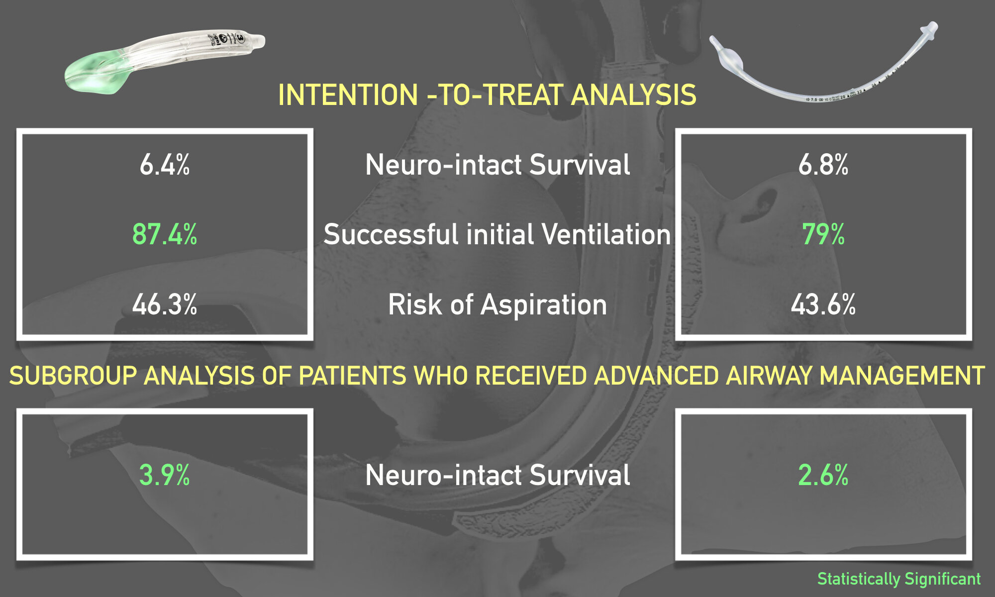
·       Favorable neurologic outcome [ mRS score (0-3)] at 30 days or hospital discharge (whichever came first): 6.4% (311/4882) in the supraglottic airway group versus 6.8% (300/4407) in the endotracheal intubation group (adjusted risk difference -0.6%; 95% CI -1.6%-0.4%)

·       In the subgroup analysis of 7576 patients who received advanced airway management (not intention-to-treat), more patients in the supraglottic airway device group had a favorable neurologic outcome (3.9%) vs. the tracheal intubation group (2.6%); risk difference, 2.1%; 95% CI, 1.2 – 2.9%

·       Successful initial ventilation: 87.4% (4255/4868) in the supraglottic airway group versus 79.0% (3473/4397) in the endotracheal intubation group (adjusted risk difference 8.3%; 95%CI 6.3% to 10.2%).

·       Rates of aspiration and regurgitation were not found to be different between the groups

Infographic.001.jpeg

Takeways:

Among patients with OHCA, advanced airway management with a supraglottic device was not associated with a favorable neurological outcome at 30 days compared to endotracheal intubation.

What This Means for EMS:

While the results of this study clearly indicated that advanced airway management with a supraglottic device was not associated with improvement in patient centered outcomes (in this case, neurological function), these devices were still associated with more successful initial ventilation without an increase in secondary complications such as aspiration/regurgitation. Furthermore, given the ease of use, less number of attempts required to obtain proficiency from a standpoint of training, and widespread availability, supraglottic devices are feasible intervention to provide airway support for patients with OHCA. Unlike endotracheal intubation which can be challenging to perform in the field with active chest compressions, supraglottic devices are easier to use in terms of temporary airway management and allow for the focus to be shifted towards measures that improve outcomes including high quality CPR and early defibrillation.

Article Bites Summary by Al Lulla, @al_lulla, Article Bites Editor

Article Bites #13: How Often Do They Get More Than One? Naloxone Redosing in the Age of the Opioid Epidemic

Klebacher R, Harris MI, Ariyaprakai N, et al. Incidence of Naloxone Redosing in the Age of the New Opioid Epidemic. Prehosp Emerg Care. 2017;21(6):682-687.

Background & Objectives:

The surging opioid epidemic has largely been combated with the use of intravenous and intramuscular naloxone administration. More recently, intranasal naloxone has been shown to be easily administered by not only EMS providers, but also law enforcement and family members to help reverse potentially fatal overdoses. Recently, mixed overdoses and ingestions with far more potent agents (such as carfentanyl) are on the rise, necessitating repeat naloxone dosing. The primary objective of this study was to determine the incidence of repeat naloxone administration for patients with suspected opioid overdose. The secondary endpoint was a more detailed descriptive and statistical analysis evaluating the precise characteristics associated with individuals who required repeat naloxone dosing.

Methods:

The investigators conducted a retrospective chart review of the electronic health record of the largest EMS service in New Jersey. Charts were searched for the presence of naloxone administration and other key words including “drug overdose”, “poisoning” and “unresponsive”. Charts were examined between April 2014 and June 2016. In order to be included in the study, patients had to be over the age of 17 years and administered an initial dose of 2mg of intranasal naloxone. Initial naloxone administration was performed by law enforcement or a BLS unit per New Jersey state regulations. Subsequent doses of naloxone were administered by ALS units.  Resolution or “response” to therapy was defined as GCS of 15. In addition, demographic data was extracted from each patient encounter. 

p2253.png

Key Results:

In total, 2,166 patients received naloxone for suspected opioid overdose during the study period) April 2014 to June 2016). The key results from the study were as follows:

  • 1,971 of 2,166 (91%) of patients had reversal of overdose symptoms after a single dose of naloxone administered by law enforcement or BLS units 

  • 195 of 2,166 patients (9%) required a second dose of naloxone by an ALS unit given failure to respond after the initial dose

  • 53 of 2,166 patients (2.4%) required a third dose of naloxone by an ALS unit

  • Patients who required a second dose of naloxone had a mean GCS of 5.3 (standard deviation of 3.7). The mean respiratory rate was 10.4 breaths per minute with a mean oxygen saturation of 86.8%. 

  • Patients who required a third dose of naloxone had similar mean GCS scores (4.9) and oxygen saturations (86.4%). Two-thirds of the 53 patients who received a third dose of naloxone improved to a GCS of 15, suggesting that the remaining 1/3 patients may have had an alternative diagnosis for their altered mental status.


Takeaways:

  • Among patients with suspected opioid overdose treated with intranasal naloxone by first responders or ALS units, 91% of patients had complete reversal of symptoms after a single dose of naloxone, with only 9% requiring repeat dosing.

  • Naloxone is overall very effective at reversing symptoms of opioid overdose after a single dose

What this means for EMS:

With the rising incidence of mixed ingestions, more potent opioids such as carfentanyl, EMS providers are faced with more complexities in the management of opioid overdose. This study suggests that, in addition to basic support of ventilations, naloxone is still the mainstay of management in these patients, and highly effective at reversing overdose symptoms. Furthermore, this study suggests that in the majority of cases, first responders including law enforcement, and BLS units may be able to safely manage opioid overdose without the need for ALS units. This may improve resource utilization in EMS systems that are already stretched very thin. 

Article Summary by Al Lulla, MD (@al_lulla)

Article Bites #12: The Profile of Wounding in Civilian Public Mass Shooting Fatalities. 

Article: Smith ER, Shapiro G, Sarani B. The profile of wounding in civilian public mass shooting fatalities. J Trauma Acute Care Surg. 2016;81(1):86-92.

Background & Objectives:

Civilian mass shootings are unfortunately on the rise and afflict the lives of many individuals and their families. Given the rising incidence and severity of these events, there has been much in the way of public initiatives at improving morbidity and mortality in individuals who have been critically wounded. Much of the prior emphasis on management of these patients in the prehospital environment has focused on external hemorrhage control with widespread education on use of tourniquets. The strong focus on civilian management of exsanguinating extremity hemorrhage during mass shootings is largely based on the blast injury patterns identified during the US operations in Iraq and Afghanistan which suggest that between 52% and 64% of injuries in combat are to the extremities. Whether these lessons translate to civilian mass shootings is unclear. The overall purpose of this paper was to precisely identify the anatomic wounding pattern, fatal wounds and incidence of potentially survival wounds in civilian mass shooting incidents. 


Methods:

The investigators conducted a retrospective study evaluating autopsy reports performed by medical examiners or coroners in 12 different mass shooting events. The investigators utilized the term “mass shooting” as defined by the FBI to mean:

  1. An incident occurring in a public space with 4 or more deaths (not including the shooter);

  2. Gunmen who select victims at random

  3. Violence without means to an end (i.e. not associated with robbery

Using reports made available by the New York Police Department and the FBI that provide detailed descriptions of civilian mass shootings dating back to 1966, the investigators identified 78 events that met the above definition for mass shooting events. 56 events that met the above definition had medical examiners or coroners that could be contacted. If the medical examiner or coroner was not listed or they could be not be contacted, the mass shooting event was eliminated from analysis. Request for official autopsy reports were sent to the respective examiners/coroners. Based on these reports, data was compiled regarding body site of wound, type of injury, probable site of fatal injury and whether wounds were potentially survivable. 

Key Results:

In total, based on responses from medical examiners, a total of 12 mass public shooting events were analyzed in the study. A total of 139 fatalities with 371 total wounds were examined by the investigators. The key results from the study were as follows:

  • There was an average of 2.7 wounds associated within the group of fatalities

  • The case fatality rate for civilian mass shootings was 44.6% (compared to approximately 10% during Operation Iraqi Freedom and Operation Enduring Freedom as reported in other studies). 

  • 58% of all victims (with fatal and non-fatal wounds) had at least one wound to head or chest/upper back

  • 20% (28/139) of all wounds were to the extremity, of which none were deemed to be fatal

  • 77% of all fatal wounds were identified in the head or chest/upper back. 

  • In total, only 9 of the 125 fatalities or roughly 7% (14 excluded given absence of autopsy data) were determined to be potentially survivable 

  • The most common survivable injury was a wound to the chest (89% of all survivable injuries) without obvious evidence of vascular or cardiac injury

  • There was 100% agreement between the reviewers of the study regarding potential survivability of injuries

Profile_mass_Shooting.jpg

Takeaways:

  • Only 7% of victims in civilian mass shootings had a potentially survivable wound. No fatalities likely occurred secondary to exsanguination from extremity hemorrhage

  • The majority of wounds in civilian mass shootings occur primarily in the head, chest/upper back compared to combat environments where the majority of wounds occur in the extremities. 

  • The case fatality rate for civilian mass shootings compared to military data was much higher, and associated with lower number of potentially survivable injuries 

What this means for EMS:

While our nation faces a crisis with the issue of gun violence at the forefront of public discourse, regardless of what stance one may take on this issue, one thing remains abundantly clear: EMS providers are front and center when it comes to management of victims of mass shootings in the field. Historically, much of the focus on managing victims of mass shootings has been based on Tactical Combat Casualty Care (TCCC) guidelines based on the US military conflicts on the battlefields of Iraq and Afghanistan. These guidelines are largely predicated on the management of exsanguinating extremity hemorrhage with the use of tourniquets. This study despite all its limitations including retrospective design, missing data, and possibility of miscategorization of survivable and non survivable injuries, calls into question the applicability of these findings to the civilian arena where body armor is not worn. Based on the results of this study, EMS providers on the front lines who bravely care for victims of civilian mass shootings may encounter patients with wounding patterns that differ significantly compared to those seen in combat. While there is no question the importance of training and implementation of easy interventions such as tourniquets for the management of extremity hemorrhage, perhaps EMS providers must have a broader implementation of other treatment strategies that more accurately reflect the injury profile seen in civilian mass shootings, such as penetrating chest trauma. The authors of the present study carried out a more recent analysis looking at the victims of the Pulse nightclub shooting in Orlando, FL. The findings of this newer study which examined this single event (versus the 12 events in the examined in the present study) identified a disproportionately higher rate of individuals with extremity wounds (90% versus 20%). In this newer study, 4 patients were determined to have preventable death secondary to extremity hemorrhage (or in this case, junctional hemorrhage in the axilla). These patients who died did not have any evidence of tourniquet application, further emphasizing the point that despite the overall low incidence of death from extremity hemorrhage in mass shootings, it remains a quick and easy intervention that has the potential to save lives. The authors further concluded that in the Pulse nightclub shooting, similar to the 12 prior incidents, the majority of fatalities were again secondary to torso injuries highlighting the need for other interventions such as decompression of tension pneumothorax, basic airway management and management of hypothermia, which likely play a critical role in improving the dismal survival rates associated with civilian mass shootings. 

References:

  1. Smith ER, Shapiro G, Sarani B. Fatal Wounding Pattern and Causes of Potentially Preventable Death Following the Pulse Night Club Shooting Event. Prehosp Emerg Care. 2018;22(6):662-668. Available at: https://www.ncbi.nlm.nih.gov/pubmed/29693490


Summary by Article Bites Editor, Al Lulla MD (@al_lulla)

Article Bites #11: Measuring the Impact of a Telehealth Program on Ambulance Transports

Article Reviewed:

Champagne-langabeer T, Langabeer JR, Roberts KE, et al. Telehealth Impact on Primary Care Related Ambulance Transports. Prehosp Emerg Care. 2019;:1-6. [PMID: 30626250]

Background & Objectives:

Prior studies have confirmed what is known by many of those who work in EMS: a high proportion of patients that are transported have non-emergent conditions. Several studies have demonstrated that between 33 and 50% of all ambulance transports are for non-emergent causes. These transports often times result in signifiant resource utilization from EMS systems that are stretched very thin.  Furthermore, these transports may often be linked with ED overcrowding problems and increased healthcare costs. The role of telehealth has already been shown to be a cost effective and beneficial approach to many aspects of healthcare, including tele-ICUs and within EMS as part of trauma, stroke and cardiovascular care. The primary objective of this study was to investigate the impact of a large-scale telehealth program that utilizes non-ambulance based transportation (i.e. taxi) and paramedic triage of non-urgent complaints on overall EMS transports.

Methods:

The investigators conducted an observational study from January 2015 to December 2017 for patients triaged by the Emergency Telehealth and Navigation Program (ETHAN) program developed by the Houston Fire Department. According to the study protocol, EMTs and paramedics were tasked with enrolling patients with non-life threatening conditions or mild illnesses. To be formally included in the study, the following criteria needed to be met:

  1. full history and physical exam with no obvious emergency

  2. age >3 months

  3. English speaking

  4. Normal vital signs; afebrile if chronically ill or over 65 years of age

  5. ability to care for self

  6. ability to be transported in a passenger vehicle. 

Patients who did not meet all inclusion criteria or who had other high risk features of their presentation suggestive of an emergency condition were excluded from the analysis. For those who qualified for the study, enrollees were connected via tablet to a board certified emergency physician who determined if the patient could be referred for follow-up with a primary care facility via versus requiring transport via ambulance. The primary variable that was studied was whether patients were transported by ambulance. Patients who were not transported via ambulance were offered transport to the ED or a primary care facility via taxi. 


Key Results:

During the study period, the investigators enrolled 15,067 patients in the telehealth program (equivalent to 2% of the overall EMS volume during this period). The key results from the study were as follows:

  • 11.2% of patients in the telehealth program were transported by ambulance

  • 75.6% of patients were transported by taxi instead of ambulance (5% of these patients were transported to a clinic instead of the ED)

  • 13.2% of patients transported themselves or were not transported at all

  • Patients were more likely to be transported by ambulance in the telehealth program if the chief complaint was abdominal pain (19.6%), low-risk chest pain (8.3%), shortness of breath (5.2%), or dizziness (3.7%)

alttransports.jpg

Takeaways:

  • Over the course of the study period, a telehealth program to identify patients with non-emergent conditions was successful in helping avoid unnecessary ambulance transports

What this means for EMS:

EMS agencies are faced with increased demand of services by the public with decreased available resources and higher costs. Telehealth in the prehospital setting is a novel approach to identify patients that may be suitable for transport via taxi and allow for EMS units to stay in service and serve other patients who present with other time critical conditions. This study did not show a significant decrease in the number of patients that were ultimately transported to the ED (only 2% of overall call volume participated & the majority of patients were still transported to the ED via taxi) and did not provide patient outcome data regarding accuracy of triage as non-emergent. However, it does demonstrate that a telehealth program is feasible within a large EMS system and highlights a promising avenue towards matching healthcare resources with patient needs and thus represents an important advancement in the field of EMS medicine.

Article Review by EMS MEd Article Bites Editor, Al Lulla MD (@al_lulla)

Article Bites #10: Delivering Right Care and Transporting to the Right Place: Medical clearance of Psychiatric Emergencies in the Field

Article: Trivedi TK, Glenn M, Hern G, Schriger DL, Sporer KA. Emergency Medical Services Use Among Patients Receiving Involuntary Psychiatric Holds and the Safety of an Out-of-Hospital Screening Protocol to "Medically Clear" Psychiatric Emergencies in the Field, 2011 to 2016. Ann Emerg Med. 2019;73(1):42-51.

Background & Objectives:

Due to a nationwide shortage of inpatient psychiatric beds, patients with psychiatric emergencies often spend long periods of time waiting in the ED for placement for psychiatric care. These long wait times are associated with more ED overcrowding, increased costs, and unfortunately sometimes inhumane conditions for patients and increased stress for staff. The vast majority of patients with involuntary psychiatric holds are brought to the ED by EMS, usually for medical clearance and evaluation for other possible non-psychiatric causes of the patient’s presentation. This study investigated the role of an EMS field protocol to allow EMS to bypass EDs and transport patients directly to a psychiatric facility. 

Methods:

The investigators conducted a retrospective review of all EMS transports in Alameda County, CA between November 1, 2011 to November 1, 2016, focusing particularly on patients receiving involuntary psychiatric holds. To assess for patients who received involuntary holds, the investigators evaluated the medical priority dispatch system code, primary impression, secondary impression and medical narrative as documented by EMS providers. According to the Alameda County EMS Agency protocol (see further reading below) patients with isolated psychiatric presentations can be transported directly to a stand alone psychiatric facility provided protocol criteria is met. Two primary outcomes were examined. First, the investigators compared “involuntary hold patients” with those patients who never received an involuntary hold to identify what specific characteristics were associated with patients receiving involuntary hold status. The second outcome that was evaluated was the safety of an EMS field protocol to screen patients for direct transport to a psychiatric facility and bypass of the ED. This measure was defined by retransport of a patient to the ED within 12 hours of transport to the psychiatric facility (AKA “failed diversion”).

Key Results:

During the study period, the investigators identified 541,731 total EMS transports (257,725 unique transports). Of the total transports, 10% (n=53,887) were for involuntary holds. The key results from the study were as follows

  • 41% (n=22,074) of transports for involuntary hold patients met protocol criteria for ED diversion and direct transport to stand alone psychiatric facility

  • Of patients who were transported to stand alone psychiatric facility, 0.3% (n=60) failed diversion and required retransport to ED within 12 hours

  • Involuntary hold patients were found to have significantly more total EMS use (24% of all encounters; n=128,003) compared to patients that never received hold status. They were also more likely to be younger, men, and have uninsured status

  • Of the patients requiring retransport within 12 hours, 54 of 60 of those patients developed new symptoms on arrival to the facility which did not manifest with their initial presentation to EMS. Reasons for retransport included traumatic injury (n=5), previously unrecognized or unreported symptom (n=13), seizure (n=8), excessive sedation (n=10), staff request for medical clearance of asymptomatic patient (n=7) new mental status change (n=5) or patient discharge from psychiatric service and self referral to EMS (n=5)

p1731.png

Takeaways:

  • Over the course of a 5 year period, an EMS field protocol to screen psychiatric involuntary hold patients for direct transport to a stand alone psychiatric facility performed safely, with only 0.3% of transported patients requiring retransport to an ED within 12 hours

  • Involuntary hold patients were usually younger and often uninsured. In addition, they had significantly higher overall EMS utilization

What this means for EMS:

This study demonstrates that implementation of an EMS field protocol can allow for safe diversion from ED directly to a psychiatric facility. The implementation of such protocol in EMS systems would likely have a significant impact on ED overcrowding and length of stay. This study highlights two important points: 1) The role of EMS with respect to hospital operations, ED operations and the health care system as a whole cannot be overstated. EMS systems nationwide may be able to build upon the lessons from Alameda County and help reduce ED overcrowding concerns as well as more rapidly direct patients to the psychiatric care they need. 2) EMS utilization by patients with psychiatric illness is significant, with roughly one-quarter of all transports in Alameda County being for “involuntary holds” during the 5 year study period. This further re-inforces the importance of both federal and local resource allocation for psychiatric illness. 

Further Reading: 

Alameda County EMS involuntary hold protocol:

https://www.annemergmed.com/cms/10.1016/j.annemergmed.2018.08.422/attachment/b3ec30ed-3ced-4eca-963b-82a60f05d663/mmc2.pdf

Review and Infographic by Article Bites Editor, Al Lulla MD (@al_lulla)

Article Bites #9: The Emergency within EMS - Risk of Suicide in EMS Compared to the General Public

Death by Suicide — The EMS Profession Compared to the General Public

Vigil NH, Grant AR, Perez O, et al. Death by Suicide-The EMS Profession Compared to the General Public. Prehosp Emerg Care. 2018;:1-6.[PMID: 30136908]


Background & Objectives:

 Suicide is a public health crisis with an estimate 45,000 individuals dying from suicide annually. Certain professions, including law enforcement and EMS are exposed to high degrees of workplace stress, therefore it is hypothesized that these individuals are more predisposed to conditions including anxiety, depression and suicidal ideation and behaviors. Survey data examined by the National Association of Emergency Medical Technicians (NAEMT) has indicated that there is very high occurrence of suicidal ideation within the EMS community. Despite this important information, the relationship between suicidal ideation and suicide attempt with the completion of suicide in EMS providers has not been well studied. The authors of this study aimed to assess the odds of death by suicide completion in EMTs compared to non-EMTs.  

 Methods:

The investigators conducted a retrospective case-control study that analyzed the electronic death registry in Arizona from January 2009 to December 2015. Only adults greater than or equal to the age of 18 were included in the analysis. Multiple variables from the death registry were examined including age gender, race, ethnicity, and most importantly for the purposes of this study: cause of death and occupation. With respect to occupation, the term "EMT" was categorized as all individuals who had EMT certification, including firefighters, EMTs and paramedics. A logistic regression model was implemented to calculate the mortality odds ratio (MOR) of suicide between EMTs (exposed group) and non-EMTs (non-exposed group). 


Key Results:

In total, 350,998 adults were analyzed who died during the above time period. The key results from the study were as follows:

· There were 1,205 EMT deaths during the study period

o   63 (5.2%) were attributed to suicide. This is compared to the non-EMT group of which 2.2% of deaths were due to suicide. 

· MOR for EMTs versus non-EMTs was 2.45; [95% CI (1.88-3.13)]

· Adjusted MOR for EMTs versus non-EMTs was 1.29; [95% CI (1.06-1.82)] adjusted for gender, age, race and ethnicity 

p1693.jpeg

· The most common mechanisms of suicide in the EMT group was firearm (67%), however there was no significant difference between death by firearm in the EMT cohort versus the non-EMT cohort. 


Takeaways:

· In the Arizona electronic death registry, there were higher odds for death by suicide in EMTs compared to the general public. 


What this means for EMS:

EMS providers are faced with significant workplace stressors. Whether it’s traumatic calls, poor sleep quality, poor compensation, long hours, or overall low job satisfaction, EMS remains one of the most challenging professions. These aspects of the EMS profession, unfortunately, contribute to a host of mental health issues including depression, anxiety, PTSD, all of which predispose individuals to developing suicidal ideation and behaviors. The time to act is now. The results of this study are extremely compelling and must serve as the impetus for change within the profession. Further studies that precisely characterize the risk factors that place EMS providers at higher risk than the general public should be examined as well. Despite this, the findings from this research still demonstrate the need for greater EMS education regarding the symptoms and warning signs of suicidal behavior, the importance of adequate resources for counseling and mental health, and improved work conditions to protect those individuals who protect our communities and our patients. 

Article Reviewed by Article Bites Editor Al Lulla, MD.

Article Bites #8: Reconsidering Priorities of care: epinephrine in out of hospital cardiac arrest

A Randomized Trial of Epinephrine in Out-of-Hospital Cardiac Arrest

Perkins GD, Ji C, Deakin CD, et al. A Randomized Trial of Epinephrine in Out-of-Hospital Cardiac Arrest. N Engl J Med. 2018. [PMID: 30021076]

Background & Objectives:

Other than early CPR and defibrillation, there are few measures that have been shown to improve outcomes for out-of-hospital cardiac arrest (OHCA). Despite this, epinephrine has been at the crux of ACLS management of patients with OHCA given the thought that it can cause peripheral vasoconstriction, increased beta adrenergic activity and augment coronary blood flow. In turn, epinephrine increase chances of return of spontaneous circulation (ROSC). While higher rates of ROSC have been confirmed in prior studies on epinephrine, unfortunately most of what we know about epinephrine suggests that it’s administration may not improve the most important clinical outcome - neurologically intact survival. The PARAMEDIC2 trial (Prehospital Assessment of the Role of Adrenaline: Measuring the Effectiveness of Drug Administration in Cardiac Arrest) was performed to assess whether epinephrine was beneficial or harmful as demonstrated by the primary outcome of 30 day survival. 

Methods:

The investigators conducted a multi agency (5 ambulance services), randomized, double-blind, placebo controlled trial in the United Kingdom from December 2014 to October 2017 in adult patients who sustained OHCA for which ACLS was provided by paramedics. Patients were excluded from the trial if they were pregnant, less than 16 years of age, had cardiac arrest secondary to anaphylaxis or asthma, or if they had administration of epinephrine prior to the arrival of EMS personnel. If initial resuscitation measures (CPR and defibrillation) were unsuccessful, patients were randomized to the intervention arm (1mg epinephrine q3-5 mins in accordance with ACLS protocols) or the control arm (normal saline placebo). As stated above, the primary outcome of the trial was 30 day survival. Secondary outcomes that were examined included rate of survival until hospital admission, length of hospital stay and ICU stay, rates of survival at hospital discharge and at 3 months, neurological outcomes at hospital discharge and at 3 months. Favorable neurological outcome was defined as modified Ranking score of 3 or less. 

Key Results:

In total, 8014 patients with OHCA were enrolled in the study over the 3 year period of which 4015 patients were in the intervention arm (epinephrine) compared to 3999 patients in the placebo arm. Both groups were well matched in terms of baseline patient characteristics.The key results from the trial were as follows:

  • 30 day survival: 3.2% in epinephrine group vs 2.4% in placebo group (OR 1.39; 95% CI 1.06-1.82, p=0.02).

  • Favorable neurological survival at 3 months (modified Rankin score 3 or less): 2.1% in epinephrine group vs. 1.6% in placebo group (OR  1.31; 95% CI 0.94-1.82.

  • Severe neurological impairment (modified Rankin score 4 or 5): 31% in epinephrine group vs. 17.8% in placebo group

  • ROSC during prehospital resuscitation: 36.3% in epinephrine group vs. 11.7% in placebo group

Takeaways:In this multi-agency, prospective, double blinded randomized placebo controlled trial, administration of epinephrine for OHCA was associated with a statistically significant higher 30 day rate of survival compared to placebo, but no difference in neurologically-intact survival.

Epinephrine Group (2).jpg


What this means for EMS:This study is the largest randomized controlled trial performed to date studying the impact of epinephrine administration on survival and neurological outcomes for OHCA. While administration of epinephrine has long been the pharmacological mainstay of prehospital (as well as in-hospital) management of OHCA, this trial calls into question its influence on patient centered outcomes (i.e. neurological intact survival). While this paper will surely be at the center of debate in the upcoming years within EMS circles around the world, one thing remains abundantly clear at this point: good quality BLS Care in the form high quality CPR and early defibrillation have the greatest impact on neurologically intact survival and should be the primary focus of resuscitation for cardiac arrest.

author_bios_lulla_sva.001.jpeg

Article Bites #7: Should Air Medical Transport administer plasma to trauma patients at risk for hemorrhagic shock?

Prehospital Plasma during Air Medical Transport in Trauma Patients at Risk for Hemorrhagic Shock

Sperry JL, Guyette FX, Brown JB, et al. Prehospital Plasma during Air Medical Transport in Trauma Patients at Risk for Hemorrhagic Shock. N Engl J Med. 2018;379(4):315-326. [PMID: 30044935]

Background & Objectives:

Hemorrhagic shock remains the most significant cause of mortality in trauma patients. In particular, coagulopathy is a significant contributor to death in this patient population and has been a focus of what is termed “damage-control resuscitation” in both civilian and battlefield arenas. Currently, there is a stronger push for resuscitation with blood-components including platelets and packed red cells in favor over crystalloid based resuscitation strategies. The premise of early damage control resuscitation in the pre-hospital environment is predicated on intervening at the point of injury and mitigating downstream complications including coagulopathy and irreversible hemorrhagic shock. Plasma administration as part of damage control resuscitation is thought directly address coagulopathy and improve chances for survival. This trial, termed the “Prehospital Air Medical Plasma (PAMPer)” trial sought to investigate the efficacy and safety of prehospital plasma administration in severely injured trauma patients. The primary outcome was the impact of prehospital plasma administration on 30 day mortality. 

Methods:

The investigators conducted a phase 3 multi-center cluster-randomized trial involving trauma  patients (blunt or penetrating) who were deemed to be at risk for hemorrhagic shock during air medical transport. Individual air medical bases were randomized to give plasma vs standard resuscitation in 1 month blocks. The intervention arm included patients who received 2 units of universal donor thawed plasma. The comparison group received standard of care resuscitation (crystalloid based resuscitation). Patients were deemed to be “at risk” for hemorrhagic shock were enrolled in the trial if they had at least one episode of hypotension (defined as systolic BP <90 mm Hg) and tachycardia (defined as HR >108 BPM) or if they had severe hypotension (defined as SBP <70 mm Hg) at any point in the prehospital phase of care. There were several exclusion criteria some of which included patients older than age 90 or younger than age 18, individuals who were pregnant, had traumatic cardiac arrest lasting longer than 5 minutes, penetrating brain injury, or refusal by family member to participate in the trial or if the patient was wearing an “opt-out” bracelet reflecting their wish not to participate in the trial, among others. 

Key Results:

There were 27 air medical transport bases that were recruited for the study that transported patients to 9 different level 1 trauma centers across the United States between 2014 to 2017. In total, 7,275 patients were transported during the study period, of which 501 patients qualified for the study. Of these patients, 230 received plasma and 271 received standard care resuscitation. Average prehospital time was 40 minutes (95% CI 33-51) and 42 minutes (95% CI 34 to 53) in the plasma and saline treated groups respectively. The key findings were as follows:

  • Mortality at 30 days was significantly lower in the plasma group (23.2%) versus standard group (33.0%)

  • Absolute reduction was 9.8% in the plasma group (95% CI 1.0 to 18.6%; P=0.03). 

  • Median INR was lower in plasma group compared to standard group (1.2 vs 1.3; p<0.001)

  • No statistically significant difference was found in secondary outcomes including 24-hr mortality, in-hospital mortality, volume of pRBC transfusion in the first 24 hrs, multi-organ failure, acute lung injury/ARDS, or transfusion-related reactions after correction for multiple comparisons.

  • Number needed to treat (NNT) was 10. 

PAMPER.jpg

Takeaways:

  • In this prospective trial administration of plasma in the prehospital aeromedical transport setting was associated with decreased 30 day mortality in trauma patients at risk for hemorrhagic shock.

What this means for EMS:

In 2015, the PROPPR trial demonstrated improved outcomes in trauma patients receiving blood products (packed red cells, platelets, plasma) compared to crystalloid resuscitation. Little research has been done on the role of plasma administration at the point of injury in the prehospital setting. This study was one of the first to show that rapid prehospital administration of plasma products is associated with improved 30 day mortality. As stated in Article Bite #4, transfusion of blood products in the prehospital setting is associated with many logistical roadblocks, including but not limited to a refrigeration, coordination with blood banks, and issues pertaining to wastage of products with a short shelf life, are all important considerations prior to routine implementation of this intervention. Given the traditional model of prehospital trauma care has focused on rapid transfer to a trauma center for definitive management, prehospital administration of plasma is a potential intervention that may lead to improved patient outcomes in some systems where distance to trauma center leads to extended prehospital times*.

Article summary and figure by Article Bites Editor Al Lulla, MD

* The COMBAT trial, which evaluated prehospital plasma transfusion for patients with signs of hemorrhagic shock within an urban system, did not find similar benefit.