EMS MEd Blog

Happy EMS Week: Pediatric Wednesday

Happy EMS Week Peds Day

EMS Strong.png

Yesterday we delved into our history to stress safety in a different form.  Today, we look to the future and say AWWWW how cute!!!  That's right we're talking about pediatric EMS today!

girl-kid-animated-gif-6.gif

Our very own triple boarded in Pediatrics, Pediatric Emergency Medicine, and EMS Dr. Joelle Donofrio (who is waaaay smarter than the average bear) went around the nation to interview fellow triple boarded Peds EMS physicians to thank YOU, our EMS providers for taking care of our kids.

Joelle Donofrio.JPG

Yes we know she's a nerd, a nerd about little ones.  But man is she passionate and so very smart about all things pediatrics!  If you ever want advice on pediatric prehospital care...go to her.  She knows her stuff!!!  But wait there's more...

but wait.gif

These stellar Pediatricians want to tell you what they are most looking forward to in the world of pediatric EMS so we can better serve our smaller communities.  So listen to this podcast and be melted by the cuteness, the passion, and the future of pediatric EMS care! 

Special thanks to Dr. Bjorn Peterson (@bjorn_peterson) for helping create our pediatric intro and conclusion!

bkpeterson.jpg

So to all our EMS providers during this EMS week, remain #EMSstrong, thank you for caring for our communities, and thank you for caring for our littlest patients. 

Cheers!


Happy EMS Week : Safety Tuesday

Happy EMS Week SAFETY TUESDAY!

EMS Strong.png

Today is SAFETY TUESDAY! I know, I know...it’s drilled into your heads.  Scene safety, scene safety, and scene safety.  Can you hear Men Without Hats?!?!?

And you know what, you’re absolutely right.  The scene SHOULD be safe.But today we’re not talking about THAT kind of safety.  We’re talking about your safety when it comes to fatigue.  Fatigue, which is the overwhelming feeling of exhaustion and tiredness, is a rampant problem in our field.  It is a pernicious symptom that leaks into every aspect of our lives affecting not only patient safety, but yours in EMS as well.

sleepy.jpg

Fatigue, which is the overwhelming feeling of exhaustion and tiredness, is a rampant problem in our field.  It is a pernicious symptom that leaks into every aspect of our lives affecting not only patient safety, but yours in EMS as well.

So we decided to delve into our archives (well not that deep) and play for you one of our favorite interviews with a sleep scientist and colleague of paramedics, Dr. Daniel Patterson and his evidence based guideline manuscript on fatigue.  To be honest, it is very intimidating to interview Dr. Patterson because he is a paramedic AND a PhD. Fortunately, Dr. Patterson’s gentlemanly southern accent and conversational tone made this interview so relaxed and informative that it felt like we were being taught by a great mentor.  

elect PattersonPhoto.jpeg

Now, Dr. Patterson made this interview great, but when we decided to branch out and hear from our ground medics around the nation about fatigue, we knew we had something special.  This episode provides a mixture of educational research with real life stories to drive the point home...that fatigue is a rampant problem in EMS and MUST be addressed for our patient’s safety and more importantly...our EMS provider’s safety.  So while on your drive home, working on your spring cleaning project, or even waiting to pick up your kid, check out this episode for Safety Tuesday.

50.gif

Thank you EMS providers for all that you do.  Happy Safety Tuesday!

Cheers and BE SAFE!!!

Happy EMS Week from NAEMSP: Education Monday

To all our first responders and paramedics out there, Happy EMS Week!  

EMS Strong.png


NAEMSP, EMS Med Blog, and the PEC Podcast team would like to say that we are so very grateful and proud of you for keeping our communities safe every...single...day.  We fully recognize that it takes a special kind of tough yet gentle person to practice prehospital medicine. You are that kind of person that the world needs more of.  You are our heroes and know that we respect and honor you not for only this week, but every day.

Gladiator.gif

As part of EMS week day 1 is dedicated to education.  Now, everyone that is part of this blog, NAEMSP, and our podcast love to teach.  It’s fun, challenging, and incredibly gratifying. Dr. Jeremiah Escajeda, my very good cat loving friend (seriously go check out his facebook page, it’s all about his cat Chester), EMS physician, and NAEMSP Education Chair obviously loves teaching.  Be it teaching about his cat Chester or prehospital cardiac arrest, Dr. Escajeda has that “special” educational gift.

Teacher gif.gif

Recently he decided to have a sit-down conversation with someone who equally loves to teach. Her name is Ginger Locke.

Ginger Locke.png
Medic Mindset.png


You may have heard of her, she is the creator of the podcast called Medic Mindset, a podcast dedicated to understanding the paramedic mind and advancing the paramedic educational agenda.  She is just as nerdy (we say this with all affection and love Ginger) as our very own Dr. Jeremiah Escajeda. Together they have a wonderfully convivial, nerd-filled conversation from why they love teaching to the joys of podcasting to what tools they learn to advance their education to who their heroes are.  They even talk about, yes you guessed it, Dr. Escajeda’s beloved cat Chester. So for your listening enjoyment in the car, in the ambulance, while on a run, or while doing chores, check out this podcast here, and be entertained by all things educational and by Dr. Escajeda’s cat, Chester :).  

And for your entertainment, here’s a picture of Dr. Escajeda with his cat in a backpack...called a catpack.  

26114094_520465565002763_570594035387832682_n.jpg


Article Bites #2: Identifying factors associated with repeated transports of older adults

Cookie_stylzed_square.jpg

Background & Objectives:

Adults aged 65 and older make up a significant proportion of the population (estimated to be approximately 20% of the total US population by 2030). In addition, this subset of patients has disproportionate utilization of EDs as well as EMS services (estimated at 38% of EMS transports). Little is known regarding the precise characteristics of this population that is associated with higher utilization of EMS. The investigators of the study aimed to investigate the proportion of older adults receiving repeat transport within 30 days and to potentially identify characteristics that were associated with repeat use of EMS. 

Methods:

The investigators conducted a retrospective analysis of EMS transports listed in the North Carolina Prehospital Medical Information Systems (PreMIS) database from 2010 to 2015. In particular, EMS encounters that were associated with 911 calls for adults aged 65 years or older that resulted in transport were examined. The primary outcome that was evaluated was repeated EMS transport within 30 days. Additional secondary outcomes that were examined included stratification of individuals by total number of EMS transports during the study period within 30 days. A multivariable logistic regression model was used to calculate adjusted odds ratios and 95% confidence intervals of repeated EMS transport within 30 days. 

Key Results:

A total of 1,719,998 transports for individuals aged 65 or greater were analyzed during the study period, of which 689,664 were for unique individuals. In this specific population, the key findings were as follows:

  • 17.7% (303,099 transports) had at least one repeated transport within 30 days
  • Odds of repeated transport within 30 days was higher in the following individuals: 

- Those from healthcare/residential facilities (OR 1.42 CI 1.38 to 1.47)

 - Black vs white (OR 1.29 95% CI 1.24 to 1.33)

 - Dispatch complaint of “sick person”, “fall”, “breathing problem”, “abdominal pain”, “diabetic problem”, “unknown person/person down”, “back pain”, “psychiatric problem”, “headache”

  • 15.6% of all repeated transports were related to falls 

Takeaways:

  • Greater than 1 in 6 EMS transports of individuals greater than the age of 65 is followed by a repeated transport within 30 days. 
  • Individuals in healthcare/residential settings and blacks (versus whites) have increased odds of repeated transport within 30 days

What this means for EMS:

Repeat transport of elderly adults is associated with significant healthcare costs and utilization of limited resources both in the hospital and pre-hospital setting. Identification of specific variables that are associated with repeated transport may assist with the development of targeted strategies to both improve patient outcomes and simultaneously decrease the demand for EMS resources that are already stretched very thin. 

Lulla.jpg

The Arresting LVAD patient: A Review of the Updated Recommendations

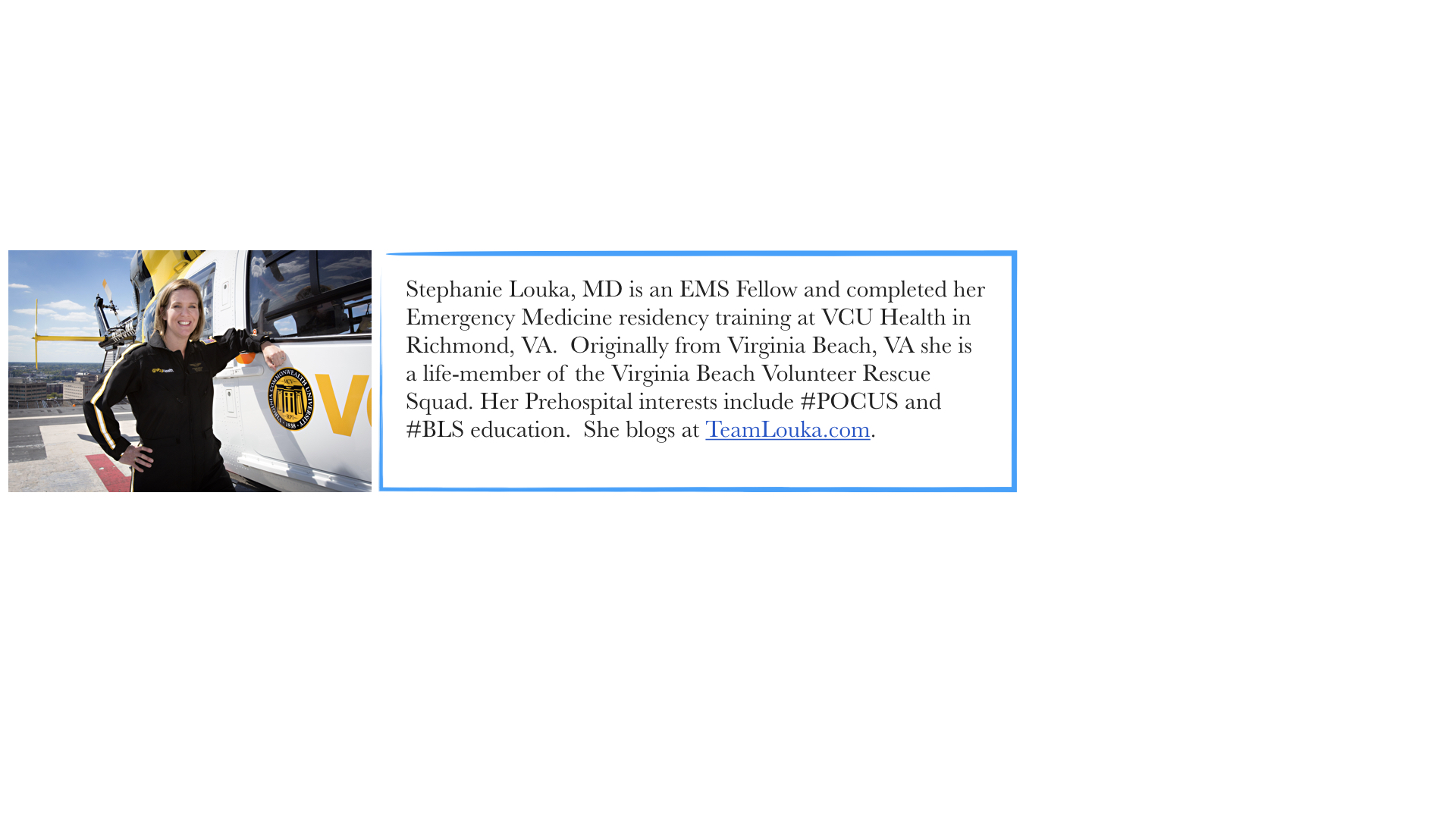
by Stephanie Louka, MD

Case Scenario:

LVAD_pic.jpg

Paramedics are called to the home of a 59 year old male patient with an LVAD who was found unconscious and unresponsive by family members.  He was last seen 2 hours earlier acting normally. There is no evidence of trauma. Family members are unaware of any recent illness or mechanical issues with his LVAD.  Your assessment is notable for warm but pale skin, lack of palpable pulses, LVAD with “low-flow” alarm but audible hum and apneic respirations. After intubation, end-tidal CO2 shows no wave form.  How should paramedics proceed?

Literature Review:

The 2015 ACLS guidelines do not address assessment and treatment of an unresponsive patient with a mechanical assist device such as an LVAD.[1]  While there has never been a reported case of LVAD dislodgement resulting from chest compressions, for years, VAD manufacturers advised against routine use of chest compressions on these patients for fear of device dislodgement and rupture of the left ventricle.  Despite a lack of evidence of dislodgment, many hospitals and EMS systems adopted this advice in management of VAD patients and withheld chest compressions even in the setting of true cardiac arrest.[2,3] Alternatively, with the risk of dislodgement theoretical and death eminent, some institutions opted to go against the manufacturer recommendation and administer chest compressions in this setting.

In 2017, the AHA published its Scientific Statement on Cardiopulmonary Resuscitation in Adults and Children with Mechanical Circulatory Support in the journal Circulation offering the first evidence-based, consensus approach to management for unconscious and/or arresting patients with mechanical assist devices.

Relying heavily on physical exam and waveform capnography for assessment, the algorithm provides a systematic approach to management of LVAD patients.  Trouble-shooting the LVAD with family members and/or LVAD coordinators is still recommended, but if the LVAD cannot be restarted or is not functioning adequately (MAP ≤ 50 mmHg and/or end-tidal CO2 ≤ 20 mmHg), external chest compressions are now recommended. 

While this AHA position was published in 2017, many EMS agencies are behind in adopting this change.  As this policy makes its way into the ACLS Provider course curriculum and manual and EMS personnel take their ACLS recertification courses, we should see broader adoption of this approach to the arresting LVAD patient.

Case Scenario Follow-up:

The Paramedic in charge contacted the regional VAD center and received online medical direction from the EMS Physician on duty who instructed the crew to administer chest compressions and transport the patient to his facility.  Firefighters on scene initiated chest compressions which resulted in a notable and sustained increase in end-tidal CO2 levels. 

The patient was transported to the Emergency Department where ROSC was obtained, and he was admitted to the ICU for continuing care.

Take Home:

Per the American Heart Association (AHA), chest compressions are now the standard of care in arresting patients with mechanical circulatory support devices (e.g. LVAD), and end-tidal CO2 <20 for whom device troubleshooting was ineffective.

louka.001.jpeg

Additional LVAD resources:

Make sure to review the EMS Field Guides provided at www.mylvad.com

Some additional LVAD Resources:

LVAD Management in the ED on the NUEM Blog

ALiEM PV Card on LVAD Management in the ED


References:

[1] American Heart Association (AHA). Advanced Cardiovascular Life Support (ACLS) Provider Manual, 16th edition. 2016.

[2] Shinar Z, Bellezo J, Stahovich M, Cheskes S, Chillcott S, Dembitsky W. Chest compressions may be safe in arresting aptients with left ventricular assist devices (LVADs). Resuscitation. 2014 May; 85(5):702-4.

[3] Mabvuure NT, Rodrigues JN. External cardiac compression during cardiopulmonary resuscitation of patients with left ventricular assist devices. Interactive CardiVascular and Thoracic Surgery. 2014 Aug; 19(2):286-289.

[4] Peberdy MA, Gluck JA, Ornato JP, et al. Cardiopulmonary Resuscitation in Adults and Children with Mechanical Circulatory Support. Circulation. 2017 Jun 13;135(24):e1115-e1134.

Dispatches from the Borderlands Part I: A Review of The Line Becomes a River, by Francisco Cantú

by Melody Glenn, MD

From 2008-2012, Francisco Cantú worked as an agent with the U.S. Border Patrol in the deserts of Texas, New Mexico, and Arizona. The Line Becomes a River is a memoir describing his experience, and like any talented memoirist, he uses his individual story to tell a bigger truth: violence has a way of seeping into the lives of those residing and working in the borderlands, and uniting over our shared humanity must be part of the solution.  His mom eloquently describes the way Cantú himself was affected by the work:

line_river_cover.jpg

You spent nearly four years on the border. You weren’t just observing a reality, you were participating in it.  You can’t exist within a system for that long without being implicated, without absorbing its poison. And let me tell you, it isn’t something that’s just going to slowly go away.  It’s part of who you’ve become.  So what will you do?  All you can do is try to find a place to hold it, a way not to lose some purpose for it all (231).

These words could also apply to those working as first responders, medics, and emergency medicine providers. In our daily work, we come face to face with horrors that other people rarely see.  We bear witness. Similarly, Cantú describes finding people in the desert in various states of extremis: extremely dehydrated and overheated, victims of brutal violence, and near the brink of death.

Although the personal vignettes he shares about those crossing are heartfelt, they alone are not what make Cantú’s memoir so powerful. It is the bigger themes that link them together into a unified narrative, the references from poetry and sociology that deepen the work, and the beauty of his writing that really make the book shine.

The memoir is split into three parts.  In the first, we see the motivation and idealism that lead him to the Border Patrol -- he wanted to gain a boots-on-the-ground understanding to go along with the book smarts he gained while studying international relations in college.  We are right there with him during his first experiences in the field, learning more about his coworkers and the migrants they apprehend. We see how he is trying to help those in a vulnerable state, even though he is arresting them. He paints his characters as multifaceted, complex individuals, illustrating the grey area of the borderlands.

 Cantú shooting and killing a yellow songbird, a metaphor for him crossing a line that cannot be undone, marks the transition to Part II.   We see how the daily violence starts to wear on his soul and seep out around the edges.  Every morning, he reviews an email briefing about the most recent brutal murders associated with the cartel.  Although we often think of the border patrol’s primary mission as stopping immigrants from crossing, they are also trying to prevent the entry of narco-traffickers and their drugs. The victims of the cartel are faceless, nameless:

The summaries included photos of human bodies that had been disassembled, their parts scattered, separated, jumbled together and hidden away or put on display as if in accordance with some grim and ancient ritual.  Victims’ faces were frozen in death, reverberating outward from the computer screen without identity or personal history, severed from the bodies they had inhabited and the human relations that had sustained them (86).

It is unknown how many people die due to the war on drugs or crossing the border, but they are often inextricably linked. Reporter Joel Millman wrote that there has been “a shift in the people-smuggling business.  A couple of decades ago, workers commonly traveled back and forth across the U.S.- Mexico border… Now, organized gangs own the people-smuggling trade” (93).  According to U.S. and Mexican police, this takeover was in part “an unintended consequence of a border crackdown” (93). As Cantú writes, “as border crossing become more difficult, traffickers increased their smuggling fees.  In turn, as smuggling became more profitable, it was increasingly consolidated under the regional operations of the drug cartels” (93).

When Mexican president Felipe Calderón took office in 2006, he declared war on the drug cartels. From 2007-2014, the Mexican government said there were more than 164,000 homicides, which does not include the estimated 25,000 missing and disappeared, or those who have died crossing the border into the U.S. while attempting to flee their violence-ridden towns. The Border Patrol recorded over 6,000 deaths between 2000 and 2016. Both Tijuana and Juarez had to increase their morgue operations to keep up with the rising demand. The numbers become overwhelming and have a tendency to turn the individual victims into an anonymous mass. As Cantú puts it:

It is difficult, of course, to conceive of such numbers in any tangible and appropriate way.  The number of border deaths, just like the number of drug war homicides… does little to account for all the ways that violence rips and ripples through a society, through the lives and minds of its inhabitants (107).

Even though Cantú was finally in the field, he was no closer to “figuring it out.”  He tells his coworker:

When I made the decision to apply for this job, I had the idea that I’d see things in the patrol that would somehow unlock the border for me, you know? I thought I’d come up with all sorts of answers.  And then working here, you see so much, you have all these experiences.  But I don’t know how to put it into context, I don’t know where I fit in it all. I have more questions now than ever before (142).

Cantú thinks the answers lie in humanizing others, in transforming the numbers and statistics into human stories, and in part III, he does just that.  This section focuses on his friend José, who is detained and separated from his family when he tries to cross back into the U.S. after visiting his sick mother in Mexico.  He takes the story from the perspective of law enforcement to that of someone victimized by the system, further allowing us to see the border from multiple angles.

Taking a cue from Cantú, I turned to various voices in healthcare to help paint a picture of the unique challenges of practicing medicine in the borderlands. My goal was to highlight their individual perspectives, as personal stories can shed light into dark spaces that numbers alone cannot reach.  Furthermore, I believe these stories can form a bridge across divided political views, bringing us to a platform of shared human experience.  Stay tuned for interviews with EMS providers, medical practitioners, and patients living and working along La Frontera, from Texas to California.

Glenn.001.jpeg

Article Bites #1: Bystander Efforts and 1-Year Outcomes in Out-of-Hospital Cardiac Arrest

Cookie_stylzed_square.jpg

Reference: Kragholm K, Wissenberg M, Mortensen RN, et al. Bystander Efforts and 1-Year Outcomes in Out-of-Hospital Cardiac Arrest. N Engl J Med. 2017;376(18):1737-1747. [PMID: 2846789]

Background & Objectives:

Bystander CPR and defibrillation has been associated with increased survival in patients with out-of-hospital cardiac arrest (OHCA). It is well studied that many patients who survive cardiac arrest have long term neurological deficits secondary to anoxic brain injury. Many of these patients require assistance with activities of daily living in the form of nursing home care. However, there is little known whether there is any improvement in functional outcomes for patients with OHCA in the setting of bystander CPR and defibrillation. The authors of this study hypothesized that bystander CPR in addition to use of an automated external defibrillator would potentially augment cerebral perfusion and reduce the extent of neurological insult by decreasing the amount of time to acquisition of ROSC. The authors sought to examine whether bystander CPR and bystander defibrillation would affect long term risk of anoxic brain damage or nursing home admission among 30 day survivors of OHCA over a 1 year period.  

Methods:

Danish investigators evaluated all 30 day survivors for OHCA aged 18 and older who were listed in the Danish Cardiac Arrest Registry from 2001 to 2012. Using nationwide registries, 1 year risk of anoxic brain injury, nursing home admission and all cause mortality was examined. Survivors of cardiac arrest were divided into four groups: 1) no bystander resuscitation 2) bystander CPR but no bystander defibrillation 3) bystander debrillation (regardless of bystander CPR status) and 4) EMS witnessed cardiac arrest. In addition, temporal changes in bystander interventions were studied (i.e. outcomes in relationship to increasing rates of bystander interventions in Denmark over the study period). 

Key Results:

Of the 34,459 individuals eligible for the study, 2,855 patients were 30 day survivors of OHCA during the 2001-2012 study period. The key findings were as follows:

  • 10.5% of patients had anoxic brain injury or were admitted to a nursing home. 9.7% of patients died within 1 year.  
  • Percentage of 30 day survivors increased from 3.9% to 12.4% over the course of the study
  • Percentage of bystander CPR in OHCA unwitnessed by EMS (n=2084) increased from 66.7% to 80.6% (p<0.001) over the course of the study. Percentage of bystander defibrillation increased from 2.1% to 16.8% (p<0.001). In concert, the rate of brain damage or nursing home admission decreased from 10.0% to 7.6% (p<0.001). All cause mortality decreased from 18.0% to 7.9% (p=0.002). 
  • Bystander CPR was associated with lower risk of brain damage or nursing home admission compared to no bystander resuscitation (adjusted hazard ratio 0.62 95% CI 0.47-0.82). Similar findings were observed with bystander defibrillation compared to no bystander resuscitation (adjusted hazard ratio 0.45 95% CI 0.24-0.84) 

Takeaways:

  • There is lower risk of anoxic brain damage, nursing home admission or death from any cause in 30 day survivors of OHCA who undergo bystander CPR or bystander defibrillation compared to those who do not receive bystander intervention.
  • Increasing rates of bystander interventions in Denmark during the course of the study period was associated with decreased rates of anoxic brain injury, nursing home admission, and all-cause mortality

What this means for EMS:

Denmark has instituted widespread initiatives including mandatory and voluntary CPR training, dissemination of automated external defibrillators throughout the country, health care professionals at emergency dispatch centers and dispatcher-assisted CPR. System wide measures and efforts to educate the lay public on BLS skills including high quality CPR and use of automated external defibrillation, is a critical step in both increasing survival rates from OHCA and improving functional outcomes for patients. 

Lulla.jpg

A Patient by Any Other Name: Approach to the "Lift Assist"

On March 3rd, we published the following case stem:

lift_image.jpg

EMS is dispatched to the home of a 75 yo female for a “lift assist”.  Reportedly, the patient slipped out of bed when getting up in the morning and needs assistance in getting off the floor.  Per dispatch, she is alert and denies other complaints.  This is not the first time EMS has been dispatched to this household for a similar complaint.  Driving to the scene, the EMS crew begins to debate whether a full Patient Care Record should be completed.  

We asked our readers three questions with regard to this scenario:

How do you define a "patient" in EMS?  

What defines a "lift assist" in your system?

What is the minimum assessment that should be performed and/or documented?


Our readers responded with a number of thoughtful comments and references.


How do you define a "patient" in EMS?  

There was general consensus that the term “patient” should be defined broadly within EMS: 

 “A patient is an individual requesting or potentially needing medical evaluation or treatment. The patient-provider relationship is established by either phone, radio, or personal contact. It is the providers responsibility to insure all potential patients regardless of the size of the incident are offered the opportunity for evaluation, treatment, and/or transport.”  - Eddie, from Wake Co EMS

 Is it safe to say any one an ems professional physically touches is a patient? - Jeremiah Escajeda

 “A patient is any member of the community served by the EMS that has a health problem requiring assessment and management by an EMT.” - Bill Lord

“My definition would be any person who needs or requests care. I have struggled with this question as an EMS Medical Director for over 30 years. My corporate legal counsel and our legal counsel for our state EMS agency gives this opinion: a person becomes a patient when they are contacted by an EMS provider. Period. No questions in their mind. My classic example is a non-injury MVC, called in by a passerby who dutifully calls 911 making the report. When asked if there are any injuries, they say "don't know", and they don't because they drove on. In our system, "don't know" gets a fire/ems/law enforcement response. If the people have no injury, did not request ems service, have a low or non-existent MOI, do not request evaluation or transport, I would think this would be a "no patient contact" response. However, as long as my legal consultants have the opinion that these folks are "patients" we will evaluate them and complete a PCR.”  - Irvin Smith, MD. Mercy Regional EMS. Paducah, Ky

Indeed, in a review of EMS liability cases (albeit from 1987 – 1992), the most common negligence allegations in patient care cases were arrival delay, inadequate assessment, inadequate treatment, patient transport delay and no patient transport. [1]  Legally speaking, proof of negligence does not require intent, but it does require that the EMS provider has a duty to act. [2] While the above definitions of patients are rather broad, they have important differences regarding interpretation of this duty to act.  The definition of patient provided by Bill Lord requires that you have a health problem requiring evaluation, while the Wake County definition considers someone with the potential to need evaluation and treatment a patient as well.  As alluded to by Irvin Smith, this becomes tricky when a third party caller requests assistance on the behalf of someone who does not want it, but it seems reasonable that there is some sort of documentation of this encounter (i.e. a person with decision making capacity refused evaluation and care) in case an accusation of negligence or abandonment were to appear in the future.


What defines a "lift assist" in your system and what is the minimum assessment that should be performed and/or documented?

Commenters generally agreed that “lift assist” was a potentially dangerous term because it puts providers in the mindset that the patient is not ill.

Heard this conversation in hospital ems room one day btw two different agency medics: “does your medical director make you get vital signs on your lift assists? Ours does!”

We should also ask what is response level in your EMS system for a “lift assist” call? Does your 911 system use the term “lift assist”

We should stop using that term. Exchange it with “fall.” A simple approach to highlighting a high risk pt population who we know are often elderly (high under triage rate [1]) and have a frequent short term representation rate with the healthcare system for what is often serious pathology[2].

1.Kodadek LM, Selvarajah S, Velopulos CG et al. Undertriage of older trauma patients: is this a national phenomenon? J Surg Research. 2015;199:220-229.
2.https://www.tandfonline.com/doi/abs/10.1080/10903127.2017.1308607.”
- Jeremiah Escajeda


There is no such term. Anyone with impaired mobility that requires assistance to mobilize requires an assessment of their health status before deciding that the patient does not require further medical assessment or referral to an agency that can implement a falls assessment and mitigation program with the patient’s consent….We know that a significant number of elderly who experience a fall but are not transported by EMTs have a subsequent fall, often resulting in a catastrophic injury such as a fractured neck of femur. The mortality rate in this cohort is also alarmingly high. http://qualitysafety.bmj.com/content/15/6/390
This reinforces the need for a focused examination in any older person who has fallen, even if they appear uninjured. Assessment should include tests of proprioception, balance and ability to mobilize. This is particularly important where the patient lives independently or has inadequate support. This suggests the need for an experienced clinician trained in this type of clinical assessment. However, there is evidence that paramedics don’t see this as their “core business”. We need to change this perception. See: https://bmchealthservres.biomedcentral.com/articles/10.1186/s12913-017-2243-y
Bill Lord


My mentor and EMS fellowship program director, Sabina Braithwaite, describes her approach to the “lift assist” the same way she describes most things: directly and logically.  Per Sabina, “When you need to call 911 to get your ass off the floor, that ain’t normal. You’ve probably got a problem and you deserve to be evaluated.”  Two studies that have examined outcomes of the “lift assist” patient have provided evidence for this clinical approach.  Cone et. al. examined the outcomes of 9-1-1 responses coded as “lift assists” from 2004-2009.  They found that these accounted for a total of 4.8% of all EMS calls and more than half of these had a return visit within 30 days.  On return visits, more than 50% were transported to the hospital. [3] More recently, Leggatt at al. published a retrospective analysis of 14-day morbidity and mortality among patients with an index lift-assist call. They found that out of 414 patients who called for a lift-assist, 21% had an ED visit, 11.6 % had a hospital admission and 1.1% died within 14 days of the index lift assist call.[4] These findings suggest that the index “lift-assist” call could have been an indicator of a functional decline, or alternatively just represent an overall sicker population. [5] While not every “lift assist” patient is a fall (e.g. the patient who needs assistance from the toilet to their bedroom), the vast majority likely fit the criteria for broad definition of patient as described above and deserve a medical assessment.

The commenters also make a more subtle and important point about the role of EMS in caring for these patients.  While few paramedics enter school with the goal of taking care of geriatric fallers, this population makes up a substantial proportion of EMS calls. [6,7]  As EMS providers find themselves spending their time on non-life-threatening work, this fosters  “low acuity fatigue”,  a “misplaced preconception before arriving at the scene that there will be nothing seriously wrong with the patient.” [6] While a substantial proportion of these patients will not require transport and should not be transported [8,9], a number of serious illnesses can lead to patient being on the floor and unable to get up (e.g. hypotension, stroke etc.).  Even in the absence of serious illness, visits to patients’ homes by EMS represent a unique opportunity for intervention for elderly patients who fall. EMS providers are able to assess multiple factors that likely affect long term health of elderly patients, such as the safety of their home environment or what their home environment conveys about their ability to care for themselves at their current level of assistance. [10]   Ideally, in cases of both transported and non-transported patients who fall, EMS can be incorporated into the integrated healthcare network that provides preventative care to this population to both decrease their risk of subsequent fall or their risk of harm from a subsequent fall. [11,12] EMS education will be essential to this process by promoting the perception that the care of the geriatric fall patient is “legitimate work” and an opportunity to do a tremendous amount of good. [6] Indeed, the health of the community and EMS provider job satisfaction stand to gain a lot from such a culture change.

Summary by EMS MEd Editor  Maia Dorsett, MD PhD @maiadorsett

1.     Ogilvie, W. (2017). EMS, Legal and Ethical Issues.

2.     Morgan, D. L., Wainscott, M. P., & Knowles, H. C. (1994). Emergency medical services liability litigation in the United States: 1987 to 1992. Prehospital and disaster medicine9(4), 214-220.

3.     Cone, D. C., Ahern, J., Lee, C. H., Baker, D., Murphy, T., & Bogucki, S. (2013). A descriptive study of the “lift-assist” call. Prehospital Emergency Care17(1), 51-56.

4.     Leggatt, L., Van Aarsen, K., Columbus, M., Dukelow, A., Lewell, M., Davis, M., & McLeod, S. (2017). Morbidity and mortality associated with prehospital “lift-assist” calls. Prehospital Emergency Care21(5), 556-562.

5.     Brown, Lawrence H. "Researching Lift-Assists: Nebulous Complexity." (2017): Prehospital Emergency Care . 670-672.

6.     Simpson, P., Thomas, R., Bendall, J., Lord, B., Lord, S., & Close, J. (2017). ‘Popping nana back into bed’-a qualitative exploration of paramedic decision making when caring for older people who have fallen. BMC health services research17(1), 299.

7.     Evans, C. S., Platts-Mills, T. F., Fernandez, A. R., Grover, J. M., Cabanas, J. G., Patel, M. D., ... & Brice, J. H. (2017). Repeated emergency medical services use by older adults: analysis of a comprehensive statewide database. Annals of emergency medicine.

8.     Williams, J. G., Bachman, M. W., Jones, A. W., Myers, J. B., Kronhaus, A. K., Miller, D. L., ... & Tibbo-Valeriote, H. (2015). Retrospective validation of a protocol to limit unnecessary transport of assisted-living residents who fall. Prehospital Emergency Care19(1), 68-78.

9.     Williams, J. G., Bachman, M. W., Lyons, M. D., Currie, B. B., Brown, L. H., Jones, A. W., ... & Myers, J. B. (2017). Improving decisions about transport to the emergency department for assisted living residents who fall. Annals of internal medicine.

10.  Weiss, S. J., Chong, R., Ong, M., Ernst, A. A., & Balash, M. (2003). Emergency Medical Services Screening of Elderly Falls in the Home. Prehospital Emergency Care7(1), 79-84.

11.  Snooks, H. A., Anthony, R., Chatters, R., Dale, J., Fothergill, R. T., Gaze, S., ... & Lyons, R. A. (2017). Paramedic assessment of older adults after falls, including community care referral pathway: cluster randomized trial. Annals of emergency medicine

12.  Wofford, J. L., Schwartz, E., & Byrum, J. E. (1993). The role of emergency services in health care for the elderly: a review. The Journal of emergency medicine11(3), 317-326.


This is why we do advocacy

by Ritu Sahni, MD, MPH, FAEMS

table.jpg

On the Friday before this past Thanksgiving, the President signed HR304, otherwise known as the Protecting Patient Access to Emergency Medications Act.  In a year in which dysfunction would have been an improvement in the political world, this important legislation was passed in a bipartisan manner.  As EMS physicians, we have a unique view.  We look at our population as a whole – not necessarily individual cases and certainly just unique disease processes and specialties.  We are not responsible for one patient at a time but an entire community. This is why advocacy matters.  We have a responsibility to do what is right for our patients and as system-thinkers, we have a unique responsibility to do what we can to enhance the system.  This is especially true when it comes to advocating on behalf of our patients and our system and this is why we helped create and advocated for HR304.

In January of 2015 I was completing my term as President of NAEMSP.  We had been discussing issues regarding the management of controlled substances in EMS for years.  The only consistency was inconsistency.  In some locales, EMS Medical Directors were required to get a separate DEA license for every location that stored controlled substances of any variety.  Some EMS agencies were required to get a distributors license because they “distributed” controlled substances among their various rigs and stations.  It was in this context that the Drug Enforcement Administration’s policy/regulatory section approached the EMS community proposing to create a set of rules specific to EMS.  We were pleased that there would possibly some consistency and excited to hear that the DEA was reaching out to the EMS community.  During the NAEMSP meeting in New Orleans we had the opportunity to meet with the DEA’s policy personnel  As we sat in my presidential suite in New Orleans it became increasingly clear that we had a problem.   The DEA’s authority comes from the Controlled Substances Act.  The CSA was written two years before Johnny and Roy premiered on television (for you youngsters – Johnny and Roy are a reason many of us ended up in EMS).   The law didn’t anticipate the use of controlled substances in a mobile environment and without a physician present.  Ultimately, the DEA stated that the CSA had some very specific guidelines as to when controlled substances could be delivered.  The crux was this, all orders for controlled substances had to be “patient-specific.”   There couldn’t be a “standing order” that allowed non-physicians to deliver controlled substances without an order given to them directly by a physician in real-time.  When we suggested that the new EMS rules could allow this, the DEA representatives appropriately pointed that they could not write a rule that was counter to the requirements of the statute.  The only way to get rules that made sense was to change the law.

NAEMSP had seen the importance of advocacy many years earlier.   Dr. Richard Hunt correctly identified that EMS had been left out in the cold when there was a large increase in preparedness funding following the attacks on 9/11.  Law enforcement and operational fire had received specific funding lines.  Medical preparedness was focused on hospitals, who controlled local distribution of federal funds.   He asked a staff member of his local congressman why was EMS left out and the answer was simple: EMS had no one at the table when decisions were being made.  NAEMSP realized that caring for our patients required being involved when policy was made.   A spot at the table requires resources, which NAEMSP was unable to afford by itself.   As a result, Advocates for EMS (AEMS) was born.

Advocates was born from a desire to be provide a “Generic EMS” advocacy arm.   NAEMSP sought to bring the “alphabet soup” of EMS organizations together to provide a patient-focused advocacy outlet separate from some of the issues that may divide us in EMS.   Early on, the National Association of State EMS Officials (NASEMSO) was a key partner.  Later on, the National Association of EMTs (NAEMT) was the major partner.  This allowed the organizations to pool resources and invest in professional lobbying along with a more strategic legislative focus.  AEMS adopted many strategies as it strove for relevance.  Early on, AEMS sought to ensure that “report language” and grant requirements included EMS.  It was successful in these endeavors and some small victories were helpful to the EMS community.   Ultimately, AEMS attempted to get more aggressive and developed the EMS Field Bill.  This bill was large and meant to be impactful.  It called for a formal Federal “Home” for EMS that was in Health and Human Services (not NHTSA).  It led to significant discussion and even controversy in the EMS community – but did not achieve passage. Ultimately, trying to run an “Association of Associations” can be difficult. Each association has a slightly different “twist” on EMS issues and more importantly, different processes when it comes to setting legislative goals.  As this became more difficult, AEMS had to come to end.  This does not mean AEMS was a failure.  In fact, it was quite the opposite.  EMS associations realized that “You must be present to win.”  Having a presence in Washington, DC is imperative or national policy will roll right over you.  Based on this experience, NAEMSP decided that it needed to invest its resources into a permanent presence in Washington.

This brings us back to the DEA.  Shortly after NAEMSP formalized its own government affairs plan by creating an Advocacy Committee and contracting with Holland & Knight as our DC representation, it became apparent that any regulations regarding controlled substances would negatively impact patient care.  This is not because regulations are inherently bad, but because the CSA was not designed for prehospital use.  Because of the lobbying experience available to us from our Holland and Knight partners, we were able to identify a Member of Congress willing to listen to us and take up our fight.  Representative Hudson from North Carolina heard us and, as a result introduced the Protecting Patient Access to Emergency Medications Act.  We tried our hand at Advocacy.  NAEMSP members starting contacting Congress.   Additionally, we quickly partnered with ACEP and NAEMT – both of whom activated their membership on the issue.   NAEMT agreed to make the bill a priority on EMS on the Hill and members of the EMS community walked the hall of Congress to advocate for a bill in which NAEMSP led the development.  Our issue almost got done in 2016 – which would have been amazing.  But politics prevailed, and the bill didn’t pass.  Representative Hudson didn’t give up and he reintroduced the bill in the House and Senator Cassidy introduced the bill in the Senate.  This time, the pieces fell into place and the bill was passed by both the House and the Senate, and signed by the President.  To some it was a small thing, but using protocols or “standing orders” for EMS to deliver controlled substances was now legal.  Presence in Washington would have a direct and positive impact on the provision of care at the patient’s side.

What next?

NAEMSP strives to continue to be a force in healthcare policy development, especially as it relates to time-critical emergencies and high quality prehospital care.  As we move forward, the issue of medical oversight and its value to the system and role in driving quality care is key.  High quality medical direction improves patient outcomes and the system should acknowledge that and fund it.   NAEMSP plans to lead this discussion.  Your membership in NAEMSP helps fund this.  Additionally, NAEMSP has decided to form a political action committee or PAC.  Unfortunately, neither George Soros or the Koch Brothers can fund this PAC.  Only NAEMSP members can fund the PAC.  Why do we do this?  It allows NAEMSP to do it what it can in an aboveboard and ethical manner to support legislators who are open and supportive to EMS.  How can you help?  Here are a couple of things:

  • Donate money to the PAC (www.naemsppac.com)
  • Attend the NAEMSP Government Relations Academy on April 10 (Space available on First come, First Serve Basis, Click here to RSVP)
  • Attend the NAEMT EMS on the Hill Day on April 11 (https://www.naemt.org/events/ems-on-the-hill-day)
  • Get involved in local politics
  • Be present at local and state meetings, especially when EMS issues arise.
  •  Serve on local and state policy committees that impact EMS
  • Here’s the crazy one – RUN FOR OFFICE.  Imagine a world in which your county commissioner is an actual EMS physician?   It could be a game changer.  We can provide information but only when holding the levers of power can you truly make change. 

In EMS, we are system-thinkers.  Our primary objective is to improve the care of patients in our entire community.  We cannot assume that lawmakers will understand the intricacies of the care we provide or the barriers we face in achieving our primary objective.  We must be at the table. 


Sahni.001.jpg

Lift Me Up Before You Go Go: Defining A Patient in EMS

lift_image.jpg

EMS is dispatched to the home of a 75 yo female for a “lift assist”.  Reportedly, the patient slipped out of bed when getting up in the morning and needs assistance in getting off the floor.  Per dispatch, she is alert and denies other complaints.  This is not the first time EMS has been dispatched to this household for a similar complaint.  Driving to the scene, the EMS crew begins to debate whether a full Patient Care Record should be completed.  


 How do you define a "patient" in EMS?  

What defines a "lift assist" in your system?

What is the minimum assessment that should be performed and/or documented?


Thank you all for all your commentary.  You can read the Discussion forum summary here.


Tranexamic Acid: Does it Have A Role In Prehospital Management of Trauma Patients?

by Carly Loner, MD

Case Scenario:

EMS is called to the scene of a motorcycle accident involving a 42 year old male.  The patient was helmeted and his head is atraumatic, but he is confused.  Breath sounds are equal bilaterally. The patient’s abdomen is diffusely tender and he has an open left femur fracture that is bleeding profusely.  A tourniquet is applied to the left proximal thigh with control of active bleeding and a pelvic binder is placed. Initial vitals are HR 132, BP 85/60, RR 28.  The patient is loaded into the ambulance and they depart towards the Level One trauma center 35 minutes away.  The ground team does not carry blood, but they have been considering adding TXA for situations such as this…

Literature Review:

thumbnail.jpg

Tranexamic acid (TXA) is a synthetic analog of the amino acid lysine that stabilizes clot formation by binding to lysine receptor sites on plasmin, thus preventing it from binding to and degrading fibrin.  It has been used in the medical arena for many years for the treatment of bleeding.  TXA is approved by the US Food and Drug Administration (FDA) only for use in heavy menstrual bleeding and for patients with hemophilia undergoing procedures, but it has a long history of off label use in the elective surgery setting [1]. More recently, it has been utilized as a therapy for the prevention and treatment of hemorrhagic shock.    TXA use to treat traumatic hemorrhagic shock became more widespread following publication of the Clinical Randomization of an Antifibrinolytic in Significant Hemorrhage-2 (CRASH-2) trial [2].  This study showed that administration of TXA within 3 hours of injury reduced mortality, but had increased mortality if given after the 3 hours time-point.  The results of CRASH-2 were substantiated by the military application of tranexamic acid in trauma emergency resuscitation (MATTERs) study.  This retrospective study of a UK combat hospital found that subjects (combat casualties receiving 1 L or more of RBCs) had improved survival due to TXA versus placebo, and this benefit was increased in patients requiring massive transfusion [3].  Another study by Gayet-Ageron et al. sought to quantify the effects of treatment delay on TXA.  Simulated models determined that the benefit of TXA decreased by 10% for every 15 min of treatment delay until the 3 hour mark, after which there was no longer a benefit of TXA administration [4].  The literature supporting the use of TXA has continued to develop and the Department of Defense’s Committee on Tactical Combat Casualty Care (CoTCCC), American college of Surgeons Committee on Trauma, European Task Force for Advance bleeding Care in Trauma all recommend administering TXA to hospitalized trauma patients as soon as possible [5, 6].

The time-dependent benefit for TXA may be due to the pathophysiology of trauma-induced coagulopathy.  Massive bleeding within trauma patients has been shown to have distinct phases.  The fibrinolytic phase shows these patient are prone to acute blood loss not only from hypovolemia but also from coagulopathy resulting from acidemia, hypothermia, shock, and hemodilution [1].    The coagulation cascade is activated immediately after a trauma with increased tissue factor and thrombin production and activation [1].  Tissue hypoxia due to hemorrhagic shock causes release of tissue-plasminogen factor [1].  The early phase of response to trauma is a fibrinolytic coagulopathy and studies have shown that this is where TXA may be most beneficial [2-4].  If given at a later stage of post-trauma coagulopathy, the fibrinolytic shut-down phase,  TXA could enhance the pro-thrombotic state and increase multi-organ dysfunction secondary to vascular microvascular occlusion [1, 11].  The coagulation/fibrinolysis states of the patient may be important for determining benefit versus detriment of administering TXA. 

There are multiple mechanisms by which TXA is thought to contribute to improved outcomes in trauma patients.  In addition to its anti-fibrinolytic role in preventing fibrin breakdown, TXA prevents trauma- induced coagulopathy by preserving the endothelial glycocalyx and thereby reducing vascular permeability and intervascular hypovolemia contributing to shock [1,6,7].  TXA also has anti-inflammatory effects that reduces post-ischemic neutropenic and mast cell activation which protects lung tissue, reduces vasopressor requirements, and reduces chest tube output [8-10].

However, prehospital TXA administration remains controversial.

On the positive side, TXA administration has a time-dependent effect on mortality reduction:  a post-hoc analysis of the CRASH-2 data suggests that the mortality benefit is achieved by administration within one hour of injury [12].  Several studies have found benefits of prehospital TXA administration. A UK prospective analysis studied the effects of prehospital administration of 1 g TXA in patients with concern of hemorrhagic shock. This study found that reduced multi-organ failure (OR 0.27, 95% CI 0.10 – 0.73) and reduced mortality (OR 0.16, 95% CI 0.03 – 0.86) in patients with shock when TXA was given by prehospital providers [13].   A more recent retrospective, propensity-matched German study of trauma patients transported by helicopter found significantly reduced 24 hr mortality (5.8 %  with TXA vs. 12.4 % without TXA), but no significant difference in overall mortality (14.7% with TXA vs. 16.3 % without).  TXA was found to prolong time to death (8.8 +/- 13.4 days vs. 3.6 +/- 4 days) [14].  Preliminary evidence from the  Cal-Pat Study suggests a non-significant trend towards decreased 24 hr, 48 hr and 28 day mortality in patients receiving prehospital TXA [15].  In Israel, TXA is given at the point of injury in both civilian and military settings [16]. In the pediatric population, Eckert et al. studied the effects of TXA in pediatric patients  injured in a combat setting in Afghanistan and found reduced mortality [18].  Multiple studies of prehospital TXA use in trauma are ongoing, including the Study of TXA During Air and Ground Medical Prehospital Transport Trial (STAAMP Trial) is a US multicenter randomized control trial investigating TXA on US Helicopter EMS services and is expected to be completed in March 2018 [17]. 

On the opposite side, TXA use is associated with side effects induce GI pain, joint pain, fatigue, visual disturbances, and of greatest concern, thromboembolic events.  However, the CRASH-2 trial found no significant difference in vascular occlusive events between TXA group and controls, but did contribute thromboembolic events with delayed administration during fibrinolytic shut down phase [2].  Multiple other studies have found no significant increase in occurrence of thromboembolic events [4, 15].    However, in the UK prospective analysis, while favorable of TXA administration did find 4-fold increase of thromboembolic events in the shock group which received TXA [13].  TXA has not been found to increase thromboembolic events in the elective setting and thromboembolic events in the setting of trauma may be due to additional factors such as stasis and surgery [3].   

Given concern for increased coagulopathy and incidence of multi-organ failure if TXA is administered during the fibrinolytic shutdown phase, some argue that TXA should be withheld until coagulation testing can be done which demonstrates hyperfibrinolysis [5,19]. However, awaiting for this testing delays time to administration and puts patients closer to the fibrinolytic shut down phase of trauma- induced coagulopathy.  A study by Stein et al. compared coagulation studies both on scene and upon arrival to the hospital between patients administered TXA versus placebo.  On-scene samples showed no significant difference but coagulation studies of the TXA group on hospital arrival demonstrated reduced hyperfibrinolysis and preserved fibrinogen levels [20].  

A 2017 review of the literature of prehospital TXA administration recommends an intermediate approach where the 1 g initial bolus of TXA is given in the field with further administration of TXA only given after coagulation testing at the hospital demonstrates continued hyperfibrinolysis [6].  There is no current evidence to support this two-step approach,  bringing up the ever-important point that prehospital and in-hospital trauma care are two points on the same continuum.  Regardless of location, prehospital implementation of a TXA protocol requires a collaborative effort with hospital emergency department and trauma services to ensure that care that is initiated is continued.  

Take Home: The use of TXA within trauma is controversial and still developing as literature expands.  However, studies do indicate that there is greater benefit to early administration of TXA versus delayed administration.  The negative effects of TXA may also be decreased if given early in the course of hemorrhagic shock.  There is growing evidence demonstrating reduced morbidity and mortality with prehospital TXA administration.  The role of TXA will likely continue to expand and it has potential to be employed in the prehospital setting to improve survival of patients in hemorrhagic shock.  

EMS MEd Editors Maia Dorsett, MD PhD (@maiadorsett) & Jeremiah Escajeda (@jerescajeda)

bio.001.jpeg


Additional Resources:

Current clinical prehospital trials of TXA use in Trauma at ClinicalTrials.gov

Enthusiasm for prehospital TXA use may be premature (JEMS)

Tranexamic acid's potentially bright future relies on collaborative data (JEMS)

Trending: More EMS Agencies administering TXA (EMS1)


References

1.         Nishida, T., T. Kinoshita, and K. Yamakawa, Tranexamic acid and trauma-induced coagulopathy. J Intensive Care, 2017. 5: p. 5.

2.         Roberts, I., et al., The CRASH-2 trial: a randomised controlled trial and economic evaluation of the effects of tranexamic acid on death, vascular occlusive events and transfusion requirement in bleeding trauma patients. Health Technol Assess, 2013. 17(10): p. 1-79.

3.         Morrison, J.J., et al., Military Application of Tranexamic Acid in Trauma Emergency Resuscitation (MATTERs) Study. Arch Surg, 2012. 147(2): p. 113-9.

4.         Gayet-Ageron, A., et al., Effect of treatment delay on the effectiveness and safety of antifibrinolytics in acute severe haemorrhage: a meta-analysis of individual patient-level data from 40 138 bleeding patients. Lancet, 2017.

5.         Chang, R., B.J. Eastridge, and J.B. Holcomb, Remote Damage Control Resuscitation in Austere Environments. Wilderness Environ Med, 2017. 28(2s): p. S124-s134.

6.         Huebner, B.R., W.C. Dorlac, and C. Cribari, Tranexamic Acid Use in Prehospital Uncontrolled Hemorrhage. Wilderness Environ Med, 2017. 28(2s): p. S50-s60.

7.         Diebel, M.E., et al., The temporal response and mechanism of action of tranexamic acid in endothelial glycocalyx degradation. J Trauma Acute Care Surg, 2018. 84(1): p. 75-80.

8.         Jimenez, J.J., et al., Safety and effectiveness of two treatment regimes with tranexamic acid to minimize inflammatory response in elective cardiopulmonary bypass patients: a randomized double-blind, dose-dependent, phase IV clinical trial. J Cardiothorac Surg, 2011. 6: p. 138.

9.         Peng, Z., et al., Intraluminal tranexamic acid inhibits intestinal sheddases and mitigates gut and lung injury and inflammation in a rodent model of hemorrhagic shock. J Trauma Acute Care Surg, 2016. 81(2): p. 358-65.

10.      Reichel, C.A., et al., Plasmin inhibitors prevent leukocyte accumulation and remodeling events in the postischemic microvasculature. PLoS One, 2011. 6(2): p. e17229.

11.         Moore, E.E., et al., Postinjury fibrinolysis shutdown: Rationale for selective tranexamic acid. J Trauma Acute Care Surg, 2015. 78(6 Suppl 1): p. S65-9.

12.         Crash-2 Collaborators. (2011). The importance of early treatment with tranexamic acid in bleeding trauma patients: an exploratory analysis of the CRASH-2 randomised controlled trial. The Lancet, 377(9771), 1096-1101.

13.         Cole, E., et al., Tranexamic acid use in severely injured civilian patients and the effects on outcomes: a prospective cohort study. Ann Surg, 2015. 261(2): p. 390-4.

14.         Wafaisade, A., Lefering, R., Bouillon, B., Böhmer, A. B., Gäßler, M., & Ruppert, M. (2016). Prehospital administration of tranexamic acid in trauma patients. Critical Care, 20(1), 143.

15.         Neeki, M.M., et al., Efficacy and Safety of Tranexamic Acid in Prehospital Traumatic Hemorrhagic Shock: Outcomes of the Cal-PAT Study. West J Emerg Med, 2017. 18(4): p. 673-683.

16.        Nadler, R., Gendler, S., Benov, A., Strugo, R., Abramovich, A., & Glassberg, E. (2014). Tranexamic acid at the point of injury: the Israeli combined civilian and military experience. Journal of Trauma and Acute Care Surgery, 77(3), S146-S150.

17.        https://clinicaltrials.gov/ct2/show/NCT02086500

18.         Eckert, M.J., et al., Tranexamic acid administration to pediatric trauma patients in a combat setting: the pediatric trauma and tranexamic acid study (PED-TRAX). J Trauma Acute Care Surg, 2014. 77(6): p. 852-8; discussion 858.

19.        La Rochelle, P., Prehospital transfer strategies and tranexamic acid during major trauma. The Lancet. 389(10079): p. 1604-1605.

20.      Stein, P., et al., The Impact of Prehospital Tranexamic Acid on Blood Coagulation in Trauma Patients. Anesth Analg, 2017.

We Gave an Inch, They Took a Mile

by Clayton Kazan, MD MS

ambulances_wall_strip.jpg

EMS Physicians need to be drivers of the EMS system and recognize that we are a Mobile Community Healthcare Provider and not providing medical direction to a fleet of glorified Ubers.  This seems like a total “no-brainer,” yet we find ourselves grappling with problems like Ambulance Patient Offload Delay (APOD, aka Ambulance Wall Time) that we should never have allowed to happen.  If, in your system, APOD is not a problem, then I suggest you stop reading this and migrate over to your Facebook account because you must be the Medical Director of the Shangri-La EMS system.  For those of you who share my system’s difficulties, I am going to blow your mind…we often blame the hospitals for APOD, but the fault lies with us because we depended on the hospitals to fix a problem that they have little incentive to address.  Meanwhile, despite the fact that EMTALA gives us firm legal ground to hold hospitals accountable, our inaction on the issue has led the problem to fester to the point of ridiculousness. 

Figure1.jpg

EMTALA is quite clear about who bears responsibility for patients that present to Emergency Departments.  The 250 yard rule has always been a bit difficult for me to understand, especially when it means that my ER is responsible for a “patient” in the Burger King Drive-Thru across the street.  Regardless, there is no question that a patient belongs to the hospital the minute the ambulance wheels stop.  So, the ambulance enters the ER doors, passes through the gauntlet of parked ambulance gurneys  a volley of offcolor remarks from our inebriates, and vomiting in stereo from our flu patients, and our patient finds their way to the triage nurse.  With the state of ED’s these days, it would be laughably unrealistic to expect them to have a space for our patient, but when did this become an EMS problem?  Our shared experience is that the triage nurse, in true pirate captain form, shanghais the ambulance crew and sentences them to hours on the wall as unpaid members of the ED staff.  Part of this comes from a mistaken belief by some that the patient remains the responsibility of the EMS crew until such a time as the ED is ready to accept the patient, and part of this is sheer desperation at paralyzed ED and hospital throughput.  But, again, when did this become an EMS problem?  If the EMS call volume was ever too high, would it be OK for us to kidnap 2 ER nurses and put them on an ambulance?  Why is the opposite any more reasonable or palatable?  Is this a game of chicken with the hospitals to see how long our crews will wait on the wall until we direct them to start leaving?

Figure_2.jpg

None of this speaks to the ethics of a formalized handoff of patient care.  I certainly understand the importance of providing critical care, and I recognize that sometimes ED’s need a few minutes to rein in their chaos.  I do not suggest that ambulance patients be placed on luggage carousels in the ambulance bay to be claimed inside (or not), but the kindness and patience of EMS crews has clearly been taken advantage of.  EMS and ED work is a team sport, but the ED has become a Kobe Bryant-like teammate, that takes all the shots and glares at any dissent.  When did 10-15 minutes of acceptable waiting become 4 hours?  When did the priorities of the ED outweigh the importance of insuring that someone shows up when communities dial 911?  Perhaps the root of the problem lies in our background as hospital workers and our sympathy to the ED. 

So, I cannot raise a problem without proposing a solution.  The answer truly is fixing hospital throughput, and I spent 4 years on various hospital committees championing just that, with uninspiring results.  How about if the hospitals hire their own EMTs to hold the wall with these patients…the standard of care is the same, but, at least the hospital bears the cost and the community gets its ambulance back.  The hospital can carve roast beef in the ambulance bay if it wants to, but their overcrowding and failure to address their throughput issues really isn’t an EMS problem.  Until we hold the hospitals’ feet to the fire, they have no incentive to fix the problem. 

Kazan.jpg

So, when people ask you how much APOD time is acceptable, the answer is zero.  This is a hospital problem that demands a hospital solution.  We wait out of courtesy and support for our ED partners, but our patience is wearing thin.  The day we start walking out when our clock runs out or when it hits the hospital’s pocket book is the day the hospitals will engage.

When Vfib is Stubborn...

wall_Vfib.strip.jpg

On December 5th we posted the following case for discussion and asked our readers to comment on the management of refractory ventricular fibrillation.  Here is the summary of comments received, both on the blog, and via twitter.

The Case….

A 56 year-old male is cleaning out his garage with his wife when she hears him fall.  She turns around to find him unresponsive on the ground.  He is making gasping breath sounds but otherwise does not respond when she shakes him and yells at him.  She is instructed to perform CPR by pre-arrival instructions after calling 911 on her cellphone.  Within minutes, the BLS Fire Department arrives and takes over.  After confirming that the patient is pulseless, they resume CPR while applying an AED.   The AED states “shock advised” for ventricular fibrillation.  After resuming CPR after one shock is delivered, the ALS ambulance arrives.  High performance CPR is continued and the patient is defibrillated three more times for persistent ventricular fibrillation.  The end-tidal CO2 is 40 mmHg.   The patient has now been pulseless for almost twenty minutes.  The paramedics plan on continuing high performance CPR, but wonder what they will do if the patient remains in ventricular fibrillation with a good end-tidal 10 minutes from now…


Improvements in the care of patients with out-of-hospital cardiac arrest have changed patient outcomes dramatically.  More uniform collection of out-of-hospital cardiac arrest data  has allowed benchmarking and the identification of high performance CPR as the key ingredient in neurologically intact survival.  We have learned that doing the basics well makes the critical difference.  Cardiac arrest management has undergone a major transition from the ‘load and go’ strategy to high-performance CPR on scene.

However, a small proportion of out-of-hospital cardiac arrest patients may benefit from more than what we typically are able to offer prehospital.  Emerging evidence suggests that patients with refractory ventricular fibrillation(VF) may be one such population.   However, as pointed out by comments made by Dr. Aurora Lybeck, this population remains ill-defined:


“One major issue/barrier to researching this in a meaningful way is that there doesn't seem to be a consensus definition of what "refractory" or "persistent" VF/pVT is. Is it 3 shocks? 5? 7? a predefined number of minutes? To date, there doesn't seem to be even a majority agreement as to how we define this.” – Aurora Lybeck


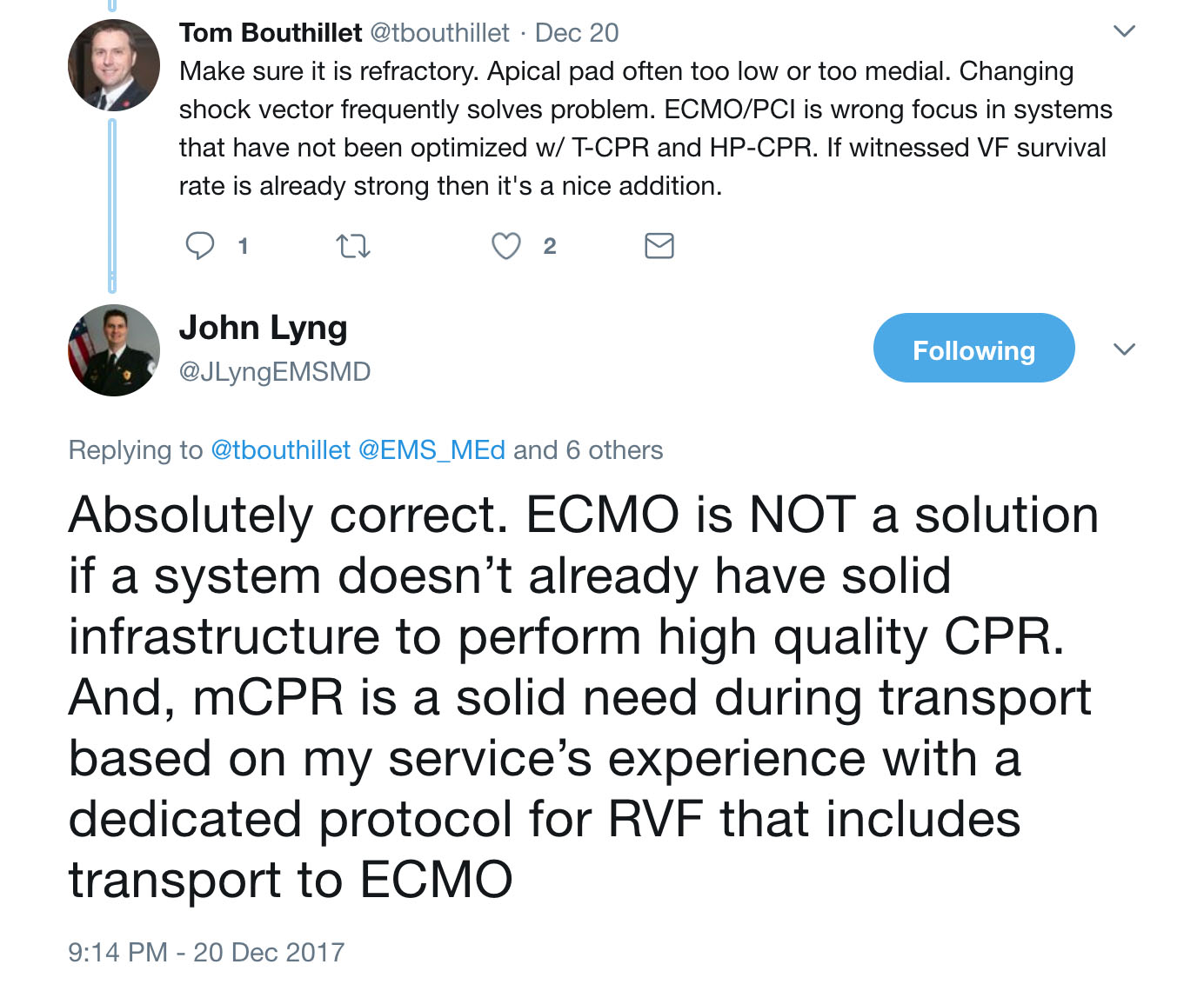
One Japanese study defined refractory ventricular fibrillation as presentation to the hospital in ventricular fibrillation after at least one out-of-hospital defibrillation [1].  In this study, refractory VF accounted for 23% of all patients with VF as a presenting rhythm and 4% of witnessed OHCA.   In other studies, refractory VF was VF that was unresponsive to at least 3 defibrillation attempts and administration of 300 mg of IV amiodarone [2], or a median of 6 defibrillation attempts and anti-arrhythmic administration [3].  As discussed over twitter by Tom Bouthillet and John Lyng, “refractory” may be considered persistent VF despite already excellent high-performance CPR and correctly performed defibrillation:

twitter_refractoryVfib.jpg

If at minimum we define refractory VF as VF unresponsive to the best we have to offer with standard high performance prehospital ALS care, what are some of the non-standard interventions that we may be able to offer?


“There is an ever- growing body of literature to help us understand at least how to get the patient OUT of VF/VT. We are better understanding therapeutic options, be it pharmaceutical, electric (shameless plug for changing pad vectors, double sequential defibrillation), or some of the more aggressive options such as ECMO, an amazing but obviously not universally feasible option” – Aurora Lybeck


Dr. Lybeck starts by mentioning pharmaceutical options and double sequential defibrillation.  Let’s review the evidence regarding these as adjuncts to high performance CPR.

Pharmaceutical options: Recently, a small number of observational studies have been published suggesting that esmolol administration should be considered for patients in refractory ventricular fibrillation.  In a paper published in 2014, Driver et. al. reviewed the cases of 25 patients with OHCA and refractory ventricular fibrillation (no ROSC despite three defibrillation attempts, 300 mg of amiodarone and 3 mg of epinephrine) and arrival in ED with persistent ventricular fibrillation [3].  They compared patients with received esmolol (n=6) to patients who did not (n=19).  Patient’s had a similar proportion of patients with VF as their presenting rhythm and with witnessed arrest. 3/6 (50%) of patients receiving esmolol survived to hospital discharge with good neurologic outcome compared with 2/19 (10.5%) of patients in the no-esmolol group. A subsequent paper published in 2016 by a Korean group (Lee et. al.) was a pre-post study of inclusion of esmolol in a treatment algorithm for refractory VF [4].  Using the same inclusion criteria as the Driver et. al. study, they compared patients who did and did not receive esmolol.  While patients who received esmolol were more likely to get ROSC [9/16 (53%) vs. 4/25 (16%)], there was no statistically significant difference in neurologically-intact survival at 30 days (18.8% in esmolol group vs. 8% in non-esmolol group).  The numbers were overall very small.

Double Sequential Defibrillation: Double Sequential defibrillation (DSD), the use of two sets of pads and defibrillators to deliver two nearly simultaneously shocks at two different vectors, has gained attention as a therapy for refractory ventricular fibrillation.  The majority of examples are case reports [5] or case series [6].  The few retrospective studies that have been published have very small numbers of patients who received the therapy. In Ohio, a retrospective study of 2428 patients with OHCA found that 12 were treated with DSD.  Of these, 9 patients were converted out of VF, with 2 surviving to hospital discharge with a good neurologic outcome (CPC 1 or 2) [7].   A subsequent retrospective review of DSD use in OHCA in London found that of 45 patients treated with DSD in an 18 month period, only 7% survived to hospital discharge.  This rate was comparable to a that in a comparator group that continued to receive standard defibrillation alone [8]. The jury on double sequential defibrillation is still undecided and a randomized control trial does not exist, but it remains something to consider in the case of refractory VF.  

Above and beyond drugs and electricity, Dr. Lybeck mentions another less available but more aggressive intervention – ECMO (aka ECPR).  The goal of ECMO is to restore oxygenation and perfusion while enabling interventions that treat the underlying etiology of the arrest.  In the case of refractory VF, it is worthwhile thinking about etiologies that are not reversible with standard prehospital ALS care.   The most common etiology for refractory Vfib in multiple studies is acute coronary syndrome, anywhere from 42.1% in a French study to 84% in a study in Minnesota [2,8].  Other less common etiologies include aortic dissection and pulmonary embolism [3].  This suggests that what a subset of patients with refractory VF need is coronary reperfusion therapy in order to re-establish a perfusing rhythm. 

ECMO/ Extracorporeal Life Support (ECLS) and coronary reperfusion therapy has been pursued in a number of EMS systems internationally, some with very impressive results.   The CHEER trial was carried out in Melbourne, Australia [10].  They utilized a combination of mechanical CPR, hypothermia, ECMO and early reperfusion for patients with refractory cardiac arrest.  Inclusion criteria included age 18-65 years, cardiac arrest due to suspected cardiac etiology, chest compressions initiated within 10 minutes by bystanders or EMS, an initial rhythm of VF and availability of mechanical CPR.  A total of 11 patients were transported over a 32 month period and 9 received ECLS.  Five of 11 (45%) transported patients survived with good neurologic outcome.  Subsequently, a larger trial has been carried out in the United States (Minnesota).  Using a more protocolized approach, patients with VF/VT as their initial rhythm, age 18-75 yrs and VF refractory to 3 EMS delivered shocks, 300 mg of IV/IO amiodarone, lack of pre-existing severe comorbidities or terminal illness, body habitus to fit within a mechanical CPR device and estimated transfer time from scene to the cardiac catheterization lab < 30 minutes were transported with mechanical CPR in progress. ECLS was initiated in the cardiac catheterization lab and patients underwent cardiac catheterization which identified coronary occlusion in 84%.   62 patients met transport criteria and 55 had ECLS initiated.  Of these, 28 (45%) survived to hospital discharge, 26 of whom (42%) had good neurologic outcome [2].  This was better than outcomes in a historical comparison group (15.3% neuro-intact survival).  These findings are consistent with a prior prospective observational study in Japan comparing outcomes for patients with refractory VF who underwent conventional cardiopulmonary resuscitation versus ECLS [11].   The authors compared neurologically-intact survival for patients transported to tertiary centers that performed ECLS on standard protocol versus those that did not. They found that patients who received ECLS had significantly high neurologically-intact survival (12.3%) than those who did not (1.5%), although these rates were overall lower than those documented in the CHEER and Minnesota trials. This trial was unable to account for differences in baseline care between ECLS and non-ECLS tertiary care centers.

ECLS, however, is a resource-intensive endeavor.  Low threshold for implementation of advanced therapies such as EPCR is not likely to lead to a high value intervention.  Can we identify patients who both require advanced therapies to convert out ventricular fibrillation and are likely to do well?

With the respect to our case, In Princess Bride-like form, Dr. Jeremiah Escajeda stated the following:  “This patient has aliveness. He should be transported …”

1477157576-0.jpg

What features of this patient’s case make it so that he is only “mostly dead”?  Dr. Escajeda goes on to share the criteria in the Pittsburgh area for transport for EPCR:

We have a prehospital alert system in place here in Pittsburgh that when providers identify a refractory organized rhythm, in a "young," healthy person, they speak with a command physician to run our prehospital ECPR checklist. If criteria are met, the patient is then expeditiously transported to our ECPR center with an alert sent to our ECMO team, ED team, Post Cardiac Arrest team and Cardiology. After the patient is placed on the circuit, next destination is cath lab.

Here is the prehospital checklist:
* Strongly suggested to place patient on LUCAS Device as soon as available
* Call attending medic command physician to run checklist

[ ] Witnessed arrest
[ ] Bystander CPR
[ ] Age ≥ 18 and ≤60
[ ] Initial shockable rhythm or PEA rate > 20 bpm
[ ] Good functional status prior to arrest (patient living independently and not from a skilled nursing facility/ LTAC and no prior neurocognitive dysfunction)
[ ] No signs of irreversible organ dysfunction (such as COPD on home O2, stigmata of liver cirrhosis or ESRD such as AV fistula or terminal cancer)
[ ] No morbid obesity (Morbid obesity defined as inability to fit into LUCAS device and/or abdominal pannus overhanging inguinal crease)
[ ] End tidal CO2 >10 mmHg with CPR
[ ] Expected time from collapse to ED arrival <= 30 mins

Hey maybe even one day we will be placing ECLS devices prehospital, has this has already been done in France https://www.ncbi.nlm.nih.gov/pubmed/28414164
and now they have impella devices that deliver 5L/min, and are the size of a pencil. Exciting future for these refractory cases
.”

Image Source/Reference:&nbsp;Reynolds, J. C., Grunau, B. E., Elmer, J., Rittenberger, J. C., Sawyer, K. N., Kurz, M. C., ... &amp; Callaway, C. W. (2017). Prevalence, natural history, and time-dependent outcomes of a multi-center North American coho…

Image Source/Reference: Reynolds, J. C., Grunau, B. E., Elmer, J., Rittenberger, J. C., Sawyer, K. N., Kurz, M. C., ... & Callaway, C. W. (2017). Prevalence, natural history, and time-dependent outcomes of a multi-center North American cohort of out-of-hospital cardiac arrest extracorporeal CPR candidates. Resuscitation.

This prehospital checklist accounts for factors that we already know are associated with favorable neurologically-intact survival from OHCA.  But a critical question (and perhaps gets back to the question of the term “refractory”) is at what time interval should we start thinking about transporting the patient? How can we identify patients who have received the maximum potential benefit of on-scene care while still retaining benefit from care escalation in the form of ECLS?  Many patients will achieve ROSC without EPCR and initiating EPCR too early may distract from continuous, high-quality chest compressions.  The Pittsburgh protocol of time of collapse to ED arrival of < 30 minutes has evidence behind it.  A retrospective study of patients with OHCA within the ROC consortium examined the probability of good neurologic outcome in patients who would be considered eligible for EPCR (met age and pre-cardiac arrest functional status data) versus duration of resuscitation [12]. They found that amongst all eligible patients, the probability of neurologically-intact survival dropped below 10% after 30 minutes of resuscitation (see Figure).  They thus concluded that mobilization towards EPCR resources should be considered after 9-20 minutes of active resuscitation. Interestingly, amongst patients who achieved ROSC, longer durations of CPR were no longer associated with impaired neurologic outcomes (See Figure).  The results of this study concurred with a prior study of consecutive patients age < 65 with witnessed arrest and initiation of CPR in < 10 minutes that concluded that “transport for ECPR should be considered between 8 to 24 minutes of professional on-scene resuscitation, with 16 minutes balancing the risks and benefits of early and later transport. Earlier transport within this window may be preferred if high quality CPR can be maintained during transport and for those with initial non-shockable rhythms.” [13]

Conclusion

In the end, every EMS system has a limited amount of time and resources for training.  The healthcare system itself is resource-limited.  After years of focus on “Airway” before “Circulation”, we have come to the understanding that we need to focus on circulation; excellent BLS care in the form of high quality CPR and early defibrillation is the cornerstone of cardiac arrest care.  However, there are a subset of patients with potential for neurologically-intact survival that may be saved by additional circulatory intervention, including extracorporeal support and coronary reperfusion therapy.  Identifying who these patients are and the best way to both provide this therapy while utilizing limited healthcare resources in a high value manner may be the future of cardiac arrest care. 

Dr. Lybeck said it best, so we’ll end with her quote:

Particularly on the topic of OHCA, it's an exciting time to be an EMS physician, many thanks to our researchers, educators, and advocates out there, keep up the great work!”

Case Summary by  Maia Dorsett,  MD PhD, @maiadorsett

References:

1.     Sakai, T., Iwami, T., Tasaki, O., Kawamura, T., Hayashi, Y., Rinka, H., ... & Kajino, K. (2010). Incidence and outcomes of out-of-hospital cardiac arrest with shock-resistant ventricular fibrillation: data from a large population-based cohort. Resuscitation81(8), 956-961.

2.     Yannopoulos, D., Bartos, J. A., Raveendran, G., Conterato, M., Frascone, R. J., Trembley, A., ... & Wilson, R. F. (2017). Coronary artery disease in patients with out-of-Hospital refractory ventricular fibrillation cardiac arrest. Journal of the American College of Cardiology70(9), 1109-1117.

3.     Driver, B. E., Debaty, G., Plummer, D. W., & Smith, S. W. (2014). Use of esmolol after failure of standard cardiopulmonary resuscitation to treat patients with refractory ventricular fibrillation. Resuscitation85(10), 1337-1341.

4.     Lee, Y. H., Lee, K. J., Min, Y. H., Ahn, H. C., Sohn, Y. D., Lee, W. W., ... & Park, S. O. (2016). Refractory ventricular fibrillation treated with esmolol. Resuscitation107, 150-155.

5.     Lybeck, A. M., Moy, H. P., & Tan, D. K. (2015). Double sequential defibrillation for refractory ventricular fibrillation: a case report. Prehospital emergency care19(4), 554-557.

6.     Cabañas, J. G., Myers, J. B., Williams, J. G., De Maio, V. J., & Bachman, M. W. (2015). Double sequential external defibrillation in out-of-hospital refractory ventricular fibrillation: a report of ten cases. Prehospital emergency care19(1), 126-130.

7.     Cortez, E., Krebs, W., Davis, J., Keseg, D. P., & Panchal, A. R. (2016). Use of double sequential external defibrillation for refractory ventricular fibrillation during out-of-hospital cardiac arrest. Resuscitation108, 82-86.

8.     Emmerson, A. C., Whitbread, M., & Fothergill, R. T. (2017). Double sequential defibrillation therapy for out-of-hospital cardiac arrests: the London experience. Resuscitation.

9.     Pozzi, M., Koffel, C., Armoiry, X., Pavlakovic, I., Neidecker, J., Prieur, C., ... & Obadia, J. F. (2016). Extracorporeal life support for refractory out-of-hospital cardiac arrest: should we still fight for? A single-centre, 5-year experience. International journal of cardiology204, 70-76.

10.  Stub, D., Bernard, S., Pellegrino, V., Smith, K., Walker, T., Sheldrake, J., ... & Cameron, P. (2015). Refractory cardiac arrest treated with mechanical CPR, hypothermia, ECMO and early reperfusion (the CHEER trial). Resuscitation86, 88-94.

11.  Sakamoto, T., Morimura, N., Nagao, K., Asai, Y., Yokota, H., Nara, S., ... & SAVE-J Study Group. (2014). Extracorporeal cardiopulmonary resuscitation versus conventional cardiopulmonary resuscitation in adults with out-of-hospital cardiac arrest: a prospective observational study. Resuscitation85(6), 762-768.

12.  Reynolds, J. C., Grunau, B. E., Elmer, J., Rittenberger, J. C., Sawyer, K. N., Kurz, M. C., ... & Callaway, C. W. (2017). Prevalence, natural history, and time-dependent outcomes of a multi-center North American cohort of out-of-hospital cardiac arrest extracorporeal CPR candidates. Resuscitation.

13.  Grunau, B., Reynolds, J., Scheuermeyer, F., Stenstom, R., Stub, D., Pennington, S., ... & Christenson, J. (2016). Relationship between time-to-ROSC and survival in out-of-hospital cardiac arrest ECPR candidates: When is the best time to consider transport to hospital?. Prehospital Emergency Care20(5), 615-622.


A Call to Action: Elizabeth Rosenthal's An American Sickness

by Melody Glenn, MD

9781594206757_custom-3801ead7ce5df3a0cb7eee5e36afe7976ff4501d-s400-c85.jpg

The voice on the other end of the phone sounds frantic and rushed, “He can’t breathe!” The palpable panic wakes the emergency dispatcher out of his post-lunch daze. He sits up a little straighter and shifts his gaze to the periphery, preparing to listen more closely.  “Okay, tell me exactly what happened.”

“My son is allergic to peanuts, and I think he accidentally ate some. He is wheezing and his voice sounds muffled.  Are you sending someone?!”

“Yes, I’m sending the paramedics to help you now. Stay on the line and I’ll tell you exactly what to do next.” As he flips to the epinephrine auto injector card, he asks, “Does he have any specific injections or other medications to treat this type of reaction?”

“He used to have an epi-pen, but we used it a few months ago.  When I went to refill his prescription, it was $600!  Please, hurry!”


We have all heard about the exorbitant price increases in epinephrine, narcan, and albuterol, and may have seen firsthand the impacts on patient care. But what is the story behind these increases? In An American Sickness, Elisabeth Rosenthal attempts to break down some of the perverse incentives that lead to rising healthcare costs, costs that quickly add up.  Healthcare bills now comprise the greatest percentage of consumer debt, and medical debt is the number one reason why Americans file for bankruptcy.

Image source:&nbsp;https://www.theatlantic.com/health/archive/2014/10/why-americans-are-drowning-in-medical-debt/381163/

Image source: https://www.theatlantic.com/health/archive/2014/10/why-americans-are-drowning-in-medical-debt/381163/

Rosenthal trained in internal medicine, worked for a few years in an ER, and then switched careers to become a journalist/editor. Although sometimes her tone is overly strong, making the writing feel like more of an OpEd than nonfiction, and some of her examples are made to appear more black-and-white than clinical medicine actually is, the overall message rings true.  Health care is a highly profitable industry with a lot of players scrambling to augment their slice of the pie.  As such, healthcare isn’t going to regulate itself.

The first half of the book delves into the problem.  Each chapter focuses on a different segment of the healthcare sector, including pharmaceuticals, insurance companies, hospitals, physicians, and medical devices, showing how each has been guilty of prioritizing their own financial gain over patient value. In our bookclub discussion, we looked at examples that seemed to hold the most relevance to EMS, the first being the shortage of generic medications.  Perhaps the most notorious example is that of Droperidol, and the almost conspiratorial series of events surrounding its disappearance (p. 119-122).

Around 2005, GlaxoSmithKline started to promote the use of Zofran for general nausea, as initially, it had just been marketed as a treatment for chemo-induced nausea. Around that same time, the FDA issued a black box warning linking Droperidol to life-threatening arrhythmias, and a major pharmaceutical company purchased and subsequently closed the plant that was producing the generic compazine. Physicians were stuck using Zofran, which cost several times that of its generic competitors.  Some doctors filed a Freedom of Information Act to obtain the documents that lead to the FDA’s warning.  In the documents, they found that the abnormal heart rhythms were only induced by administering Droperidol in very high quantities -- 50-100 times greater than the standard amount -- and that the same arrhythmias could be provoked by high doses of other anti-nausea drugs (including Zofran). Since then, Glaxo has reached a $3 billion settlement with the U.S. Department of Justice for a variety of misdeeds, but Zofran continues to earn a profit. 

Rosenthal also addresses the potential for conflicts of interest in the creation of clinical guidelines (p.200-204). When specialists write their own guidelines, they have financial incentives to promote their own procedures. For example, urologists recommended that all men be screened with a PSA level, radiologist’s advised yearly mammograms, and orthopedists encouraged arthroscopy to help with knee pain. These recommendations have since been revised based on new evidence questioning their benefit.  Although the majority of EMS and emergency medicine position statements and guidelines  address common conditions and do not encourage expensive diagnostics or treatments, we still get paid for doing more to our patients.  It remains true that very often we work harder to do less, both in terms of time for shared decision making, patient discussion and documentation, and get paid less to so. In EMS, the system aligns financial incentives with transport, which is our default mode, whether or not this is best for the patient.

 In Chapter 9, Rosenthal suggests that the trend towards hospital consolidation often leads to increased costs and decreased quality.  Although initially billed as a way to obtain lower rates through economies of sale, this isn’t necessarily what occurs (p.207).  When one network owns all of the local hospitals and clinics in an area, and employs the majority of physicians, there is no longer any competition.  This effectively gives the conglomerate the leverage needed to demand high rates from corporations, insurers, and HMO’s (p. 207). They can also use their proprietary electronic medical record system as a tool to keep competitors out, which is antithesis to the original intent of moving towards EMR systems (211). Although Rosenthal doesn’t mention EMS in this context, I wonder if the same cautions apply.

Rosenthal only devotes a few pages to EMS (p.157-160), and they don’t seem particularly well-informed. She seems longful for the days when ambulance services were free (p.158). She doesn’t seem to understand that quality has improved and that EMS is now predominantly staff by medical professionals and considered a medical subspecialty. Instead, she attributes the rise in cost to the use of professional billing companies (p.159). Also, she doesn’t address the cost of preparedness. It takes time, preparation, training, and money to be ready to handle any emergency at any time.

Part II of the book provides various strategies that individuals, institutions, and policymakers can implement in order to fix the rising costs of healthcare. She suggests that individuals ask their clinicians how much various diagnostic and treatment options cost. I imagine many would not know, as we are purposefully not taught this information in medical school or residency because of the notion that cost should not drive our decision whether or not a treatment is clinically necessary. Also, there is little price transparency in our medical system, so it is hard for us to even get this information from our own hospital finance departments.  Therefore, she also suggests that patients do their own research. In Appendices A and B, she shares various websites where patients can find the “fair price” for a large number of procedures and medications and see how your hospital compares. Of course, this is less useful in emergency situations.

From the policy perspective, she gives several concrete suggestions, including that:

·       Limits be placed on economic damages in malpractice lawsuits

·       Medical practices offer warranties and guarantees (reducing the need for malpractice suits)

·       Drug companies obtain approval before ceasing a drug’s production

·       The FDA reform their drug patent process to promote low-cost generics

·       The US government work harder to negotiate and set national drug prices

·       A public option of Medicare be available

·       There is more regulation over health insurance companies

·       The Federal Trade Commission become more involved in using antitrust law to break up large conglomerates

Although these solutions, much like the rest of the book, are not written specifically for emergency medicine or EMS, the overall ideas are still relevant. The book’s strong tone is likely designed to serve as a call to action.  We should take this an an opportunity to ask ourselves -- how can EMS be involved in reforming our country’s healthcare system? 

Glenn.001.jpeg

Special thanks to John Brown, MD, MPA, the medical director of the San Francisco EMS Agency and program director of the UCSF-ZSFG Fellowship in EMS and Disaster Medicine, and Thomas Sugarman, MD, FACEP, FAAEM, Secretary-Treasurer of the Alameda Contra Costa Medical Association, Co-Chair of the East Bay Safe Prescribing Coalition and a Past-President of the California chapter of the American College of Emergency Physicians, for adding their expertise to the bookclub discussion.


When Vfib is Stubborn…

wall_Vfib.strip.jpg

A 56 year old male is cleaning out his garage with his wife when she hears him fall.  She turns around to find him unresponsive on the ground.  He is making gasping breath sounds but otherwise does not respond when she shakes him and yells at him.  She is instructed to perform CPR by pre-arrival instructions after calling 911 on her cellphone.  Within minutes, the BLS Fire Department arrives and takes over.  After confirming that the patient is pulseless, they resume CPR while applying an AED.   The AED states “shock advised” for ventricular fibrillation.  After resuming CPR after one shock is delivered, the ALS ambulance arrives.  High performance CPR is continued and the patient is defibrillated three more times for persistent ventricular fibrillation.  The end-tidal CO2 is 40 mmHg.   The patient has now been pulseless for almost twenty minutes.  The paramedics plan on continuing high performance CPR, but wonder what they will do if the patient remains in ventricular fibrillation with a good end-tidal 10 minutes from now…

As we have improved the care of patients in out-of-hospital arrest, many agencies are now facilitating advanced therapies for patients with refractory ventricular fibrillation.

How does your EMS system manage these patients?  Please share your comments below.  A summary of discussion points will posted to the blog at the end of the month.

Challenging the Dogma of “All Clear”: Is Hands-On Defibrillation the Next Step in Reducing the Peri-shock Pause?

by Brandon Bleess, MD EMT-T and Jeremy Cushman, MD, MS, EMT-P, FACEP, FAEMS

Case:

halfcolor.jpg

A 54-year-old male is working in his yard when he collapses to the ground and his wife calls 911.  The call is dispatched as a cardiac arrest and the patient's wife is instructed to perform CPR per pre-arrival instructions.  Upon EMS arrival, the patient is found to be  apneic and pulseless.  EMS relieves the patient’s wife and compressions are continued while the patient is connected to the monitor.  The monitor is charged prior to the next pulse check where the patient is noted to be in ventricular fibrillation.  Everyone takes the time to drop contact with the patient as the operator declares “everyone clear” before shocking and resuming compressions and ventilation.   

When debriefing the case, one of the paramedics brings up the idea of hands-on defibrillation that he attended a lecture on at a recent conference.  Is it safe to defibrillate a patient while CPR is actively being performed?

The Evidence For:

There is little question that high quality, continuous compressions improve neurologic outcomes in patients with out-of-hospital cardiac arrest.  The choice of outcome here is important: compressions are really to maintain some oxygenated blood flow to the brain in order to keep it alive long enough to get the heart started again.  All too often, ROSC is achieved but the patient has anoxic brain injury which can be a result of extended down time or ineffective compressions. 

Thus the goal in performing compressions is to minimize interruptions, for every time compressions are stopped it takes a significantly longer time to return to the flow state that existed just prior to stopping them [1-5].  With the goal of increasing compression fraction and thus improving neurologic outcomes,  the American Heart Association (AHA) began recommending charging the defibrillator during chest compressions in 2005 [6].  Multiple studies have since demonstrated that charging during compressions decreases both post- and peri-shock pauses [7-9].  These included the Resuscitation Outcomes Consortium (ROC) PRIMED trial which found that the median peri-shock pause was reduced from 21 seconds to 9 seconds with compressions during charging. This reduction in peri-shock pause lead to a significant increase in mean chest compression fraction  (0.77 vs 0.70, 95% CI: 0.03-0.11), an independent factor in increasing survival [9, 10, 11].  Thus, the traditional analyze – charge - shock pattern should be changed to charge – analyze- shock.

But can we go one step further and continue CPR during defibrillation?  After decades of “I’m clear, you’re clear, we’re all clear,” it might be time to let that go the way of the backboard in the closet of EMS dogma.  Traditionally, external defibrillation has been considered a safety hazard to rescuers and clearing the patient is an almost universal practice, but there is little data to support this notion in the age of adherent defibrillation pads [12].  Indeed, there is now evidence to suggest that hands-on defibrillation is actually safe.

How much energy is potentially transferred to the compressor during hands-on defibrillation? Defibrillators generally deliver 30-40 A of current with each shock, and the threshold for perception is 2.5-4.0 mA, and exposure becomes painful at 6-10 mA.  In healthy adults 200-500 mA of current is thought to be needed to induce ventricular fibrillation with incidence correlating linearly to increasing current [12].

Lloyd et. al. found that the amount of "leakage" (the amount of current going to the provider) in an ideal setting (outpatient cardiac electrophysiology evaluations) was well below the allowable minimum [13].  Keep in mind, this study was looking at microamps of current.  The allowable being 3,500 microamps and the leakage was an order of magnitude less than that.  To put this into further perspective, when pacing patients with transcutaneous pacing generally 60-80 mA is required for capture.  Compared to the study’s mean leakage of 283 µA or 0.28 mA, transcutaneous pacing uses over 200 times the amperage to create capture.

Neumann et al. in 2012 followed up this study by inducing ventricular fibrillation in a swine model and comparing hands-on versus hands-off defibrillation.  They found that in the hands-on group, chest compressions were interrupted for 0.8% versus 8.2% of the total CPR time (P=0.0003) and coronary perfusion pressure was restored earlier to its pre-interruption level (P=0.0205). They also found that not only was the defibrillation shock imperceptible, but the compressor wearing a cardiac monitor had no arrhythmias noted [14].

Insulating the provider from current leakage is probably one of the easiest ways to protect the compressor from harm.  This was studied by Deakin et al. in 2015 using Class 1 electrical insulating gloves while simulating hands-on defibrillation.  They found that the median current leakage was 20 μA from the 61 shocks studied, and even at 360 J the median current leakage was 27 μA.  The highest recorded leakage was 28 μA, all below the 1 mA threshold they set [15].  More recently in 2016, Wampler et al. published a study looking at perception of shocks using multiple insulating barriers, including nitrile gloves, firefighting gloves, a neoprene pad, and a manual compression/decompression device.  Out of the 100 shocks with no barrier device, all but 1 shock was not detected.  Out of 500 shocks, only 5 were detected by the compressor - none causing harm, and most importantly the CPR puck prevented any detection [16].  

The Evidence Against:

Closer evaluation of these studies may give some rescuers pause.  In the 2008 Lloyd study, 8 of the 72 phases (36 shocks, 11.1%) exceeded the 500 µA threshold [13].  In the Neumann study of 2012, the compressors wore 2 pairs of polyethylene gloves which is outside the standard practice of many providers [14]. While Deakin’s study presents some very convincing data in relation to the safety, the compressor was wearing Class 1 electrical insulating gloves.  According to the Occupational Safety and Health Administration (OSHA) regulation 29 CFR 1910.137, Class 1 gloves are rated for a maximum usage of 7500 V AC and are proof tested to 10,000 VAC and 40,000 VDC [18].  Defibrillators typically fall well below this at around 2700 V for a biphasic defibrillator. 

In 2014, Lemkin et. al. published a cadaver study where they measured the differential of the voltages at various points on the body with several skin preparations (bare, water, saline, ultrasound gel).  This allowed them to collect the resistance and exposure voltage in relation to anatomic landmarks to create a map of the providers’ exposure.  From there, they derived a formula to measure the rescuer-received dose (RRD) to represent the proportion of energy the rescuer could receive from a shock to a patient.  Their results demonstrated the rescuer-exposure could exceed 1 J in any location at some energy level, and reached as high as 9.4J on the anterior chest wall [17].  The argument made is that 1 J could potentially cause a provider to be shocked into ventricular fibrillation themselves.  Since this does not include any barriers, it would apply if there was a large tear in the glove or the rescuer was contacting the patient without any gloves.

Conclusion:

While the literature suggests that hands-on defibrillation is safe, it should occur at the discretion of the compressor who should consider wearing two pairs of gloves or utilize both gloves and a CPR-feedback device if performing the procedure.  There are no studies that have measured the effect of this on clinical outcome, although it is postulated that increased compression fraction is a useful surrogate.  At minimum, the charge-analyze-shock method should be used during every defibrillation to minimize the hands-off time and increase the compression fraction.

author_bios.001.jpeg

For additional Resources, REBEL EM published the following review of Hands-On-Defibrillation: CPR Hands-On or Hands-Off Defibrillation

EMS MEd Editor:  Maia Dorsett (@maiadorsett)

References:

1.     Berg RA, Sanders AB, Kern KB, et al. Adverse hemodynamic effects of interrupting chest compressions for rescue breathing during cardiopulmonary resuscitation for ventricular fibrillation cardiac arrest. Circulation. 2001 Nov 13;104(20):2465-70.

2.     Cunningham LM, Mattu A, O’Connor RE, Brady WJ. Cardiopulmonary resuscitation for cardiac arrest: the importance of uninterrupted chest compressions and cardiac arrest resuscitation. Am J Emerg Med. 2012 Oct;30(8):1630-8.

3.     Paradis NA, Martin GB, Rivers EP, et al. Coronary perfusion and the return of spontaneous circulation in human cardiopulmonary resuscitation. JAMA. 1990 Feb 23;263(8):1106-13

4.     Hazinski MF, Nolan JP, Aicken R, et al. Part 1: executive summary: 2015 International Consensus on Cardiopulmonary Resuscitation and Emergency Cardiovascular Care Science With Treatment Recommendations. Circulation. 2015;132(16)(suppl 1).

5.     Neumar RW, Shuster M, Callaway CW, et al. Part 1: executive summary: 2015 American Heart Association Guidelines Update for Cardiopulmonary Resuscitation and Emergency Cardiovascular Care. Circulation. 2015;132(18)(suppl 2).

6.     2005 American Heart Association Guidelines for Cardiopulmonary Resuscitation and Emergency Cardiovascular Care. Circulation. 2005;112:IV1–IV203.

7.     Perkins GD, Davies RP, Soar J, Thickett DR. The impact of manual defibrillation technique on no-flow time during simulated cardiopulmonary resuscitation. Resuscitation. 2007;73:109–114.

8.     Edelson DP, Robertson-Dick BJ, Yuen TC, et al. Safety and efficacy of defibrillator charging during ongoing chest compressions: a multi-center study. Resuscitation. 2010;81(11), 1521-1526.

9.     Cheskes S, Schmicker RH, Verbeek PR, Salcido DD, Brown SP, Brooks S, Menegazzi JJ, Vaillancourt C, Powell J, May S, et al. The impact of peri-shock pause on survival from out-of-hospital shockable cardiac arrest during the Resuscitation Outcomes Consortium PRIMED trial. Resuscitation. 2014 Mar; 85(3):336-42.

10.  Christenson J, Andrusiek D, Everson-Stewart S, et al. Chest compression fraction determines survival in patients with out-of-hospital ventricular fibrillation. Circulation. 120 (2009), pp. 1241-1247.

11.  Cheskes S, Schmicker RH, Christenson J, et al. Peri-shock pause: an independent predictor of survival from out-of-hospital shockable cardiac arrest. Circulation. 124 (2011), pp. 58-66.

12.  Brady W, Berlat JA. Hands-on defibrillation during active chest compressions: eliminating another interruption. Am J Emerg Med. 2016 Nov;34(11):2172-2176.

13.  Lloyd MS, Heeke B, Walter PF, Langberg JJ.  Hands-on defibrillation: an analysis of electoral current flor through rescuers in direct contact with patients during biphasic external defibrillation. Circulation. 2008 May 13;117(19):2510-4.

14.  Neumann T, Gruenewald M, Lauenstein C, Drews T, Iden T, Meybohm P. Hands-on defibrillation has the potential to improve the quality of cardiopulmonary resuscitation and is safe for rescuers—a preclinical study. J Am Heart Assoc. 2012 Oct;1(5):e001313.

15.  Deakin CD, Thomsen JE, Løfgren B, Petley GW. Achieving safe hands-on defibrillation using electrical safety gloves—a clinical evaluation. Resuscitation. 2015 May;90:163-7.

16.  Wampler D, Kharod C, Bolleter S, Burkett A, Gabehart C, Manifold C. A randomized control hands-on defibrillation study- Barrier use evaluation. Resuscitation. 2016 Jun;103:37-40.

17.  Lemkin DL, Witting MD, Allison MG, Farzad A, Bond MC, Lemkin MA.  Electrical exposure risk associated with hands-on defibrillation. Resuscitation. 2014 Oct;85(10):1330-6

18.  https://www.grainger.com/content/qt-electrical-safety-gloves-inspection-262


EMS: The Best Kept Secret in Healthcare

by Maia Dorsett, MD PhD

cover_photo.jpg

At the end of my third year of residency, I was in the process of solidifying my decision to pursue a fellowship in EMS.   I was on rotation in the Medical ICU and we were having an informal conversation about plans following residency.   When I stated that I planned to pursue an EMS fellowship, the ICU Attending asked what it was. 

My response?

EMS is the subspecialty of medicine that encompasses provision care beyond the borders of the hospital, at the level not only of individual patients but the entire community.  That the care was not limited to 911 response in the traditional sense, but also public health, community education, disaster preparedness, provision of continuity of care following hospital discharge and in fact, to every critically ill patient transferred to his very own ICU.   From then on, I pulled up available EMS records on every admission to point out the critical and often life-saving interventions provided to patients before they entered the hospital borders. My mission was to highlight the scope and importance of care provided by EMS providers. 

I am not sure if this ICU attending – and the countless others who stated that they have never heard of an ‘EMS fellowship’ - were unaware of what EMS stands for.  I think that they did not recognize the term in the context in which it was presented; they did not recognize it as a physician subspecialty, let alone a practice of medicine.  I’m sure that those who did not recognize the term ‘EMS fellowship’ would expect a prompt and competent medical response if they were to call 911 from their living room or public place.  In the grand scheme of things, EMS is relatively new.  Accidental Death and Disability, which spurred the development of both EMS and Emergency Medicine, was only published a half century ago.   EMS was only approved as a physician subspecialty in 2010, with the first board certifying examination offered in 2013.   Like many developments that are also relatively young– the internet, cellular data network - EMS has become an assumption of peoples’ lives.   Much like the delayed knowledge translation window between quality research and change in practice, moving the behemoth of the house of medicine to change the way it thinks is a long, arduous and inefficient process.

When it comes to recognition of EMS as a practice of medicine, we need to speed things up.  Advocacy for our specialty is advocacy for our patients.  

The reason? 

Failure to recognize EMS as a practice of medicine stunts the growth of the specialty towards the model set out in the EMS Agenda for the Future:

“Emergency medical services (EMS) of the future will be community-based health management that is fully integrated with the overall health care system. It will have the ability to identify and modify illness and injury risks, provide acute illness and injury care and follow-up, and contribute to treatment of chronic conditions and community health monitoring. This new entity will be developed from redistribution of existing health care resources and will be integrated with other health care providers and public health and public safety agencies. It will improve community health and result in more appropriate use of acute health care resources. EMS will remain the public’s emergency medical safety net.”

While small steps have been made, the defacto situation is that EMS is reimbursed as a taxi service, mobile integrated healthcare programs are stunted, EMS providers are disrespected and underpaid, national certification of EMS providers fails to be 100% nationally accepted, EMS research is still underperformed and underfunded, hospitals fail to share outcome data and operational metrics rule assessments of EMS quality. 

There are many different approaches to changing the status quo.  EMS physicians and providers with significantly more experience and knowledge than me are pursuing those routes.  But as someone new to EMS (a lab nerd turned emergency physician who caught the EMS bug mid-residency), I can tell you that part of every approach needs to be explaining the specialty of EMS not only to the public and lawmakers, but to our colleagues in medicine.  I have now given talks on the principles of and barriers to Mobile Integrated Healthcare in a limited number of venues – three different EM residencies and a conference on healthcare overuse.  In every situation, audience members have been surprised and inspired by how EMS can be used to provide patient-centered care with decreased healthcare utilization.  They have been similarly frustrated by the payment by transport model.  They have shared in our vision for a truly integrated healthcare system. 

For our healthcare system to meet its potential to improve the health of our communities, it must be transformed.  Many of us became EMS physicians because we wanted to be part of this transformation.

Those of us who took the EMS Boards in September are anxiously awaiting exam results.  Many if not most of us put ourselves through the exam not for better pay or a new position, but because of dedication to the specialty – to the knowledge that we can positively influence the lives of an incredible number of people by improving the quality of care they receive on a system-wide level.  It’s time that our colleagues in medicine understood what we actually do.

Untitled 2.001.jpeg

A debate of supraglottic proportions... the conclusion

supraglottic_cover.jpg

The Case

It’s a brisk fall afternoon when an ALS unit is dispatched to the home of a 62 yo female in respiratory distress.  She lives on the third story of an apartment building.  The team carries their gear upstairs to find a woman in severe distress.  She is obese, tripoding and beginning to get combative.  The medics are setting up their CPAP and calling for assistance when the patient stops fighting and becomes apneic.  Two-person BVM is initiated but the patient is difficult to ventilate and oxygen saturations remain poor.  The medic decides to attempt to intubate, but is unsuccessful after one attempt and they decide to move on to a supraglottic airway.

Which supraglottic airway should they be using?

What supraglottic airway is available within your EMS system?  Why is it preferred and what are its advantages over the alternatives?

Our readers shared their comments - and there were a lot of them.  A number of important perspectives were voiced by our readership regarding their preferred supraglottic airway.  


Supraglottic Airways: A Brief Review

First developed as an operating room adjunct, supraglottic airways have now been widely adopted in prehospital care. There are a wide-variety of subtypes [1], but our readers described prehospital use of predominantly two subtypes:

Laryngeal Mask Airway (LMA): The LMA was invented in 1988 by a British Anesthesiologist, Dr. Archie Brain.  It involves a mask component connected to a ventilation tube and is designed to sit in the patient’s hypopharynx and cover the supraglottic structures. Most LMAs are elliptical in shape with an inflatable cuff.  The i-gelTM, a modification of the LMA device, was invented in 2003.  Rather than having an inflatable cuff, the i-gelTM is made from a thermoplastic elastomer that conforms to the pharyngeal and laryngeal anatomy. (Figure 1A).  Most widely used LMA models, such as the LMA-SupremeTM   or the i-gelTM , have ports to allow for gastric decompression.

Laryngeal-Tube (LT): The laryngeal tube was first introduced in the US in 2003 by King Systems.   The LT is designed to intubate the esophagus.  The device has two cuffs, a distal esophageal cuff and a proximal oropharyngeal cuff, which inflate with a single inflation port (Figure 1B).   It is available with (LTS-D) and without (LT-D) a gastric decompression port.


Figure 1: Ideal positioning of LMA-type and LT supraglottic airways.&nbsp;

Figure 1: Ideal positioning of LMA-type and LT supraglottic airways. 


Ease of Use

The majority of commenters used or preferred an LMA (and the i-gel TM   specifically) because they felt that it was easy to use and lead to a high first pass success rate:

“Igel! Easy to use, quick...” – Phil


“We having been using igel for several years and very happy. Success first pass greater than 90% easy to switch to et no cuffs.” – Peter


“From a moderate-sized, midwestern, single county urban system: Our system (paramedics) began using the igel over a year ago and we are just now starting to work with select first response agencies to incorporate the device in their tool box. We have had better first pass success rates with the igel than the King. There are less "moving parts" and it seems to be easier to troubleshoot and replace if necessary due to size/fit issues.”  - Dena Smith


We formerly used King LT, but switched to LMA Supreme a year or two ago system wide. Providers prefer the LMA.” – Ian Smith


“King but now looking at switching to I-gel as easy to use and have good feedback in field trials.” – Jeff Rabrich


“We have been using the LMA Supreme for about 5 years with great success”. – Gary McCalla


“WE like the IGel.
Easier to train on and use than the prior King.
Capital City Fire & Rescue, Juneau, AK” - Quigley Peterson


“We use Igel.
easy, quick. no cuff to inflate, no syringes, soft material and hard to inflict airway trauma.
We have first pass success rate for igel on cardiac arrest patients of around 93% (50% placed by BLS and 50% placed by ALS)....no difference in success rate between ALS/BLS.
this is after several hundred deployments over past 1.5 years.
vomit is issue, but it is issue with any airway including ET tubes.
moved to Tube Tamers to secure (can accommodate ET or supraglottics) (using OR models).” – Ryan Jacobsen


“Approximately one year into Igel (replacing King) with summary stats not yet available. Choice was made based upon some evidence for more rapid placement, Greater size range, Some local events of balloon failures with King (perhaps, technique related), and a theoretic concern regarding carotid blood flow in humans lead to our decision to use the Igel as our 'rescue' airway.”- Jim Nania M.D., Spokane County EMS, Washington State


“King! Easy to use. Some evidence to support ease of use and successes. https://www.ncbi.nlm.nih.gov/m/pubmed/17907036/?i=5&from=king%20airway%20guyette”  - - -Jeremiah Escajeda


What do published studies say about first and second pass success rate?

A number of studies have been performed to either evaluate a supraglottic placement success in live patients.  There are relatively few randomized trials, and all were in elective surgical cases.  Overall, the first pass placement success rates of the devices are variable: King LTD (53-92%), LMA supremeTM  (72-96%), and i-gelTM (74-97%) and the variability in operators and clinical setting make it difficult to determine if there are any clinically significant differences in device-placement success (Table 1), [2-18].

supraglottic_success_v2.jpg

While the table above by no means represents a rigorously-derived summary such as would be included in a systematic review, one simple observation is that each device has predominantly been evaluated in the clinical setting for which it was initially developed: LMA devices (i-gelTM and LMA-SupremeTM) in the operating room and LT in out-of-hospital studies.   This difference in intended setting was the topic of a subset of comments left on the discussion forum post:


“We switched to igel for a one year trial. So far results are mixed. Our medical director doesn't like the long list of manufacturers contraindications. i.e. Non-fasted patients for routine and emergency anaesthetic procedures. Patients with an ASA or Mallampati score of III and above. Trismus, limited mouth opening, pharyngo-perilaryngeal abscess, trauma or mass.If this device is a rescue airway for an unsuccessful ETI. If it was an unsuccessful intubation attempt due to difficulties or trauma, an igel is contraindicated. Not to say the king was any better or worse but it didn't have the box contraindications. Would love some more provider insight.” - Joshua


“This is a great discussion and where we need to focus on the context of the airway's application. First, the Mallampati scale holds virtually no relevance to emergency situations out of hospital. By definition, many of our patients will be Class III and above due to the presence of an acute, life threatening condition. In fact, the lack of visualization of the posterior oropharynx (Mallampati 4) might serve as an actual indication for these devices!” – Ben Lawner


“Let me start with a disclosure. I'm an anesthesiologist and paramedic. Our system in Upstate NY is a King system and I would like us to become an iGel system. The King was designed as a rescue airway tool for EMS. LMAs in general are a hospital tool that has come over and been adapted to EMS as a rescue device. I'm just guessing here, but my hospital system probably uses > 500 disposable LMAs for every one King airway that comes into our Level 1 quaternary academic medical center. The market for LMA manufacturers is dramatically bigger than the market for King airways.
If we polled physicians that manage airways in the US and asked what type of airway is available to them as a SGA for rescue purposes, what percentage would say King airways? I am currently not aware of any hospitals that purchase King Airways as rescue devices and they certainly don't buy them as primary airway devices. In fact, the only reason most anesthesiologists know about King airways is because we are occasionally called up the EM department to switch them out to an ETT.
 I'm not aware of any direct evidence comparing the two. It's pretty obvious to me though that less cuffs and balloons means less chance of malfunction in a uncontrolled environment. The iGel can be switched out to an ETT in a much safer manner once oxygenation has been achieved. The iGel has nothing that breaks or tears. I could see the King being preferred in a patient who required very high peak inspiratory pressures to achieve adequate ventilation, but that's pretty nuanced for a rescue device. 
Joshua, your medical director won't see those contraindications change anytime soon. EMS is likely the King airway's near total business line. EMS is a tiny portion of the iGel's manufacturers business, so they probably won't bother to make that investment. I can assure you that LMAs are used as rescue devices in CICO or CICV (cannot intubate, cannot oxygenate/ventilate) situations in hospitals around the world on a daily basis. LMAs have saved more than a few patient's lives in my practice and will continue to do so. I'll let you know when I start using the King, but don't hold your breath.”  – Christopher Galton

Although not have been developed specifically for prehospital use, the LMA-devices are widely used as rescue devices in emergency and prehospital settings, including in cases of severe facial trauma [19,20]


Facilitation of intubation:

In his commentary above, Dr. Galton brings up an important point that was echoed by other commenters – the ability to intubate through the device rather than needing to remove the device to intubate.

“IGel and King used in SW Ohio. Prefer IGel...it’s easier to use and capable of exchange for ETT in Hospital without removing the device. King has to be removed for patient to be intubated. I’m intrigued, though, by the Intubating King assuming it eventually becomes available in US.” – Josh B


 “We're an iGel shop. I moved us from the King several years ago and we've been pleased. I disliked the airway maceration I saw in the ED when I eventually swapped out the King for an ET. – Jeff Jarvis

With its current design, the LT must be removed in order for endotracheal intubation to occur.  One concern for any airway manipulation that occurs prior to endotracheal intubation is whether a supraglottic device may cause enough perilaryngeal tissue trauma to make subsequent endotracheal intubation more difficult.  In a randomized comparison of the i-gelTM, LMA SupremeTM and LTS-D devices in the operating room, the authors evaluated the incidence of “airway morbidity” caused by each of the devices [3]. They assessed how often the device had blood on the outside of it after removal and whether patients later complained of sore throat or dysphagia.  In comparison with the i-gel (13%) or LMA-supreme (13%), the LTS-D more often had blood on the outside of the device (37.5%, p=0.006).  This correlated with a significant increase in the incidence of subsequent sore throat or dysphagia.  Whether this type of “airway morbidity” has any predictive value at all for increased difficulty in airway securement after device removal is unclear.

The ventilation port of i-gelTM airway is large enough to facilitate subsequent endotracheal intubation through the device [21].  This ideally should be performed using fiber-optic guidance as blind endotracheal intubation through an i-gel has a low success rate at least in a manikin study [22].  Exchange of a King-LT over a gum-elastic bougie should not be pursued; in one cadaver, this led to penetration of the right aryepiglottic fold by the bougie which subsequently ended up in the soft tissues of the neck [23].   Intubation around the King airway using video laryngoscopy and a gum-elastic bougie has been described [24]. 


Safeguards are key.

One group of commenters made the important point that no matter which supraglottic was used, correct placement and adequate oxygenation and ventilation must be ensured:

 “Either as long as you use waveform capnography to confirm placement! No airway is foolproof....must be confirmed!” – Veer Vithalani


“Veer is spot on about requiring EtCO2 just like we do for intubation (great paper!).” – Jeff Jarvis


“I have both King and iGel at my agencies. Both are widely used and accepted by my crews.
We have slowly moved toward the iGel for a few reasons:
1. No balloon to inflate
2. No added pressure (from a balloon) in the hypopharynx which doesn't impede carotid flow (pig and cadaver studies)
3. Gastric port (12 Fr) can be inserted into the stomach (except for size 1)
 Downsides of the iGel:
1. No gastric port for the size 1
2. Packaging for the iGel consumes a lot of space compared to king
3. Cannot use commercial tube holders to stabilize the pediatric sizes.
a. Smaller sizes do not have the strap - adult sizes do.
b. Without the strap the iGel may "pop" out ever so slightly and the provider may not realize it
4. Intersurgical requires that the agency sign a waiver since the product was not intended for field airway use
 My overall feeling is that iGel is preferred, yet I like the King and agree with what Veer said in his comment.”- Peter Antevy


“Agree with the comments about the absolute need for capnometry. Our first responders are using a colormetric device and our paramedics use waveform capnometry. We do have prolonged resuscitation times (we generally do not transport unless we have ROSC and have stabilized the patient). As with any device there are considerations, however training and feedback to providers seem to increase the success of its use.” – Dena Smith


As voiced in the commentary by Dr. Vithalani, as with endotracheal intubation, supraglottic airways should always be confirmed with continuous in-line capnography to confirm both initial placement as well as safeguard against unrecognized dislodgment of the device.  Supraglottic airways should be secured, as they will dislodge with similar force to an endotracheal tube [24]. Vithalani et. al. studied the incidence of unrecognized failed airway management using a supraglottic airway device (King LTS-D) within their EMS system [25].  They retrospectively reviewed continuous capnography tracings of 344 the supraglottic airway attempts.  Objective successful airway placement was defined as a sustained 4-phase end-tidal waveform greater than or equal to 5 mmHg for the duration of patient care, while subjective successful placement was defined as documentation of successful placement by the EMS provider.  They found that only 85.1% of subjectively successful SGA placements met objective criteria for successful placement.  Conversely, 4 of 28 (14%) of SGA airways that were removed because they were deemed failed by the providers actually met the objective criteria for success.  The main conclusion of this paper is an important one:


“This study points to the critical necessity for objective measurement of airway management utilizing a supraglottic airway device… adequate education, training and quality assurance processes must be in place to ensure appropriate use and interpretation of continuous waveform capnography by EMS providers.”


Agreement on type of device, adequate system-wide training on its use and subsequent quality review to ensure that it is used with proper indications and quality controls remain both barriers and requirements for effective implementation or system-wide change:


“We've used intubating LMAs (disposable), Kings and now Air Qs. All work fairly well. In my opinion the most important thing is train intensively, QA thoroughly and make sure your paramedics have a healthy respect for the difficult airway.” - Marc Restuccia


“Currently using King LT, which we switched to from Combi-tube a number of years ago. Contemplating a switch to iGel, based on reported simplicity of use and reported good results. One challenge is getting 2 EMS medical directors and 13 EMS agencies to come to agreement for a system-wide change.” – Paul Rostykus


Patient-centered outcomes

“In terms of evidence base, there's really not a lot when it comes to the best "backup airway" decision based upon patient centered outcomes. The supraglottic airways can certainly temporize a difficult situation, but I struggle with evidence based recommendations. The King Airway seems quite popular, but I've encountered more than a few problems with dislodgement and ineffective ventilation. In terms of tried and true airways, the "LMA advantages" include: ease of insertion, quick deployment, and relative lack of side effects. LMAs have been used successfully for quite some time and are arguably the most well studies. Granted, we adapt airways for prehospital use, and there really is no "one size fits all" when it comes to the airway management of sick patients in the out of hospital setting.” – Ben Lawner


As voiced by Dr. Lawner, the supraglottic debate is similar to many clinical situations in prehospital care where there are few evidence-based recommendations to support clinical decision making based on patient-centered outcomes.  Many aspects discussed with respect to supraglottic airways – such as of ease of use and successful placement or effect on carotid blood flow – may be useful surrogates for patient-centered outcomes but fall very short of where we as a specialty need them to be.  Ease of use is basically an operational outcome, but quality in medicine is really about patient outcome and from this perspective, the debate of supraglottic proportions continues. 


Take Home

The most commonly used devices amongst our readers are the i-gelTM, King LT, and LMA-SupremeTM.  Current data regarding overall ease of use find overall high success rate within two attempts for all devices.  Regardless of which device is used, there must be rigorous training not only on placement, but continuous end-tidal capnography as a means to ensure initial placement and prevent unrecognized device dislodgment.  


Summary of discussion comments by EMS MEd Editor, Maia Dorsett MD, PhD (@maiadorsett)


For an excellent review and more in-depth discussion of supraglottic airways, we highly recommend Darren Braude’s talk available through the EMS Medicine Live site:  http://www.ems-medicine.com/single-post/2016/05/31/Extraglottic-Airways-Updates-Controversies


References

1.     Ostermayer, D. G., & Gausche-Hill, M. (2014). Supraglottic airways: the history and current state of prehospital airway adjuncts. Prehospital Emergency Care18(1), 106-115.

2.     Gatward, J. J., Cook, T. M., Seller, C., Handel, J., Simpson, T., Vanek, V., & Kelly, F. (2008). Evaluation of the size 4 i‐gel™ airway in one hundred non‐paralysed patients. Anaesthesia63(10), 1124-1130.

3.     Russo, S. G., Cremer, S., Galli, T., Eich, C., Bräuer, A., Crozier, T. A., ... & Strack, M. (2012). Randomized comparison of the i-gel™, the LMA Supreme™, and the Laryngeal Tube Suction-D using clinical and fibreoptic assessments in elective patients. BMC anesthesiology12(1), 18.

4.     Fenner, L. B., Handel, J., Srivastava, R., Nolan, J., & Seller, C. (2014). A Randomised Comparison of the Supreme Laryngeal Mask Airway with the i-gel During Anaesthesia. J Anesth Clin Res5(440), 2.

5.     Francksen, H., Renner, J., Hanss, R., Scholz, J., Doerges, V., & Bein, B. (2009). A comparison of the i‐gel™ with the LMA‐Unique™ in non‐paralysed anaesthetised adult patients. Anaesthesia64(10), 1118-1124.

6.     Weber, U., Oguz, R., Potura, L. A., Kimberger, O., Kober, A., & Tschernko, E. (2011). Comparison of the i‐gel and the LMA‐Unique laryngeal mask airway in patients with mild to moderate obesity during elective short‐term surgery. Anaesthesia66(6), 481-487.

7.     Mitra, S., Das, B., & Jamil, S. N. (2012). Comparison of Size 2.5 i-gel™ with ProSeal LMA™ in anaesthetised, paralyzed children undergoing elective surgery. North American journal of medical sciences4(10), 453.

8.     Jagannathan, N., Sommers, K., Sohn, L. E., Sawardekar, A., Shah, R. D., Mukherji, I. I., ... & Seraphin, S. (2013). A randomized equivalence trial comparing the i‐gel and laryngeal mask airway Supreme in children. Pediatric Anesthesia23(2), 127-133.

9.     Theiler, L. G., Kleine-Brueggeney, M., Kaiser, D., Urwyler, N., Luyet, C., Vogt, A., ... & Unibe, M. M. (2009). Crossover comparison of the laryngeal mask supreme™ and the i-gel™ in simulated difficult airway scenario in anesthetized patients. Anesthesiology: The Journal of the American Society of Anesthesiologists111(1), 55-62.

10.  Das, B., Mitra, S., Jamil, S. N., & Varshney, R. K. (2012). Comparison of three supraglottic devices in anesthetised paralyzed children undergoing elective surgery. Saudi journal of anaesthesia6(3), 224.

11.  Bamgbade, O. A., Macnab, W. R., & Khalaf, W. M. (2008). Evaluation of the i‐gel airway in 300 patients. European Journal of Anaesthesiology (EJA)25(10), 865

12.  Wharton, N. M., Gibbison, B., Gabbott, D. A., Haslam, G. M., Muchatuta, N., & Cook, T. M. (2008). I‐gel insertion by novices in manikins and patients. Anaesthesia63(9), 991-995.

13.  Middleton, P. M., Simpson, P. M., Thomas, R. E., & Bendall, J. C. (2014). Higher insertion success with the i-gel® supraglottic airway in out-of-hospital cardiac arrest: A randomised controlled trial. Resuscitation85(7), 893-897.

14.  Hagberg, C., Bogomolny, Y., Gilmore, C., Gibson, V., Kaitner, M., & Khurana, S. (2006). An evaluation of the insertion and function of a new supraglottic airway device, the King LT™, during spontaneous ventilation. Anesthesia & Analgesia102(2), 621-625.

15.  Gahan, K., Studnek, J. R., & Vandeventer, S. (2011). King LT-D use by urban basic life support first responders as the primary airway device for out-of-hospital cardiac arrest. Resuscitation82(12), 1525-1528.

16.  Wyne, K. T., Soltys, J. N., O’Keefe, M. F., Wolfson, D., Wang, H. E., & Freeman, K. (2012). King LTS-D use by EMT-intermediates in a rural prehospital setting without intubation availability. Resuscitation83(7), e160-e161.

17.  Frascone, R. J., Wewerka, S. S., Griffith, K. R., & Salzman, J. G. (2009). Use of the King LTS-D during medication-assisted airway management. Prehospital Emergency Care13(4), 541-545.

18.  Guyette, F. X., Wang, H., & Cole, J. S. (2007). King airway use by air medical providers. Prehospital Emergency Care11(4), 473-476.

19.  Baratto, F., Gabellini, G., Paoli, A., & Boscolo, A. (2017). I-gel O 2 resus pack, a rescue device in case of severe facial injury and difficult intubation. The American Journal of Emergency Medicine.

20.  Häske, D., Schempf, B., Niederberger, C., & Gaier, G. (2016). i-gel as alternative airway tool for difficult airway in severely injured patients. The American journal of emergency medicine34(2), 340-e1.

21.  Michalek, P., Hodgkinson, P., & Donaldson, W. (2008). Fiberoptic intubation through an I-gel supraglottic airway in two patients with predicted difficult airway and intellectual disability. Anesthesia & Analgesia106(5), 1501-1504.

22.  Michalek, P., Donaldson, W., Graham, C., & Hinds, J. D. (2010). A comparison of the I-gel supraglottic airway as a conduit for tracheal intubation with the intubating laryngeal mask airway: a manikin study. Resuscitation81(1), 74-77.

23.  Lutes, M., & Worman, D. J. (2010). An unanticipated complication of a novel approach to airway management. The Journal of emergency medicine38(2), 222-224.

24.  Klein, L., Paetow, G., Kornas, R., & Reardon, R. (2016). Technique for exchanging the King Laryngeal Tube for an endotracheal tube. Academic Emergency Medicine23(3).

25.  Carlson, J. N., Mayrose, J., & Wang, H. E. (2010). How much force is required to dislodge an alternate airway?. Prehospital Emergency Care14(1), 31-35.

26.  Vithalani, V. D., Vlk, S., Davis, S. Q., & Richmond, N. J. (2017). Unrecognized failed airway management using a supraglottic airway device. resuscitation119, 1-4.

A debate of supraglottic proportions...

supraglottic_cover.jpg

It’s a brisk fall afternoon when an ALS unit is dispatched to the home of a 62 yo female in respiratory distress.  She lives on the third story of an apartment building.  The team carries their gear upstairs to find a woman in severe distress.  She is obese, tripoding and beginning to get combative.  The medics are setting up their CPAP and calling for assistance when the patient stops fighting and becomes apneic.  Two-person BVM is initiated but the patient is difficult to ventilate and oxygen saturations remain poor.  The medic decides to attempt to intubate, but is unsuccessful after one attempt and they decide to move on to a supraglottic airway.

Which supraglottic airway should they be using?

What supraglottic airway is available within your EMS system?  Why is it preferred and what are its advantages over the alternatives?

Read the Case Conclusion here.

Quality Assurance in Innovation: Drug Shortages, Cost and the Tale of Check & Inject NY

By Melinda Johnson, EMT-B

thumbnail.001.jpeg

One could say that MacGyver is the patron saint of EMS.  Prehospital professionals pride themselves on innovative solutions to patient care.  Most frequently this takes the form of the work that goes into delivering a packaged patient to the right hospital in a timely manner no matter where, what time of day, and in what situation they originally presented.  Less frequently, but no less importantly, this takes the form of innovative solutions to patient care on a system-level.  Occasionally, this requires modification of a scope of practice limitation caught under statute or regulation. 

Anaphylaxis is a potentially lethal multi-system allergic reaction triggered by an exaggerated immune response.  The signs and symptoms of anaphylaxis include bronchospasm, urticaria, pruritis, angioedema, gastrointestinal symptoms (diarrhea, nausea, cramping), cardiac arrythmmias and hypotension [Figure 1].  These symptoms occur on a clinical continuum and can develop over time.   Most anaphylaxis occurs in the prehospital environment.

Source:&nbsp;Simons, F. E. R., Ardusso, L. R., Bilò, M. B., El-Gamal, Y. M., Ledford, D. K., Ring, J., ... &amp; Thong, B. Y. (2011). World allergy organization guidelines for the assessment and management of anaphylaxis.&nbsp;World Allergy Organiza…

Source: Simons, F. E. R., Ardusso, L. R., Bilò, M. B., El-Gamal, Y. M., Ledford, D. K., Ring, J., ... & Thong, B. Y. (2011). World allergy organization guidelines for the assessment and management of anaphylaxis. World Allergy Organization Journal4(2), 13.

Many studies have demonstrated that the treatment of choice for anaphylaxis is epinephrine [1]. Death from anaphylaxis occurs either through respiratory compromise or circulatory collapse.  The treatment of anaphylaxis is about as time-critical as it gets.  In a study of 202 patients who died of anaphylaxis in the UK from 1992 to 2001, onset of symptoms to death took 10-20 minutes for medications, 10-15 minutes for insect stings, and 25-35 minute for food exposures [2].  In two cases series comparing near-fatal and fatal anaphylactic reactions, a > 5 minute delay in epinephrine administration from time of onset of significant symptoms was closely associated with death [3,4]

Ideally, epinephrine is self-administered by patients via auto-injector as soon as severe symptoms occur.  However, in many cases, patients either do not have their auto-injector or are having a first allergic reaction and need EMS to provide this potentially life-saving intervention.  In the National Scope of Practice model, EMTs are allowed to help patients administer their own medication, but administration of IM epinephrine is left to the AEMT level. Studies published after these guidelines demonstrated that EMTs can administer epinephrine under appropriate circumstances given adequate training [5].  In 2011, NAEMSP published a position statement supporting administration of epinephrine by BLS providers, citing that it “is imperative that EMS providers have the capability to administer epinephrine in a timely fashion.” [6]  

While the majority of states allow BLS providers to administer epinephrine, they require that it be administer in the form of an epinephrine auto-injector (EAI).  Here in New York, the exponentially increasing cost in epinephrine auto injectors made it difficult for agencies to keep them stocked on their emergency vehicles.  Despite the financial challenge posed by EAI, we knew that we couldn’t absolve such a lifesaving drug from our medical supplies.  It would be unethical and harmful for our patients.  We needed a solution (pun intended).

The Check & Inject NY project was born out of an increasing need for an alternative to the epinephrine auto injector.  Several other states had used a lower-cost solution to the epinephrine auto-injector problem: syringe injectable epinephrine. A 2016 survey of all 49 states (excluding Texas because of variability in practice within the state) identified 13 states that allowed BLS providers to draw up epinephrine from an ampule and administer it by syringe [7].  At the time of the survey, 7 other states (including New York) were considering instituting training programs.

The idea of having basic EMTs draw up epinephrine seemed to be the best solution to the auto injector price hike.  After reviewing the syringe-injectable epinephrine project developed by King County Medic One in Seattle Washington, we decided to have a specialized syringe manufactured to prevent dosing issues.  We worked with CODAN Medical ApS, a company based in Denmark to develop a syringe with just two gradations on it, one for pediatric patients and the other for adult [Figure 2].  With this simple change, we avoided dosing errors throughout our project.

syring_kit_pics.001.jpg

We quickly realized that given the distribution size of this project, it was unrealistic to put together and distribute all these kits ourselves.  We entered a partnership with Bound Tree Medical to keep up with the demand for our kits state-wide.  Through this partnership, we also provided a seamless transition to prevent further delay for our agencies to obtain the cost effective Check & Inject kits. 

In EMS (and medicine in general), an intervention is only as useful as the training and quality assurance that accompanies it. The Check & Inject NY team created several different tools to be able to make this project successful.  We created an entire training program for each agency participating to ensure each provider was refreshed on the use of epinephrine and when to choose adult over pediatric dosing.  This training included a skills station in which each provider familiarized themselves with the syringe, the process of drawing up epinephrine, and the process of intramuscular administration. Additionally, students were provided pre and post tests, whose purpose was to evaluate the training and not necessarily the provider’s knowledge of the learning objectives.  This was to ensure completeness of the training program so that we were able to provide uniform education not just to local participating agencies, but to agencies statewide. 

As quality assurance was a key component to our pilot program, we established a physician phone line that enabled us to have an on-call physician 24/7 for each administration during the pilot program.  Once the provider used the syringe epinephrine kit, they were to call this phone line to discuss with the physician about the administration process as well as potential concerns.  The physicians then entered the data to a Research Electronic Data Capture database (REDCap) allowing the agency to maintain HIPPA compliance.  Additionally, the phone alert itself, triggered a replacement kit to be sent to that agency at no additional cost.

In the active demonstration project phase, 638 agencies participated across the State.  There were 83 administrations of check & inject epinephrine.  All administrations were deemed indicated by physician consultation and none resulted in injuries for the patients or providers.  It was found that kit usage was also utilized for asthma exacerbation.  This lead our team to add asthma exacerbation as another Check & Inject kit indication.  Interestingly, a provider reported that a patient stated that the syringe epinephrine kit was a less painful than an EAI as a method of receiving the medication.

On May 24, 2017, the project was formally adopted by the New York State Department of Health Bureau of EMS and Trauma (BEMSAT) with the support of the Commissioner of Health through the issuance of Policy 17-06.  This was a tremendous achievement and expansion of the BLS scope of practice in New York State. The Check & Inject NY demonstration project is the largest of its kind ever undertaken in the State’s history, requiring collaboration on the part of many individuals. Many other states across the nation have inquired about our project and are looking to start ones of their own.

Patient care starts with basic life support and should not be limited by the outrageous and unnecessary hikes in medication cost.  Rising drug costs, shortages, and evidence-based medicine require us to change our practice in order to do what is best for our patients.   The importance of training and quality-control cannot be underestimated as we advance practice to ensure that our best-intentions are realized.

Mindy_bio.001.jpeg

References

1.     Kemp, S. F., Lockey, R. F., & Simons, F. E. R. (2008). Epinephrine: the drug of choice for anaphylaxis--a statement of the World Allergy Organization. World Allergy Organization Journal1(2), S18.

2.     Pumphrey, R. (2004). Anaphylaxis: can we tell who is at risk of a fatal reaction?. Current opinion in allergy and clinical immunology4(4), 285-290.

3.     Sampson, H. A., Mendelson, L., & Rosen, J. P. (1992). Fatal and near-fatal anaphylactic reactions to food in children and adolescents. New England Journal of Medicine327(6), 380-384.

4.     Yunginger, J. W., Sweeney, K. G., Sturner, W. Q., Giannandrea, L. A., Teigland, J. D., Bray, M., ... & Helm, R. M. (1988). Fatal food-induced anaphylaxis. Jama260(10), 1450-1452.

5.     Rea, T. D., Edwards, C., Murray, J. A., Cloyd, D. J., & Eisenberg, M. S. (2004). Epinephrine use by emergency medical technicians for presumed anaphylaxis. Prehospital Emergency Care8(4), 405-410.

6.     Jacobsen, R. C., & Millin, M. G. (2011). The use of epinephrine for out-of-hospital treatment of anaphylaxis: resource document for the National Association of EMS Physicians position statement. Prehospital Emergency Care15(4), 570-576.

7.     Brasted, I. D., & Dailey, M. W. (2017). Basic Life Support Access to Injectable Epinephrine across the United States. Prehospital Emergency Care, 1-6.


EMS MEd Editor: Maia Dorsett, MD PhD (@maiadorsett)