EMS MEd Blog

Synthetic Opioid Overdose: Practical Considerations for Emergency Medical Services

by Aurora Lybeck, MD and M. Riccardo Colella, DO

Abstract: First responders, including Emergency Medical Services (EMS), fire departments, and law enforcement officers (LEOs), are often the first to respond to suspected opioid overdoses. The heroin epidemic has been worsening throughout the US and Canada, from coast to coast and in every state and province. The recent increase in synthetic opioid abuse and heroin contaminants can raises additional safety concerns for first responders and strains local resources. We suggest an emphasis on provider safety including personal protective equipment (PPE) and awareness of potential first responder exposure. Patient care for suspected overdoses should focus on respiratory support, transporting patients refractory to initial scene care, and ensuring appropriate naloxone dosage and adequate supply on each responding unit. Synthetic opioids in a local area can create a surge in overdose calls that has the potential to overwhelm available emergency resources and supplies, akin to a mass casualty event. EMS systems may mitigate potential strain on local resources by awareness and monitoring of local epidemiologic patterns, preparation, and collaboration with local agencies.  

Background: The number of heroin related deaths and overdoses have significantly increased over the past decade. Overdose deaths involving heroin more than tripled in the US from 2010 to 2014, and are anticipated to be even higher given the rapidly changing epidemic [1, 2, 3]. Numerous federal organizations such as The Center for Disease Control (CDC), Drug Enforcement Administration (DEA), National Drug Early Warning System (NDEWS), and the Canadian Centre on Substance Abuse (CCSA), continue to gather and report on these data as this epidemic burgeons. In 2016, the DEA declared prescription drugs, heroin, and synthetic opioids, such as fentanyl, the most significant drug related threat to the US.. Outbreaks of fentanyl and other synthetic opioids have contributed to surges in overdoses rates and deaths. In 2016 and early 2017, several EMS agencies experienced significant increases in overdose related call volumes. Multiple cities have witnessed overdose “outbreaks” which can overwhelm local EMS resources, akin to a mass causality event. For instance, in February 2017, EMS agencies in Louisville, Kentucky received 151 overdose calls within four days, with 52 of those calls occurring within 32 hours. [4] Medical examiner data from similarly affected areas reflects similar surges in overdose deaths, raising suspicion that fentanyl and other synthetic opioids may be at least partially to blame. [5, 6].  High-potency opiates require higher doses of naloxone for reversal.  A retrospective study of NEMSIS data from 2012-2015 found that among patients receiving prehospital naloxone, the percent of patients receiving multiple doses increased from 14.5% in 2012 to 18.2 % in 2015, anoverall increase of 25.8 % suggesting increased infiltration of the opiod market by high-potency synthetics [7].

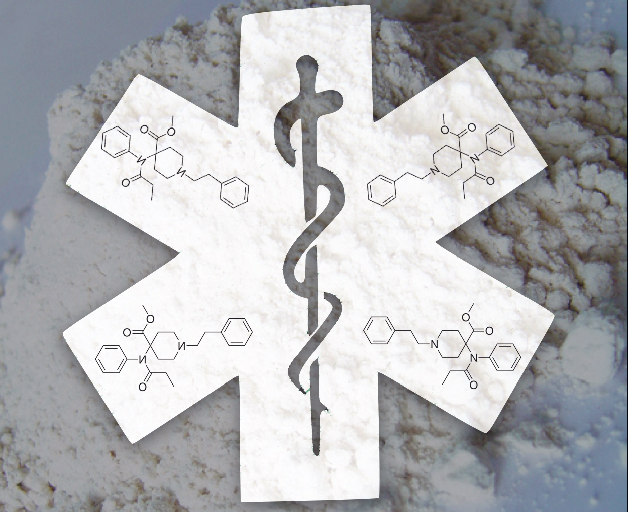
Fentanyl is considered 100 times more potent than morphine and 600 times more lipid soluble, subsequently increasing brain absorption. Illicitly produced synthetic opioids include non-pharmaceutical fentanyl, fentanyl analogs, and novel synthetic opioids [8]. Synthetic opioid overdose outbreaks have occurred historically on a smaller scale, as evidenced by the China White (3-methylfentanyl) in the US in the 1980’s [9] and several hundred fentanyl deaths across the US in the mid 2000’s [10,11]. The CDC reports a marked increase in deaths involving synthetic opioids since 2013 [2, 12]. Since Pharmaceutical fentanyl prescription rates (primarily fentanyl patches) remained relatively stable in comparison, the etiology of this surge is not prescription fentanyl. [12, 13]. Synthetic opioids are often found as a heroin contaminant or are sold in pill form [14, 15]. Hundreds of thousands of counterfeit pills have been entering the US and Canadian drug market, many of which contain fentanyl and other synthetic opioids, some at lethal doses in a single pill [12-18].

The fentanyl analog carfentanil is considered 10,000 times more potent than morphine and 100 times more potent than fentanyl [5, 6, 19], and is reported in medical examiner overdoses cases across the US and Canada. In 2002, an unknown aerosolized “gas” was used by Russian special operations forces in an attempt to rescue 800 hostages held in a theater by Chechnan rebels. Sadly, at least 125 hostages were killed, and years later carfentanil and remifentanil were positively identified as likely agents in post-mortem samples [20]. Other synthetic opioids including various fentanyl analogs, (such as MT-45, AH-7921 and an isomer U-47700) are present in the illicit marketplace and have all been confirmed in deaths in the US, Canada, and Europe [21-24]. Once novel synthetic drugs appear in the market, there is often a significant time delay to the development of an assay for identification in post-mortem samples. By the time the chemical is identified with reliability, either from post mortem samples or seized drug samples, the synthetic drug manufacturers often have already flooded the market with a new compound. Consequently, first responders may encounter either an affected patient or a drug exposure in the field before it has been identified.

Provider Safety and Personal protective equipment (PPE)

The most commonly used PPE for first responders includes nitrile gloves and occasionally eye protection, escalating to other types of PPE as situationally indicated. In a 2017 descriptive surveillance study, data collected from 572 EMS workers who sought treatment in emergency departments (EDs) between 2010-2014 demonstrated that exposures to harmful substances were the second leading occupational injury (behind strains and sprains). The authors recommended new and enhanced efforts to prevent EMS worker injuries and exposures to harmful substances [25]. The concept of evolving and improving our use of PPE is not new to healthcare providers. Diseases such as Ebola and SARS have spurred additional training and use of PPE, and the use of PPE should be continually readdressed in the face of new threats to provider safety.

The DEA released a warning in June 2016 to the police and public regarding fentanyl exposures after two law enforcement officers (LEOs) experienced overdose symptoms after exposure to airborne particulate from a “tiny amount” of white powder. This warning was later expanded to include all first responders, with a guide for prehospital providers released in Jun 2017 [26, 27]. Heroin found in the white powder form (predominantly, with some regional variability) and is visually indistinguishable from most synthetic opioids (1). Fentanyl and other synthetic opioids in the white powder form may put first responders at risk by mucous membrane contact, inhalation of airborne particulate, and potentially skin contact. Toxic doses of carfentanil in particular pose a significant risk to first responders in small amounts, some as little as a few grains of salt. The Acting DEA Administrator stated in reference to synthetic opioids: “I hope our first responders- and the public- will read and heed our health and safety warning. These men and women have remarkably difficult jobs and we need them to be well and healthy” [19].

With increasing concern for first responder safety and exposures, the Justice Institute of British Colombia, in conjunction with the Royal Canadian Mounted Police (RCMP), British Colombia Emergency Health services, and Vancouver Fire Rescue services, and other collaborating services created an innovative website at www.fentanylsafety.com. Included are resources for all levels of first responders outlining specific considerations for LEOs, firefighters, EMS, and Hazmat personnel. Within the EMS recommendations is a guide specific for fentanyl/synthetic overdose education, treatment guidelines, field risk assessment, donning and doffing PPE including N95 masks, and protocol recommendations for self-administration of naloxone in the event of symptomatic exposure. NIOSH, the National Institute for Occupational Safety and Health, provides similar PPE recommendations for first responders, found at www.cdc.gov/niosh/topics/fentanyl/risk.html. Though exposures may be uncommon, the prehospital environment can be unpredictable and providers may unintentionally encounter substances while caring for patients. First responders should be intimately aware of risk for contact with, disturbing, or aerosolizing any powders on patients, their clothing, and surroundings. If such substances are on or near the patient, first responders should at minimum add respiratory precautions such as facemasks to routine PPE, and consider NIOSH recommendations for full PPE. As always, scene safety remains paramount.

Respiratory support

Synthetic opioids overdoses should be treated initially the same as all patients with respiratory failure and/or suspected opioid overdoses with a pulse, primarily with effective Bag-Valve-Mask (BVM) ventilations. Ensuring an open airway, providing respiratory support, and monitoring circulation remain cornerstones of patient care for all levels of EMS providers. Nasal or oral airway adjuncts and oxygen administration to correct hypoxemia should also be used when indicated.  Almost one quarter of synthetic opioid overdose patients described in one hospital based case series required advanced respiratory support for persistent hypoxemia despite high doses of naloxone, and repeat respiratory arrest after cessation of naloxone infusion [14]. While the current focus for lay persons and law enforcement is rapid naloxone administration for suspected opioid overdoses, EMS providers are experts in the “ABCs”. A full patient assessment, high quality basic skills such as use of airway adjuncts, BVM, and respiratory support is imperative.

Naloxone

Naloxone is a high affinity mu-opioid receptor antagonist which acts on the central nervous system. It can be given to suspected opioid overdoses to reverse respiratory depression in the prehospital setting. For routine suspected opioid overdose, there are no definitive studies that have determined the optimal dose of naloxone to administer. Recommendations for initial dose can vary 10-fold based on reference and medical specialty. In general, emergency medicine and anesthesia references suggest higher doses (0.4mg) while medical toxicology and general medicine references suggest lower doses (0.04mg) [28]. The naloxone nasal atomizer devices currently in use by most first responders and bystanders is delivered in either 2mg or 4mg single dose sprays. Intranasal (IN) naloxone has an approximate bioavailability of only 4%, significantly lower than intramuscular (IM) and intravenous (IV) naloxone [29].  One prehospital study found that 2mg IN naloxone was not inferior to 0.4mg IV naloxone at reversing opioid induced apnea or hypopnea [30].

There is some concern that following naloxone administration and reversal of opioid overdose, patients may have adverse events such as pulmonary edema, precipitation of withdrawal symptoms, vomiting, or aspiration pneumonitis. In a one year prospective study in Norway, EMS witnessed vomiting in only 9% of patients who received prehospital naloxone [31]. A retrospective study in Pittsburgh found a lower rate of adverse events following naloxone administration with vomiting in only 0.2% of patients [32]. Novel synthetic opioid overdose cases have demonstrated a variety of adverse events during or after naloxone administration, including pulmonary edema and diffuse alveolar hemorrhage [14, 21], though the exact incidence is not known. EMS and first responders should also be aware of the potential safety risk of unmasking of other drug intoxications (methamphetamine, cocaine, or other stimulants) leading to behavioral disturbances. LEO training in naloxone administration has been generally well received and initial studies also report low rates of adverse events [33-35]. Overall, prehospital naloxone administration for suspected opioid overdose is considered safe, though the impact of synthetic opioids on the safety profile is unknown.

Naloxone administered to patients with synthetic opioid overdoses may require multiple doses. During known fentanyl overdose outbreaks, patients have required up to 14mg of naloxone to reverse respiratory and CNS depression [10, 36, 37]. In the prehospital environment, resources can be limited, and a single responding unit or even an entire region may not regularly stock large quantities of naloxone for single patient use. Naloxone supply can be particularly strained during events such as multiple overdose calls for a single responding unit or a multi-day drug overdose outbreak within a single system or area. In a case series of 18 patients, many of whom required naloxone infusions after exposure to fentanyl-adulterated pills, an entire hospital supply of naloxone was depleted and required emergency delivery to replete supply. 83% of patients who had either witnessed or themselves overdosed in the preceding six months reported that two or more doses of naloxone (most commonly used 2mg IN) were required before any response in suspected fentanyl overdoses [38]. Since novel synthetic opioids may have varied street concentrations, potencies, and receptor affinities, the actual dosage of naloxone that may be required to restore adequate spontaneous respirations may be unpredictable. This further emphasizes the role of respiratory support and transport particularly for patients who do not respond adequately to initial attempts at reversal on scene.

Transport

While some studies have failed to demonstrate increased mortality after opioid overdose reversal in the field [39, 40], those who may have overdosed with known or suspected long acting opioids, synthetic opioids, or those requiring repeated doses of naloxone benefit from transport to the nearest emergency department [8, 14]. The safety of refusal of transport after a suspected synthetic opioid overdose has not been established. If spontaneous respiratory drive has not returned after initial resuscitation attempts, EMS should consider transport rather than staying for extended scene times to administer repeated doses of naloxone. En route patient care should include continued reassessment, monitoring, and ongoing respiratory support with repeated administration of naloxone titrated to return of adequate spontaneous respirations, per local protocols.

Awareness of local patterns, collaboration, and community engagement

Synthetic opioids have the potential to overwhelm available emergency resources and supplies, akin to a mass casualty event. EMS systems may mitigate potential strain on local resources by monitoring local epidemiologic patterns, preparing for outbreaks, and collaborating with local agencies, including call centers, law enforcement agencies, EMS/fire departments and EDs. EMS data is particularly well suited for surveillance of suspected overdose patterns as it is geographically indexed and can be collected in near real time [41, 42]. Initial suspicion and tracking of synthetic opioid patterns can involve local EDs, medical examiners, toxicologists, and law enforcement. By sharing knowledge of local trends, all collaborators can stay abreast of the epidemic.

EMS and other first responders are well positioned for a critical role in intervention and prevention, and can provide a unique perspective for community engagement. On-scene interaction with the patient and/or family provides a potentially impactful opportunity to provide resources and support. EMS systems should consider novel strategies for combating the underlying opioid epidemic, including providing on-scene resources, take home naloxone rescue kits, encouraging any local resource utilization including medication assisted treatment (MAT), role of community paramedicine, and other opioid reduction programs.
 

Additional Resources:

The DEA video with LEO personal accounts of their exposure

Canada's fentanyl safety website

NIOSH recommendations for prehospital PPE

DEA: "Fentanyl, a briefing guide for first responders"


Special thanks to Brooke Lerner, PhD and Jill Theobald, MD

References:

1.     United States Drug Enforcement Administration National heroin threat assessment summary—updated. (June 2016). Retrieved Feb 25, 2017. www.dea.gov/divisions/hq/2016/hq062716attach.pdf

2.     Rudd RA, Seth P, David F, Scholl L: Increases in Drug and Opioid Involved Overdose Deaths-United States, 2010-2015. December 30, 2016 MMWR Morb Mortal Wkly Rep. 2016;65(50-51):1445-1452

3.     Deaths Involving Fentanyl in Canada, 2009–2014. Canadian Centre on Substance Abuse. http://www.ccsa.ca/Resource%20Library/CCSA-CCENDU-Fentanyl-Deaths-Canada-Bulletin-2015-en.pdf. Published August 2015. Accessed February 25, 2017

4.     Sonka, Joe. (2017). Fentanyl was main driver of Louisville’s surge in drug overdose deaths in 2016. [news release] Insider Louisville. February 28, 2017. Accessed May 24, 2017. https://insiderlouisville.com/metro/fentanyl-was-main-driver-of-louisvilles-surge-in-drug-overdose-deaths-in-2016/

5.     Ohio Hamilton County Heroin Coalition: Public Health Announcement: Synthetic Opioid Carfentanil Found in Local Drugs. December 5, 2016. Retrieved Feb 25, 2017 www.hamiltoncountyhealth.org

6.     Medical Examiner Public Health Warning Deadly Carfentanil Has Been Detected in Cuyahoga County [news release]; Cuyahoga County Medical Examiner; August 17, 2016. Accessed Feb 25, 2017, http://executive.cuyahogacounty.us/en-US/ME-Public-Health-Warning.aspx

7. Faul, M., Lurie, P., Kinsman, J. M., Dailey, M. W., Crabaugh, C., & Sasser, S. M. Multiple Naloxone Administrations Among Emergency Medical Service Providers is Increasing. Prehospital Emergency Care, 2017; 1-8

8.     Lucyk SN, Nelson LS. Novel Synthetic Opioids: An Opioid epidemic within an opioid epidemic. Ann Emerg Med. 2017 Jan;69(1):91-93

9.     Martin M, Hecker J, Clark R, et al. China White epidemic: an eastern United States emergency department experience. Ann Emerg Med. 1991;20(2):158-164

10.     Schumann H, Erickson T, Thompson T et al. Fentanyl epidemic in Chicago, Illinois and surrounding Cook County. Clin Toxicol (Phila). 2008;46(6):501-506

11.   Boddinger D. Fentanyl-laced street drugs “kill hundreds”. Lancet 2006;368:569-570

12.   Gladden RM, Martinez P, Seth P. Fentanyl law enforcement submissions and increases in synthetic opioid involved deaths-27 states, 2013-2014. MMWR Morb Mortal Wkly Rep August 26, 2016;65:837-43

13.   Fentanyl and Fentanyl Analogs. National Drug Early Warning System (NDEWS) Special Report. https://ndews.umd.edu/sites/ndews.umd.edu/files/NDEWSSpecialReportFentanyl12072015.pdf Published December 7, 2015. Accessed February 25, 2017

14.   Sutter ME, Gerona R, Davis M, et al. Fatal fentanyl: One pill can kill. Acad Emerg Med. 2017 Jan;24(1):106-113

15.   Armenian P, Olson A, Anaya A, Kurtz A, Ruegner R, Gerona RR. Fentanyl and a Novel Synthetic Opioid U-47700 Masquerading as Street “Norco” in Central California: A Case Report. Ann Emerg Med 2017 Jan;69(1):87-90

16.   United States Drug Enforcement Administration Counterfeit prescription pills containing fentanyls: A global threat. (July 2016). Retrieved Feb 25, 2017, from www.dea.gov/docs/Counterfeit%20Prescription%20Pills.pdf.

17.   Novel Synthetic Opioids in Counterfeit Pharmaceuticals and other Illicit Street Drugs. Canadian Centre on Substance Abuse. http://www.ccsa.ca/Resource%20Library/CCSA-CCENDU-Novel-Synthetic-Opioids-Bulletin-2016-en.pdf Published June 2016, Accessed February 25, 2017

18.   CDC health update: influx of fentanyl-laced counterfeit pills and toxic fentanyl-related compounds further increases risk of fentanyl-related overdose and fatalities. (Aug. 25, 2016.) U.S. Centers for Disease Control and Prevention. Retrieved Feb 25, 2017, from https://emergency.cdc.gov/han/han00395.asp.

19.  Drug Enforcement Administration Report: DEA Issues Carfentanil Warning to Police and Public. September 22, 2016. Accessed Feb 25, 2017. https://www.dea.gov/divisions/hq/2016/hq092216.shtml

20.   Riches JR, Read RW, Black RM, Cooper NJ, Timperley CM. Analysis of clothing and urine from Moscow theatre siege casualties reveals carfentanil and remifentanil use. J Anal Toxicology 2012 Nov-Dec;36(9):647-56

21.   Helander A, Backberg M, Beck O. Intoxication involving the fentanyl analogs acetylfentanyl, 4-methoxybutyrfentanyl and furanylfentanyl: results from the Swedish STRIDA project. Clinical Toxicology 2016;54(4):324-32

22.   Mounteney J, Giraudon I, Denissov G, Griffiths P. Fentanyls: Are we missing the signs? Highly potent and on the rise in Europe.  International Journal of Drug Policy (2015) 26:626-631

23.   Mohr AL, Friscia M, Papsun D et al. Analysis of Novel Synthetic Opioids U-47700, U-50488 and furanyl fentanyl by LC-MS/MS in Postmortem Casework. J Anal Toxicol 2016;40(9):709-717

24.   Coopman, V, Blanckaert P, Van Parys G, et al. A case of acute intoxication due to combined use of fentanyl and 3,4-dichloro-N-[2-(dimethylamino)cyclohexyl]-N-methylbenzamide (U-47700). Forensic Sci Int 2016; 266-68-72

25.   Reichard AA, Marsh SM, Tonozzi TR et al. Occupational Injuries and Exposures among Emergency Medical Services Workers. Prehosp Emerg Care 2017 Jan 25 [Epub ahead of Print]

26.   Drug Enforcement Administration Report: DEA Warning to Police and Public: Fentanyl exposure Kills. June 10, 2016. Accessed Feb 25, 2017. https://www.dea.gov/divisions/hq/2016/hq061016.shtml

27.   Drug Enforcement Administration: Fentanyl, A Briefing Guide for First Responders. Accessed June 12, 2017. http://dig.abclocal.go.com/wls/documents/DEA_Fentanyl_Publication.pdf

28.   Connors NJ, Nelson LS. “The Evolution of Recommended Naloxone Dosing for Opioid Overdose by Medical Specialty.” J Med Toxicol. 2016 Sep;12(3):276-81.

29.   Dowling J, Isbister GK, Kirkpatrick CM, Naidoo D, Graudins A. “Population pharmacokinetics of intravenous, intramuscular, and intranasal naloxone in human volunteers.” Ther Drug Monit. 2008 Aug;30(4):490-6.

30.   Merlin MA, Saybolt M, Kapitanyan R et al. Intranasal naloxone delivery is an alternative to intravenous naloxone for opioid overdoses.

31.   Belz D, Lieb J, Rea T, Eisenberg MS. “Naloxone use in a tiered-response emergency medical services system.” Prehosp Emerg Care. 2006 Oct-Dec; 10(4):468-71.

32.   Buajordet I, Naess AC, Jacobsen D, Brørs O. Adverse events after naloxone treatment of episodes of suspected acute opioid overdose. Eur J Emerg Med. 2004 Feb;11(1):19-23.

33.   Ray B, O’Donnell D, Kahre K. Police officer attitudes towards intranasal naloxone training. Drug Alcohol Depend 2015 Jan 1;146:107-10

34.   Purviance D, Ray B, Tracy A, Southard E. Law enforcement attitudes towards naloxone following opioid overdose training. Subst Abus 2016 Aug 11:1-6 [Epub ahead of print]

35.   Fisher R, et al. Police officers can safely and effectively administer intranasal naloxone. Prehosp Emerg Care 2016;20(6):675-80.

36.   Boyer EW. Management of opioid analgesic overdose. New England Journal of Medicine. 2012;367(2):146-155

37.   Solomon, Ranee. A 20-year-old woman with severe opioid toxicity. Journal of Emergency Nursing, April 2017 [Epub ahead of Print]

38.   Somerville NJ, O’Donnell J, Gladden RM, et al. Characteristics of Fentanyl Overdose- Massachusetts, 2014-2016. MMWR Morb Mortal Wkly Rep 2017;66:382-386

39.   Wampler DA, Molina DK, McManus J, et al. No deaths associated with patient refusal of transport after naloxone-reversed opioid overdose. Prehosp Emerg Care. 2011;15(3):320–324.

40.   Kolinsky D, Keim SM, Cohn BG, Schwarz ES, Yealy DM. Is a prehospital treat and release protocol for opioid overdose safe? The Journal of Emergency Medicine. 2017;52(1):52-58.

41.   Garza, A and Dyer, S. EMS Data Can Help Stop the Opioid Epidemic. JEMS, Nov 2016. Accessed Feb 25, 2017.

42.   Moore, PQ, Weber J, Cina S, Aks S. Syndrome surveillance of fentanyl-laced heroin outbreaks: Utilization of EMS, Medical Examiner, and Poison Center databases. Am J Emerg Med 2017 May 8


When You're Stuck in the Middle: Caring for Residents During Major Events.

by Tom Grawey, DO

Case Scenario: It is the first couple hours of your shift on the rig while you're working on some chores at the station.  Your partner mentions that his wife is running her first marathon today which happens to pass through your coverage area.  While some of your colleagues are scattered throughout the race path, you are ready to respond to a call from those not participating in today's activities.

A few minutes later a call comes in from the dispatcher for a 68 y/o male who is having chest pain a few blocks away.  While on any other day this would be a pretty routine occurrence, given the race, you and your partner take a few extra minutes to map out a route to the scene with the current road closures in place.  With all of the detours and traffic congestion you realize it will probably take an extra 5-10 minutes to get to the scene.  A few questions start to run through your head:  

Do residents living in the area of a mass gathering event receive a different level of care when an event is taking place?

How much is caring for this population discussed during the planing stages of the event?

What are some strategies that can be used to mitigate the challenges of providing medical care to non-participants who experience illness during a mass gathering in their neighborhood?

Literature Review:

During a mass gathering the daily EMS needs of the community do not stop.  Undoubtedly one of the biggest challenges when planning an event is trying to have as little impact on the residents living in the area as possible.  Road closures, eliminated parking spots, large amounts of pedestrians walking through a neighborhood and a redirection of the attention of first responders are just some inconveniences that residents experience during a marathon or similar activity.

A particular focus of our profession is the effect that mass gatherings have on access to prehospital medicine.  As people who are helping plan the emergency response to an event, it is responsibility of EMS to ensure that participants, attendees and residents of the community all have appropriate access to medical care should they need it.  A recent study was published in the New England Journal of Medicine entitled “Delays in Emergency Care and Mortality during Major U.S. Marathons” evaluated whether there are delays in care for nonparticipants with a medical emergency who live close to marathon routes. [1]

This study analyzed Medicare data to identify patients who were hospitalized for acute myocardial infarction (AMI) or cardiac arrest among Medicare beneficiaries in 11 cities that hosted large marathons over a 10 year period from 2002-2012.  The idea was that this cohort would be unlikely to be running the marathon.   For this population, 30 day mortality was compared among three groups - those near the race hospitalized on the same day as the marathon, those near the race hospitalized on the same day of the week either 5 weeks before or 5 weeks after the marathon, and those with these conditions who were hospitalized on the same day as the marathon but in a surrounding zip code that was not affected by the race path.  In addition to patient outcomes, the study reviewed ambulance transport data to answer two questions - whether transport times varied before or after noon on marathon days, and whether transport times varied on marathon vs non marathon dates. 

In total, the study examined 1145 hospitalizations for AMI or cardiac arrest on marathon dates in affected hospitals compared to 11074 on non marathon dates in the 10 weeks surrounding the event.  Patient age, sex, race and past medical history were statistically similar on all dates.  Despite the race taking place there was no difference in daily frequency of hospitalizations for these complaints between marathon and non-marathon dates.

The research team found that 30 day adjusted mortality was higher among those admitted to marathon-affected hospitals on marathon dates than on non-marathon dates (28.6% [95% CI, 26.1 to 31.3] vs 24.9% [95% CI 24.1 to 25.6], absolute adjusted risk difference 3.7% [95% CI, 1.1 to 6.4]).  In control hospitals, it was found that adjusted mortality was similar on marathon (25% [95% CI, 23.6 to 26.4]) and non marathon dates (24.7% [95% CI, 24.3 to 25.2]).  Transport times increased by an average of 4.4 minutes on marathon vs non marathon dates (95% CI, 1.3 to 7.5 p=0.0005) even though mileage traveled was similar.

When trying to account for possible causes of increase in mortality, further analysis revealed that frequency of hospitalizations, distribution of home zip codes of all patients, CABG, PCI or those receiving circulatory support did not differ in either group and it was concluded that differences in morality were not attributed to out-of-towners, hospital staffing or patients forgoing care.  Of note, a high percentage of patients presenting with AMI in regions affected by a marathon had concurrent cardiac arrest on race than on non race days (5.1% vs 2.6%, absolute difference, 2.5%; 95% CI, 1.4 to 3.5; P<0.001) while this was not a significant finding in control hospitals.

A Discussion of the case raised a few key points:

 "A well planned marathon route should interrupt as little of city traffic as possible for as short a time as possible. For example, the route on one of our events, the Milwaukee Running Festival, was recently modified because it landlocked a section of the city for too long. I would imagine that any large event or other disruption (construction, parades, arena sporting events, Summerfest) disrupt the flow of traffic enough to delay emergency medical care, but I would argue that the health and lifestyle benefits of these events outweighs the negatives. Nevertheless, the lesson on the importance of careful planning to ensure as little community disruption as possible is noted.” – Ben Weston, MD

Dr. Weston mentions something that isn’t discussed in the case or the NEJM Article – the benefits of the race to the participants.  The decrease in heart disease associated with regular exercise is well known and published countless times in the literature.  Certainly training for a marathon far exceeds the current CDC recommendations of just over 20 minutes a day of moderate-intensity aerobic activity and 2+ days a week of muscle strengthening activities. [2] While the personal choice (and health limitations) preventing some from participating in a race should not be held against them there is a greater good in mind when a community plans a marathon and as with any medical decision a risk/benefit approach must be used when planning an event.

Take home: Any event large enough to cause road closures and an influx of people is likely to cause delays in care to nonparticipants.  This study shows that in the case of large marathons, these obstacles may worsen outcomes for residents living in race affected areas.  EMS physicians and race medical directors must remain vigilant to ensure that a large event can accomplish its goals while creating as few interruptions and delays in medical care to nonparticipants as possible.

Grawey_bio.001.jpeg

References:

1. Jena AB, Mann NC, Wedlund LN, Olenski A. Delays in emergency care and mortality during major US marathons. N Engl J Med. 2017;376:1441-1450. doi: 10.1056/NEJMsa1614073

2. Physical Activity Guidelines Advisory Committee. Physical Activity Guidelines Advisory Committee Report, 2008. Washington, DC: U.S. Department of Health and Human Services, 2008.

Articles in Review: The 2018 LLSA for EMS

The 2018 EMS Subspecialty LLSA Articles have been released by ABEM.   The Communications Committee has put together the following review of all 12 articles to keep you up to date and ready for the test when it eventually comes out...

1. Brown JB et al. Not all prehospital time is equal: influence of scene time on mortality.J Trauma Acute Care Surg Jul 2016;81(1):93-100.

 Reviewed by Maia Dorsett, MD PhD, @maiadorsett

Background & Objectives: Given that trauma is a time –sensitive disease, minimizing prehospital time may be beneficial to trauma patients.  Most studies have not linked increased prehospital time with worse trauma outcomes.  With the overall goal of identifying modifiable prehospital factors that may improve trauma outcomes, the objective of the study was to evaluate the association of prehospital time patterns with mortality.

Methods: The study was a retrospective review of data from the Pennsylvania Trauma Registry.  Inclusion criteria were all patients 16 years and older who were transported by EMS with a total prehospital time (TPT) of 20 minutes or longer between Jan 2000 – June 2013.  Patients were excluded if they were transported from another hospital or if prehospital time data were missing. Total prehospital time was divided into Response (notification to arrival), Scene (arrival to leaving for hospital), and Transport (scene to hospital).  In order to control for the variability of “raw” prehospital times due to EMS system characteristics, the authors evaluated the relative proportion that each interval contributed to the TPT. A time interval was classified as prolonged If it contributed to > 50% of TPT.   

After matching for TPT, a logistic regression model was used to determine the association of mortality with PH time pattern, controlling for confounders such as age, sex, race, co-morbidities, mechanism, transport mode, PH provider level (ALS vs. BLS), IVF volume, PH and admission VS, ISS, severe head injury, and blood transfusion in the ED.

Key Results:  Out of 164,471 patients included in the study, only 2% had a prolonged response time, while 19% had a prolonged scene time and 31% had a prolonged transport time.  Prolonged scene time was associated with a 21 % increase in mortality (AOR 1.21, 95% CI, 1.02-1.44, p = 0.03).  Prolonged response time and transport time were not associated with increased mortality [AOR 1.16, 95% CI 0.83-1.3, p = 0.38 for response; AOR 0.82, 95% CI 0.65 – 1.04, p = 0.11 for transport].  This was consistent for patients with both blunt and penetrating trauma, although was more pronounced for patients with penetrating trauma who more commonly required emergent operative intervention.  The effect was also more pronounced when overall prehospital time increased.

To further break down contributing factors, the authors evaluated the contribution of intubation and extrication to mortality.  They found that prehospital intubation was associated with an increased risk of mortality (AOR 4.49, 95% CI, 3.48-5.78) and contributed on average 6 min 22 seconds to scene time.  Extrication was also associated with mortality (AOR 1.40, 95% CI 1.19-1.65, p < 0.01) and extended scene time by 4 min 30 seconds on average.  Together, they mediated 60.5% of the total effect of prolonged scene time on mortality in the risk-adjusted model.  However, the more extrications or intubations an EMS agency performed, the less dramatic the effect on mortality.  In patients with GCS < 8, there was an association between prehospital intubation and mortality if patients were transported by ground (AOR 2.52, 95% CI, 1.95 – 3.26, p < 0.01) but not if they were transported by helicopter (AOR, 1.26; 95% CI 0.92 – 1.73).

Take Home: Prolonged scene time is associated with increased mortality of trauma patients.  Prehospital intubation and extrication mediate this effect significantly, although less so when EMS agencies have more experience in either procedure.


2. Weaver MD et al. An observational study of shift length, crew familiarity, and occupational injury and illness in emergency medical services workers. Occup Environ Med Nov 2015;72(11):798-804.

Reviewed by Catherine Counts, MHA (@CatherineCounts)

Background& Objectives:  EMS is high-risk work where extended shifts and lack of familiarity between teammates is common.   

Extended shifts: While OSHA defines a normal work shift as 8 hours a day, 5 days a week with at least 8 hours of rest between shift, EMS providers often work an extended shift which may increase the risk of “adverse events, medical errors and attentional deficits.”

Crew Familiarity: Data from the airline industry has shown that a lack of familiarity between pilots is linked to more errors. The average EMS provider will have 19 different partners annually; some will have as many as 50 in a year and thus lack of familiarity may lead to more errors amongst EMS providers.

Given the high rate of extended shifts in EMS, as well as lack of consistency between EMS partners, the objective of this study was to  “examine the relationship between shift length and occupational injury while controlling for relevant shift work and teamwork factors.”

Methods: This was a retrospective study using administrative data from 14 EMS agencies with 37 base sites.  Agencies provided historical shift schedules and OSHA reports which were matched by date.  The primary outcome of interest was OSHA-reported illness or injury, defined as an injury that required medical treatment beyond basic first aid “or [resulted] in loss of consciousness or an inability to perform normal duties without restriction.”  The exposure of interest was shift length; the main analysis stratified continuous shift length variable into sections: less than 8 hours, 8-12 hours, 12-16 hours, 16-24 hours, and 24+ hours.  Secondary analysis included shift length as a dichotomous variable (yes/no 12+ hours, yes/no 10+ hours), as well as a continuous variable.

The authors also evaluated the effects of the following independent variables of interest:

·       Partner Familiarity - Assigned via number of shifts with partner within past 8 weeks, categorized by quartiles.

·       Recovery period – time between end of prior shift and start of shift with injury, treated as continuous variable with 1 hour increments

·       Consecutive shift - if less than 2-hour break between shifts

·       Overnight shift – Yes/No

·       Part time – Work less than 34 hours a week

·       Number of workers at agency – estimated using unique number of workers during middle four weeks of study period (workforce size is associated with injury reporting)

Results were analyzed using multi-variable mixed-effects logistic models using both fixed and random effects.

Key Results: Fourteen agencies at 37 sites participated over 1 to 3 year period.  OSHA reports were matched on date, location and employee. Shifts were excluded when the assigned job role described a non-clinical task.  After removing non-clinical and incomplete shift records, the authors included 966, 082 total work shifts from 4,382 employees in their analysis.

Shifts < 8 hours as well as overnight shifts are associated with fewer injuries [relative risk 0.70, 95% CI 051-0.96, p = 0.029 for < 8 hr; RR 0.78, 95% CI 0.65-0.93, p = 0.005 for overnights. 16-24 hour shifts are associated with more injuries when compared with the reference category of 8-12 hour shifts (RR 1.6, 95% CI 1.22-2.10, p=0.001). Familiarity, agency workforce size, part-time status and hours of recovery were not associated with occupational injury or illness. Consecutive shifts also did not significantly alter the risk of occupational injury or illness.  Shifts > 24 hours had nearly a three-fold increase in risk (RR 2.88, 95% CI 1.74 – 4.77, p < 0.001), while shifts over 12 hours had a 38% increase in risk of injury (RR 1.38, 95% CI 1.12-1.70, p = 0.002).

Take Home: There is not a one-size-fits-all model for EMS scheduling, but this study further builds on literature that shift length serves as a contributing factor for employee wellbeing.  Before any organizational changes are made “trials of novel, minimally intrusive, intra-shift and inter-shift safety management interventions in the EMS setting are needed.”


3. Berkhemer OA et al; MR CLEAN Investigators. A randomized trial of intraarterial treatment for acute ischemic stroke.  N Engl J Med Jan 2015;372(1):11-20.

Reviewed by Maia Dorsett, MD PhD, @maiadorsett

Background & Objectives:  Intravenous alteplase  (tPA) therapy has a narrow therapeutic window and a large number of contraindications.  Moreover, it is less effective at opening proximal occlusions of the major intracranial arteries which account for a third of anterior circulation strokes.  Initial trials of endovascular approaches to reperfusion were largely negative, but there were a number of concerns about these trials including lack of imaging to confirm proximal vascular occlusion prior to intervention, long time interval to intervention, and use of earlier versions of mechanical thrombectomy devices. 

Given the concerns regarding these early trials, the objective of the MR CLEAN trial was to determine whether “intraarterial treatment plus usual care would be more effective than usual care alone in patients with a proximal arterial occlusion in the anterior cerebral circulation that could be treated intra-arterially within 6 hrs of symptom onset.”

Methods: MR CLEAN was a multi-center randomized clinical trial carried out in the Netherlands.   Patients were randomized to treatment-group assignments, underwent open-label treatment, and blinded end-point evaluation. 

Inclusion criteria included:

1. > 18 years of age with acute ischemic stroke

2. intracranial occlusion of the distal intracranial carotid artery, middle cerebral artery (M1 or M2), or anterior cerebral artery (A1 or A1) confirmed on CT or MR angiography

3. NIHSS > 2

The treatment consisted of intra-arterial treatment of the occlusion.  The method of reperfusion (thrombolytic agent, mechanical thrombectomy, or both) was left to the discretion of the interventionalist.  If eligible, patients were treated with alteplase or urokinase prior to intervention.

The primary study outcome was modified Rankin scale at 90 days.  Secondary outcomes included NIHSS at 24 hrs and at 5 to 7 days post discharge, acivities of daily living as measured by the Barthel index, and health related quality of life as measured by the EuroQol Group 5 Dimension Self-Report Questionnaire at 90 days.

Key Results: The study included 500 participants, 233 (46.6%) were assigned to the intervention group and 267 patients (53.4%) were assigned to the control group.  The large majority of patients received IV alteplase prior to intra-arterial intervention (87.1% in intervention group and 90.6% in control group), with a median time to initiation of 85-87 min. 196 of 233 participants in the treatment group received intra-arterial therapy.

For the primary outcome of modified Rankin score, there was a shift in favor of the intervention with an adjusted common odds ratio of 1.67 (95% CI 1.21-2.30).  Patients who received intra-arterial treatment were more likely to be functionally independent (modified Ranken score 0 to 2; AOR 2.16; 95% CI, 1.39-3.38) with an absolute increase of 13.5% (32.6% vs. 19.1%, NNT 7.4).   Patients receiving intra-arterial treatment also scored higher on the Barthel index (AOR for score 19 or 20 at 90 days 2.1, 95% CI 1.4 – 3.2).  There was no significant difference in QQ-5D score (69% vs. 66%).

There was no significant difference in severe adverse events between the two groups during the 90 day follow-up period.  Procedure-related complications included embolization into new vascular territories downstream of the occluded vessel (8.6% of patients) with evidence of new ischemic stroke in a different vascular territory in 5.6% patients in the intervention group.

Take Home:  There is a functional benefit of intra-arterial therapy for patients with acute ischemic stroke with NIHSS  > 2 and confirmed proximal intra-arterial occlusion in the anterior circulation who are able to receive treatment within 6 hrs.


4. Drennan IR, Lin S, Sidalak DE, Morrison LJ. Survival rates in out-of-hospital cardiac arrest patients transported without prehospital return of spontaneous circulation: an observational cohort study. Resuscitation. 2014;85(11):1488-93.

Reviewed by Jeremiah Escajeda, MD, @ JerEscajeda

Background & Objectives:  Cardiac arrest is a prevalent disease entity encountered in EMS systems. In 2009, Morrison et al. derived and validated the prehospital Universal Termination of Resuscitation (TOR) Guideline to help direct appropriate termination efforts for both basic life support (BLS) and advanced life support (ALS) prehospital services. To satisfy the TOR Guideline, a cardiac arrest patient must have an 1) unwitnessed arrest by EMS personnel 2) no shock delivered and 3) no return of spontaneous circulation (ROSC) [1]. Implementation of the TOR has been inconsistent and some prehospital services use sole criteria for termination of resuscitation based on no ROSC achieved. The authors sought to report the survival rates of patients without prehospital ROSC who still met transportation criteria based on the TOR Guideline, such as those still who have had either witnessed arrest by EMS personnel or prehospital shock delivered.

Methods: This was a retrospective observational study of the Toronto site Resuscitation Outcomes Consortium (ROC) database from April 1, 2007 –March 31, 2013.  Subjects included for analysis were adult patients (≥18 years of age) with cardiac arrest suspected to be cardiac etiology (no trauma, drowning, overdose or asphyxia). EMS services included both BLS (supraglottic device capable) and ALS services. Both EMS and inhospital records were reviewed. 20,207 patients met inclusion criteria.

Key results: Of the 20,207 adult cardiac arrest patients included, 3,374 (16.4%) did not have prehospital ROSC but met the Universal TOR guideline for continued resuscitation and transport. Of these, 551 (16.3%) obtained ROSC in the ED and 122 (3.6%) survived to hospital discharge.

In adjusted multivariable logistical regression, survival to discharge was associated with younger age (OR 0.98; 95% CI 0.97-0.99), initial shockable VF/VT (OR 5.07; 95% CI 2.77-9.30), EMS witnessed arrests (OR 3.51; 95% CI 1.73-7.15), bystander-witnessed arrests (OR 2.11; 95% CI 1.18-3.77) and public locations (OR 1.57; 95% CI 1.02-2.40).

Take home:  In this study, using absence of prehospital ROSC as sole determinant for TOR misses an unacceptably high number of potential survivors (3.6%). This is well above the 1% defined threshold for medical futility. Also, the optimal minimum time to obtain prehospital ROSC has not been established. Adherence to all of the Universal TOR criteria results in more accurate prehospital identification of futility in cardiac arrest patients. 

1. Morrison LJ, Verbeek PR, Zhan C, Kiss A, Allan KS. Validation of a universal prehospital termination of resuscitation clinical prediction rule for advanced and basic life support providers. Resuscitation. 2009;80(3):324-8.


5. Nichol G et al.; ROC Investigators. Trial of continuous or interrupted chest compressions during CPR.  N Engl J Med Dec 2015;373(23):2203-14.

Reviewed by Tom Grawey, DO @EMtgDO

Background & Objectives:  Standard CPR consists of manual chest compressions with positive-pressure ventilation until ROSC is obtained.  Traditionally chest compressions are interrupted by ventilation, though interruptions reduce circulation of blood and potentially reduce the effectiveness of CPR.  One way to reduce time off the chest in CPR is to provide breaths to the patient while continuing CPR.  The objective of this study was to determine the effect of continuous chest compressions at a rate of 100 per minute with ventilation provided concurrently at a rate of 10 per minute (experimental group) compared to interrupted compressions with a 30:2 compression:breath ratio (control group) during CPR on the rate of survival, neurologic function, or the rate of adverse events.

Methods: This was a cluster-randomized trial with crossover; 114 EMS agencies across 8 sites were grouped into 47 clusters were randomly assigned to performed continuous or interrupted compressions to all cardiac arrests to which they responded.  Twice yearly the cluster was switched to the other resuscitation strategy. Both protocols were followed for a total of 6 minutes, at which point an advanced airway was placed and all patients underwent continuous compressions with breaths provided at a rate of 10 per minute.  CPR quality was measured in both groups. The primary outcome was survival to hospital discharge with secondary outcomes including neurologic function at discharge (using modified rankin scale based on the clinical record), adverse events and hospital-free survival (number of days alive and permanently out of the hospital during the first 30 days after the arrest).

Key Results: 1129 of 12,613 patients (9%) in the continuous compressions group and 1072 of 11035 (9.7%) in the 30:2 group survived to hospital discharge.  In patients with available data on neurologic status, 883 of 12,560 patients (7%) in the intervention group and 844 of 10,955 (7.7%) in the control group survived with a modified Rankin scale score of 3 or less.  Hospital-free survival was significantly shorter in the intervention group than in the control group (mean difference, -0.2 days; 95% CI, -0.3 to -0.1; P=0.004) however this is arguably not clinically significant.

Take home: The study authors conclude that among patients with out of hospital cardiac arrest, continuous chest compressions did not result in significantly higher rates of survival or favorable neurologic status when compared to a 30:2 compression to ventilation ratio.  Despite the differences in CPR protocol, the mean difference in chest compression fraction (defined as the proportion of each minute during which compressions were given) between the intervention and control groups was small (0.83±0.14 and 0.77±0.14 ; p<0.001 respectively) in this study which may be contributing to similar outcomes and may emphasize the importance of minimizing compression interruptions.   It is unclear whether ventilation quality and strategy contributed to patient outcomes as it was not measured.


6. Fisher R, et al. Police officers can safely and effectively administer intranasal naloxone. Prehosp Emerg Care 2016;20(6):675-80.

Reviewed by Aurora Lybeck, MD, @AuroraLybeck

Background & Objectives:  Law enforcement officers (LEOs) are increasingly utilizing naloxone administration for suspected opioid overdose patients prior to EMS arrival, though limited literature is available on the safety and efficacy of such programs. This publication provides an example of training and implementation of LEO administered naloxone program, and aimed to describe indication, response, and disposition of the patients to whom LEO naloxone was administered.

Methods: This is a retrospective case series describing LEO administered naloxone within a large urban police department and included 126 occurrences over 18 months of data collection. They described a 30-minute training program for all LEOs and academy trainees. Instruction topics included signs and symptoms of suspected opioid overdose, naloxone and atomizer devices with hands-on training and instruction to administer 2mg IN naloxone, and required patient transport to the hospital if LEO naloxone was administered. The data was gathered through standard police run reports and an additional naloxone administration data form, using the narrative to complete data when possible.  Data elements included:

1)    number of times naloxone was administered by police

2)    indications for naloxone administration

3)    basic patient demographics

4)    patient response to naloxone

Other data elements collected included time to EMS arrival on scene and whether the patient was voluntarily or involuntarily transported to the hospital. If patient refused transport, LEO placed patient on involuntary hold and transported them to hospital.

Key Results: The most common indication for LEO administered naloxone was “unconscious/unresponsive” (n=117, 92.9%). Most patients receiving LEO naloxone were white (92.9%), male (59.5%), with average age of 32.8 years.  After LEO administration of naloxone, most patients regained consciousness (n=82, 65.1%) or regained spontaneous respirations (n=71, 56.3%), though some demonstrated no response (n=22, 17.5%). Of those with no response to a single dose of LEO administered naloxone, 18 of the 22 outcomes were reported: 3 were fatalities, 3 were non-opioid overdoses, and 12 responded to an additional dose of naloxone by EMS. EMS arrived within 5 minutes in 90% of the calls. 96.8% were transported to the hospital voluntarily. One patient became agitated but later went voluntarily to the hospital. No significant adverse effects noted.

Limitations of the study included selection based only on LEO naloxone administration, potentially missing those opioid overdoses cases not recognized by the LEO, 28 reports with missing EMS follow-up data, lack of toxicological confirmation tests confirming opioid as primarily intoxicant.

Take Home: Trained law enforcement officers can correctly identify an opioid overdose and effectively administer naloxone without significant adverse effects. Further research is needed regarding outcomes and system impact.


7. Rostykus P et al. Variability in the treatment of prehospital hypoglycemia: a structured review of EMS protocols in the United States. Prehosp Emerg Care 2016;20(4):524-30.

By Hawnwan P. Moy, MD, @PECpodcast

Background & Objectives: Hypoglycemia is a frequent emergency situation in many prehospital systems.  The traditional approach has been to treat hypoglycemia intravascularly (IV) with fifty milliliters of a 50% solution of glucose containing 25 grams of glucose (commonly known as an amp of D50).  However, this treatment is not without risk.  D50 is known to over-treat hypoglycemics to inappropriate hyperglycemic levels.  Mathematically speaking an amp of D50 is 5 times the amount of glucose in a normal adult and a common pediatric dose of 0.5-1 g/kg (at more dilute concentrations of D25, D12.5, or D10) of glucose provides 6-11 times the normal amount of glucose in a normal child’s blood.  Excessive glucose can cause complications in the brittle diabetic and has been known to have detrimental effects in medical conditions such as acute stroke and post cardiac arrest patients.  The hypertonic nature of D50 may also cause tissue necrosis should extravasation occur.  Finally, dilution and adjustment for the hypoglycemic pediatric population is highly prone to error.

In response to a national drug shortage of D50 and potential harmful side effects, many EMS systems adjusted their treatment of hypoglycemia to now include a 10% dextrose containing solution (D10) instead of D50.  Initial studies on D10 indicate that there is a similar time to reversal of hypoglycemia, less post treatment hyperglycemia, and less risk of tissue necrosis given the lower hypertonicity of D10 compared to D50.  As a result, this objective of this study was to determine how many EMS systems utilize D10 in their treatment protocols in place of D50.  Secondary outcomes were to describe initial and subsequent dextrose treatments, routes of administration, availability of glucagon to treat hypoglycemia, recommendation for post treatment monitoring, and non-transport policies for treated patients. 

Methods: The authors performed a structured review of EMS protocols from 50 of the largest populated cities in the United States as well as EMS protocols from http://www.emsprotocols.org.  The following data points were manually abstracted by trained investigators: the concentration of glucose recommended for the parenteral reversal of hypoglycemia in adult and pediatric patients, clinical treatment thresholds, dose recommendations, follow-up care, glucagon use, and non- transport policies.

Key Results: The authors collected a total of 185 protocols.  From those protocols, 70% had D50 as the only treatment for hypoglycemia, 8% mandated D10 as the only treatment, and 22% allowed either D50 or D10.  For the pediatric population, two thirds of the EMS protocols called for 0.5 g/kg of hypertonic glucose with the rest varying between 1 g/kg to less than 0.5 g/kg.  The most common initial dose for neonates was 0.5 g/kg.  Additionally, IV and intraosseous (IO) routes of administration were allowed in two-thirds of protocols, and a third allowed only IV.  Other major differences included post treatment guidelines (only a third of protocols had this) and less than half of all protocols had a non-transport policy of patients with corrected glycemic status.  


Take Home: Despite the fact that the administration of D50 has the known side effects of supratherapeutic blood sugar, a majority of EMS systems still utilize D50 for treatment of hypoglycemia.  Additionally, this manuscript demonstrates a wide variability in treatment of hypoglycemia in not only the dosage of medication used, but also blood glucose indication for treatment, subsequent dosage of glucose treatment, potential routes, post treatment monitoring, and non-transport policies for treated patients.  In many industries, reducing variabilities in protocols reduces errors.  Although no two EMS systems are alike, if there is no physiologic or scientific basis for protocol differences, treatment standardization may result in fewer errors and enhance patient safety.  


8. Prekker ME et al. Pediatric Intubation by paramedics in a large emergency medical services system: process, challenges, and outcomes.  Ann Emerg Med Jan 2016;67(1):20-9.

Reviewed by Maia Dorsett, MD PhD, @maiadorsett

Background & Objectives:  In many EMS systems, pediatric intubation is considered a core paramedic skill.  However, proficiency in pediatric intubation is hampered by inadequate training, infrequent opportunities to perform the skill, as well as anatomic and equipment differences.  The largest clinical trial of pediatric intubation by paramedics found that pediatric intubation and bag-mask-ventilation had comparable survival and neurologic outcomes while intubation delayed transport to the hospital [1].  Evaluating the process used by paramedics in systems in which pediatric intubation is performed is a starting point for quality improvement.

The study had two objectives:

1. Estimate “the incidence of out-of-hospital pediatric intubation within the study community and the EMS system”.

2.  Describe “the process used by paramedics in their attempt to intubate these children, with focus on the specific challenges to successful intubation, the corrective actions taken after a failed intubation attempt, and potential procedural complications.”

Methods: The study is a retrospective cohort study of patients < 13 years old who underwent at least one intubation attempt treated by the EMS system serving King County, WA between September 2006 and December 2012.  Paramedics in the King County system receive standardize training in airway management and perform an average of 6 pediatric and 40 adult intubations prior to graduation from paramedic school They are also required to perform 12 successful intubations per year.

Data collection involved retrospective chart review of EMS, ED and hospital reports as well as review of a mandated detailed airway report that paramedics must complete for each attempted intubation.

Key Results: In 651,194 calls over 6.3 years, only 299 encounters included an attempted pediatric intubation (0.05% of all calls).  Among the 123 paramedics who had at least one pediatric intubation attempt, on average paramedics had no more than one pediatric intubation every 2.6 years.  Cardiac arrest was the most common reason(44%) and most commonly involved infants.  Preschool and school age children were most commonly intubated for neurologic emergencies, including seizures and trauma. 

Amongst all intubation attempts, first pass success was only 66%.  First pass success was lower in infants (53%) and children in cardiac arrest (53%).  Second attempt success rate was 69% (cumulative success after two attempts was 89%), 57% on 3rd attempt (cumulative success 95%) and 64% after > 4 attempts (cumulative success 97%).  The most commonly noted challenge to successful intubation was body fluids (33%).  Paramedics changed their approach after failed intubation by suctioning the airway (32%), repositioning the patient (27%), or changing the operator or using a bougie after a failed third attempt.  Among the 8 patients who were not successfully intubated out-of-hospital, 5 were intubated in the ED, 1 died prior to arrival and 2 did not end up requiring intubation.  Of note, 9% of pediatric patients who were intubated out-of-hospital were extubated in the ED and 1% were discharged home.

The most frequent complication was right mainstem intubation (19%).  Major complications (peri-intubation arrest, ET tube dislodgement, injury to the respiratory tract, or bradycardia) occurred in 11% of cases.  There were no un-recognized esophageal intubations documented, but endotracheal tube placement could not be retrospectively determined in 50 cases in which the resuscitation was terminated in the field.

Take Home:  Pediatric intubation is rarely performed. Even in a system where it is considered a core skill, repeated attempts at intubation were required in a third of patients, although 97% were successfully intubated eventually.  Evaluating the process and complications step by step will help determine whether out-of-hospital pediatric intubation is appropriate for an EMS system and opportunities for improvement.

1. Gausche, M., Lewis, R. J., Stratton, S. J., Haynes, B. E., Gunter, C. S., Goodrich, S. M., ... & Seidel, J. S. (2000). Effect of out-of-hospital pediatric endotracheal intubation on survival and neurological outcome: a controlled clinical trial. Jama, 283(6), 783-790.


9. Schlapbach LJ et al. High-flow nasal cannula (HFNC) support in interhospital transport of critically ill children Intensive Care Med Apr 2014;40(4):592-9.

Reviewed by Maia Dorsett, MD PhD, @maiadorsett

Background & Objectives:  Escalating respiratory support during transport is inherently difficult; interhospital transport teams have therefore historically had a low threshold for intubating patients prior to transport.  However, intubation and invasive ventilation are not without risk.  In Pediatric Intensive care, high-flow nasal cannula (HFNC) therapy is increasingly used for respiratory support in critically ill infants and has reduced the need for intubation.  The use of HFNC during interhospital transport had not yet been studied.  The objective of this study was to evaluate the safety of HFNC during interhospital transfer of critically ill children < 2 years of age and determine the impact of its use on intubation rate and successive PICU management.

Methods: This was a single center, retrospective, pre-post study of critically ill children under 2 years of age requiring interhospital transport by a specialized tertiary pediatric retrieval team and consecutively admitted to the PICU of Mater Children’s Hospital in Brisbane, Australia between 1/2005 and 12/2012.   The authors compared the use of respiratory support modes [low flow oxygen/room air, HFNC, NIV (CPAP/BiPAP), or invasive ventilation] and complication rates [need for intubation, pneumothorax, cardiac arrest, need for invasive ventilation in the first 24 hrs of PICU admission] during the 48 month period before and after HFNC was made available as a standard treatment for interhospital transport.

Key Results: 331 children were transported in the pre-HFNC and 462 in the post-HFNC period, with a mean duration of transport of 1.4 hours.  After introduction of HFNC, 33% of all patients were transported on HFNC.  Intubation rates decreased between the pre (49%) and post(35%) periods.  This was in part due to decrease rate of intubation by the referring hospital (36% pre/28% post), but fewer patients were intubated by the retrieval team as well (13% pre/7% post). 

In the subgroup of patients with bronchiolitis, there was a significant decrease in the proportion of patients with invasive ventilation initiated by the retrieval team (33% vs. 15%, p = 0.001).  Among patients receiving HFNC, bronchiolitis was the most common condition requiring transport (77%).

There was no increase in adverse events post introduction of HFNC, including no cases of patients requiring intubation during transport.  There was no significant difference between need for intubation during the first 24 hrs of PICU admission in the pre/post period.

Take Home: HFNC therapy during transport of critically ill infants appears to be safe and may have the potential to reduce need for intubation and invasive ventilation.


10. Clemency BM, Bart JA, Malhotra A, Klun T, Campanella V, Lindstrom H. Patients immobilized with a long spine board rarely have unstable thoracolumbar injuries. Prehosp Emerg Care 2016;20(2):266-72.

Reviewed by Brandon Bleess, MD @BBBleess

Background & Objectives: Because of concern that patients with unstable spine injuries are at risk for secondary mechanical injury if exposed to significant movement, long spine boards and cervical collars were widely adopted as a mainstay of treatment for patients with suspected spine injury.  However, multiple studies have demonstrated potential harms of spine board use, including respiratory compromise, pain, tissue ischemia, and unnecessary imaging.  In order to objectively compare the relative risk and benefit of spinal immobilization, the objective of this research was to “determine the prevalence of unstable thoracolumbar spine injuries among patients receiving prehospital spine immobilization.”

Methods: This was a retrospective study of prehospital and hospital records of patients cared for by a single, large, private EMS agency in Western New York from January 1, 2010 through December 31, 2013. The corresponding hospital data was obtained from a single, Urban, academic Level I Trauma Center that served an 8 county region with approximately 65,000 emergency department visits per year.  During the study, EMS operated under a single statewide “Suspected Spinal Injury” protocol requiring complete spinal immobilization when spinal injury was suspected.  Inclusion criteria included age > 18 years old, documented spinal immobilization from the scene, and transport to the study hospital.

Hospital records were reviewed for mechanism of injury, imaging of the thoracolumbar spine, and presence of any acute fractures, dislocations, or subluxations.  For patients with injuries identified on spinal imaging, performance of thoracolumbar spine surgery during hospitalization was used as a marker for unstable thoracolumbar spine injury.  The primary outcome was the percentage of patients with unstable thoracolumbar injury who underwent prehospital spine immobilization following blunt trauma.  Rate of imaging and injuries by mechanism were secondary outcomes.

Key Results: Immobilization was documented on 5,593 patients transported to the ED.  97% of prehospital records were successfully linked with the corresponding hospital record. Spinal imaging was ordered in 82.5% of subjects and thoracolumbar imaging specifically was ordered in 51.3%of subjects.  An acute thoracolumbar fracture, dislocation, or subluxation was present in 4.3% of cases.  An unstable injury was present in 0.5% cases (n=29). No unstable injuries were found among the 951 patients that were immobilized following ground level falls.  Falls from heights greater than 20 feet had the greatest chance of causing any fractures and unstable injuries with 10% (8/80) having unstable fractures.

Take home: While spinal imaging is commonly performed on blunt trauma patients who undergo prehospital spinal immobilization, unstable thoracolumbar injury is a rare occurrence.


11. Newgard CD et al.; Western Emergency Services Translational Research Network (WESTRN) Investigators. Improving early identification of the high-risk elderly trauma patient by emergency medical services. Injury Jan 2016;47(1):19-25.

Reviewed by Melody Glenn, MD, @MGlennEM

Background & Objectives: Existing field triage guidelines often fail to identify serious injuries in elderly trauma patients.  This is partially due to the increased likelihood of sustaining injury from low-velocity mechanisms (e.g. ground-level falls) and their different physiologic responses to injury amongst the elderly. Under-triage leads to a greater proportion of seriously injured elders being transported to non-trauma hospitals, where their needs may outstretch hospital capabilities. Although the CDC added a “special consideration” section relating to adults >55 into their 2011 field triage guidelines for injured patients, little evidence exists that similar EMS protocol changes have decreased under-triage.

The objectives of the study were to:

  1. Define the high-risk injured older adult using prognostic differences associated with different injury patterns
  2. Derive alternative field trauma triage guidelines that mesh with current national guidelines to improve identification of high-risk elderly trauma patients.

Methods: This was a retrospective cohort study of 33,298 injured adults 65 years or older who were transported by 94 EMS agencies to 122 hospitals in 7 western regions in the United States from 2006 - 2008. Only patients with matched hospital records were included. The researchers used Abbreviated Injury Scale (AIS) scores or need for surgery to create 5 definitions/categories of “serious injury,” including: Injury Severity Score (ISS) ≥ 16, serious traumatic brain injury (TBI), serious chest injury, serious chest injury, serious abdomen-pelvic injury, and serious extremity injury. They considered in-hospital mortality as a marker of prognosis to compare definitions.

They derived an alternative set of field triage guidelines to identify high-risk older adults, using 60% of the sample to derive and cross-validate their decision tree. The remaining 40% were used to validate the tree.

Key Results: 80% of the elders in their sample of 32,298 patients were injured by falls. Out of their cohort, 13,401 met their definition for serious traumatic injury (4.5% with ISS ≥ 16, 4.7% with TBI, 3.4% w serious chest injury, 1.7% with Abdomen-Pelvis injury, and 3.1% with in-hospital mortality). Patients with isolated serious extremity injuries had the lowest mortality.

Based on having any positive triage criterion from the 23 field trauma triage criteria currently in existence at the study sites, 3,299 serious trauma patients would have been identified, resulting in a sensitivity of 75.9% (95% CI 72.3-79.2%) and a specificity of 77.8% (95% CI 77.1-78.5%).

Their alternative triage guidelines included: any positive triage criterion from the current guidelines, GCS ≤ 14, and abnormal vital signs. Adding these triage criteria resulted in identifying an additional 4,744 patients with serious traumatic injuries, yielding a higher sensitivity of 92.1% (95% CI 89.6-94.1%) and a lower specificity of 41.5% (95% CI 40.6-42.4%).

Take Home: Elderly-specific triage guidelines can be applied to the current national triage guidelines, and will result in greater identification of those with serious traumatic injuries. However, such changes would also result in over-triage.

Further studies should evaluate additional variables/assessment tools that could be used to increase sensitivity without decreasing specificity, the cost and resource implications of adopting new, less-specific triage guidelines, and whether or not there is a survival benefit associated with the treatment of elderly trauma patients at major trauma centers.


12. Scerbo MH, Mumm JP, Gates K, Love JD, Wade CE, Holcomb JB, Cotton BA. Safety and Appropriateness of Tourniquets in 105 Civilians. Prehosp Emerg Care. 2016 Nov-Dec;20(6):712-722.

 Reviewed by Scott Goldberg, MD MPH, @EMS_Boston

Background & Objectives: Tourniquets have been commonly used in the military environment for some time. However, tourniquet use in the civilian setting is still controversial and wide variations exist in recommendations for tourniquet use in the civilian population. The objective of this study was to examine tourniquet application in the civilian setting as well as evaluate the safety and efficacy of application by prehospital and emergency department (ED) providers.

Methods: This was a single center retrospective cohort study including all trauma activations from October 2008 through May 2013 in which a tourniquet was applied in the field or in the ED. All tourniquets used in this system were Combat Application Tourniquets (CAT).   The authors defined tourniquet use as absolutely indicated if it met any of the following criteria: urgent or operative intervention for limb injury within 2 hrs of arrival or a vascular injury requiring repair or ligation.  Tourniquet use was considered relatively indicated if there was documentation of significant blood loss at the scene or a major musculoskeletal/soft tissue injury requiring a non-emergent or urgent operation (between 2 and 8 hours of hospital arrival).

Key Results: Over the study period there were 105 tourniquet applications included for analysis, of which 14 were placed in the ED with the remainder placed in the field. Nine patients had a tourniquet placed in the field and an additional tourniquet placed after arrival to the ED.  Approximately an equal number of tourniquets were placed for penetrating and blunt trauma.

Ninety percent of tourniquet placements met the apriori definition of indicated for placement. All of the 10 non-indicated tourniquets were placed in the field. However, it is important to differentiate not indicated from inappropriate. All of the tourniquet applications in this cohort were placed with the intent to control major hemorrhage, and while on final determination of the patient’s injury the tourniquet may have been deemed not indicated, its use was nevertheless felt to be appropriate by the field provider.

There were no significant differences in patient ages, transport times, injury severity scores, or vital signs between indicated and non-indicated tourniquet placement. Thirty percent of patients underwent limb amputation, none of which were related to tourniquet use. Eighteen percent of patients had a complication potentially related to tourniquet use, including amputation, renal failure, compartment syndrome, nerve palsy, or venous thromboembolism. However, after review of each of these cases, none of the complications were felt to be related to use of the tourniquet, with the majority of complications resulting from the injury itself.

Take Home: Tourniquet application was safe and effective in this civilian patient population. The complications seen in the cohort were related to the injuries sustained and not attributable to tourniquet use. Tourniquets should therefore be considered as a potentially life-saving measure for patients suffering from hemorrhage from blunt and penetrating trauma.

Prefer a pdf version?  Download one here.


California’s Quest for Evidence-Based EMS Protocols: Chest Pain

by P. Brian Savino, MD; Melody Glenn, MD; Karl A. Sporer, MD

Paramedics respond the scene of a 65-year-old male complaining of chest pain. EKG is suspicious for STEMI. They give an aspirin and start to administer oxygen, even though the patient’s SpO2 is 97%. Based on their county protocols, medics transport the patient to the nearest STEMI receiving center, bypassing the nearest hospital. The patient receives emergency PCI within 60 minutes.

Meanwhile, another set of paramedics are also responding to a 65-year-old male, also complaining of chest pain. They give nitrates, decide not to perform an EKG, and transport the patient to the nearest hospital. The physician performs an EKG and sees that it is concerning for STEMI.  The patient is then transferred to a STEMI receiving center, but doesn’t receive his PCI until 120 minutes later. 


Who is providing better care? Although certain practices are considered commonplace in the prehospital management of chest pain, are they based on evidence? Or is it just dogma?

A team from California sought to find out. They modified the process that the American College of Emergency Physicians (ACEP) uses to create their clinical policies in order to assign levels of evidence and grade the strength of their recommendations. The California team reviewed relevant studies (summarized in an electronic appendix) and assigned LOE based on the study design, including features such as data collection methods, randomization, blinding, outcome measures and generalizability. LOE I consisted of randomized, controlled trials, prospective cohort studies, meta-analysis of randomized trials or prospective studies, or clinical guidelines/comprehensive review. LOE II consisted of nonrandomized trials and retrospective studies. LOE III consisted of case series, case reports, and expert consensus.

SUPPLEMENTAL OXYGEN THERAPY

Case: Paramedics respond the scene of a 65-year-old male complaining of chest pain. EKG is suspicious for STEMI. They start to administer oxygen, even though the patient’s SpO2 is 97%.

Clinical Question: Does giving oxygen to every patient with chest pain, even if they are not hypoxic, improve outcomes?

Summary of Current Evidence: There have been few randomized controlled studies that have attempted to answer this question. A study in 1976 by Rawles et al. randomized patients to oxygen or air and found more deaths in the oxygen group, although not clinically significant [1].  A more recent trial addressing this question was in 2012 by Ranchord et al. showing no difference in mortality between a titrated oxygen group and a high-flow oxygen group, but with a very small sample size [2].  The ILCOR ACS guidelines in 2010 do not find sufficient evidence to support the use of oxygen in suspected ACS, but do not find evidence of harm [3].  A meta-analysis by Cochrane review (updated in 2013) showed no evidence of benefit and, in fact, showed possible harm with routine oxygen administration in suspected ACS, but noted that the analysis lacked the power to substantiate or refute the use of oxygen in these cases [4]. A recent multicenter, prospective, randomized, controlled trial compared prehospital oxygen (8L/min) with no supplemental oxygen in patients with ST segment myocardial infarction (STEMI) and oxygen saturation of 94% or greater [5]. The authors demonstrated that supplemental oxygen in this group increased early myocardial injury and was associated with larger myocardial infarct size and a higher rate of re-infarction. The DETO2X-AMI trial is ongoing and will examine this question among patients with suspected ACS and should be adequately powered to address this question [6].

Conclusion: Routine oxygen administration to patients with chest pain who are not hypoxic does not improve outcomes. In fact, patients who are experiencing a true cardiac event could even be harmed by excess oxygen. Based on the level of evidence, a Level A recommendation can be made against routine oxygen use for chest pain patients who are not hypoxic. However, if a patient is hypoxic or shows sign of shock, oxygen is recommended.


NITRATES

Case: Paramedics respond the scene of a 65-year-old male complaining of chest pain. EKG is suspicious for STEMI. They start to administer oxygen, even though the patient’s SpO2 is 97%. They give nitrates and ASA and transport to a STEMI center.

Clinical Question: Does giving nitrates such as sublingual nitroglycerin, topical nitroglycerin or nitroglycerin spray improve outcomes in prehospital patients with chest pain?

Summary of Current Evidence: There have been no trials to specifically evaluate the usefulness of nitrates in the field or in the emergency department (ED) among patients with chest pain of suspected ACS [3].  A reduction in infarct size (using creatinine kinase as a surrogate measure) was noted in those treated within three hours of symptoms in three studies of intensive care unit patients [12-14].  There have been two trials showing that combined treatment with nitroglycerin and fibrinolytics may have a detrimental effect on reperfusion [15,16].  There is currently not enough evidence to suggest clinical benefit or harm of nitroglycerin use in the prehospital setting.

Conclusion: There is not sufficient evidence to either support or refute the use of nitroglycerin in chest pain. A level C recommendation can be given that if nitroglycerin is used, contraindications should include erectile dysfunction medications, hypotension and inferior/right sided infarct.


ASPIRIN

Case: Paramedics respond the scene of a 65-year-old male complaining of chest pain. EKG is suspicious for STEMI. They give an aspirin and transport to the nearest hospital.

Clinical Question: Does giving Aspirin to patients with chest pain improve outcomes?

Summary of Current evidence: There is high-quality evidence demonstrating benefit of aspirin administration (162.5mg) in improving mortality among patients with an acute myocardial infarction (MI) [3-9]. This reduction in long-term mortality is greatest when the aspirin is administered early [10,11].

Conclusion: Unsurprisingly, a Level A recommendation was given to early aspirin administration in the field. There is extensive literature praising its beneficial effects on morbidity and mortality in ACS. 


12-LEAD ECG

Case:  Paramedics respond the scene of a 65-year-old male complaining of chest pain. EKG is suspicious for STEMI, but they don’t have a way to transmit it to the ED.

Clinical Question: Does using prehospital ECG for patients with chest pain increase the ability to recognize patients with STEMI in the field?

Summary of Current Evidence

Several studies have demonstrated that prehospital 12-lead ECGs can improve the recognition of STEMI with reasonable sensitivity and specificity [17-23].  Repeat prehospital or ED 12-lead ECGs may be helpful [18,24].  The timely notification of the STEMI center is helpful in reducing door-to-intervention times [25].  The research on computer-interpreted electrocardiography has been mixed but seems to be generally accurate and had a greater influence on non-expert performance [3,26].  There has been limited research in the effectiveness of transmission of the 12-lead ECG [27].

Summary: The literature clearly supports the use of ECG in the field; a level A recommendation of benefit was assigned. Level B recommendations were added to repeat prehospital 12-lead ECG’s to improve diagnostic accuracy of STEMI and to notify receiving centers of potential STEMI, as this has been shown to potentially decrease door-to-intervention time [4,16,24].


REGIONALIZATION OF STEMI CARE

Case: Paramedics respond the scene of a 65-year-old male complaining of chest pain. EKG is suspicious for STEMI. They give an aspirin and transport the patient to the nearest STEMI receiving center, bypassing the nearest hospital. The patient receives emergency PCI within 60 minutes.

Clinical Question: Does the development of STEMI regionalization (bypassing basic hospitals to approved STEMI receiving centers) lead to decreased times to cardiac catheterization and improved patient outcomes in patients with STEMI?

Summary of Current Evidence

It has been shown in multiple studies that primary PCI is the ideal method of reperfusion in patients presenting with STEMI [28]. Timely PCI leads to decreased morbidity and mortality in this patient population [29,30].  Current AHA recommendations call for a first medical contact to intervention time of less than 90 minutes and additionally note that the EMS system can play a large role in decreasing not only D2B time, but “total ischemic time,” as well [9,31,32]. The AHA also recognizes that PCI is not always available and in these cases thrombolytics may be required [9].  Regionalization of STEMI care does lead to decreased door-to-intervention times [33,34].  The evidence for improvements in mortality and other clinical outcomes among STEMI patients are less well studied.

Rapid inter-facility transfers of patients with STEMI presenting to a non-PCI hospital can reduce time to treatment. STEMI systems should include an organized inter-facility transfer process that includes inter-hospital agreements and ambulance dispatch protocols designed to minimize transfer time.

Conclusion: A level A recommendation was given in favor of STEMI regionalization, as research shows a benefit in decreasing door-to-intervention times. A level A recommendation was also given to transporting patients to centers capable of cardiac catheterization unless this service is not available within 90 minutes.  Current research has not yet demonstrated improved mortality or morbidity from regionalization.

The above evidence-based guidelines and explanations were initially published in the following article:

Savino, P. B., Sporer, K. A., Barger, J. A., Brown, J. F., Gilbert, G. H., Koenig, K. L., ... & Salvucci, A. A. (2015). Chest Pain of Suspected Cardiac Origin: Current Evidence-based Recommendations for Prehospital Care. Western Journal of Emergency Medicine, 16(7), 983.

References:

1. Rawles JM, Kenmure AC. Controlled trial of oxygen in uncomplicated myocardial infarction. Br Med J. 1976;1(6018):1121–3. [PMC free article] [PubMed]

2. Ranchord AM, Argyle R, Beynon R, et al. High-concentration versus titrated oxygen therapy in ST-elevation myocardial infarction: a pilot randomized controlled trial. Am Heart J. 2012;163(2):168–75.[PubMed]

3. O’Connor RE, Bossaert L, Arntz HR, et al. Part 9: Acute coronary syndromes: 2010 International Consensus on Cardiopulmonary Resuscitation and Emergency Cardiovascular Care Science With Treatment Recommendations. Circulation. 2010;122:S422–65. [PubMed]

4. Cabello JB, Burls A, Emparanza JI, et al. Oxygen therapy for acute myocardial infarction. Cochrane Database Syst Rev. 2010;(6):CD007160. [PubMed]

5. Stub D, Smith K, Bernard S, et al. Air Versus Oxygen in ST-Segment Elevation Myocardial Infarction. Circulation. 2015 [PubMed]

6. Hofmann R, James SK, Svensson L, et al. Determination of the role of oxygen in suspected acute myocardial infarction trial. Am Heart J. 2014;167:322–8. [PubMed]

7. Bosson N, Gausche-Hill M, Koenig W. Implementation of a titrated oxygen protocol in the out-of-hospital setting. Prehosp Disaster Med. 2014;29:403–8. [PubMed]

8. Randomised trial of intravenous streptokinase, oral aspirin, both, or neither among 17,187 cases of suspected acute myocardial infarction: ISIS-2. ISIS-2 (Second International Study of Infarct Survival) Collaborative Group. Lancet. 1988;2:349–60. [PubMed]

9. O’Gara PT, Kushner FG, Ascheim DD, et al. 2013 ACCF/AHA guideline for the management of ST-elevation myocardial infarction: executive summary: a report of the American College of Cardiology Foundation/American Heart Association Task Force on Practice Guidelines. Circulation. 2013;127:529–55.[PubMed]

10. Freimark D, Matetzky S, Leor J, et al. Timing of aspirin administration as a determinant of survival of patients with acute myocardial infarction treated with thrombolysis. Am J Cardiol. 2002;89:381–5.[PubMed]

11. Barbash I, Freimark D, Gottlieb S, et al. Outcome of myocardial infarction in patients treated with aspirin is enhanced by pre-hospital administration. Cardiology. 2002;98:141–7. [PubMed]

12. Bussmann WD, Passek D, Seidel W, et al. Reduction of CK and CK-MB indexes of infarct size by intravenous nitroglycerin. Circulation. 1981;63:615–22. [PubMed]

13. Charvat J, Kuruvilla T, al Amad H. Beneficial effect of intravenous nitroglycerin in patients with non-Q myocardial infarction. Cardiology. 1990;35:49–54. [PubMed]

14. Jugdutt BI, Warnica JW. Intravenous nitroglycerin therapy to limit myocardial infarct size, expansion, and complications. Effect of timing, dosage, and infarct location. Circulation. 1988;78:906–19. [PubMed]

15. Ohlin H, Pavlidis N, Ohlin AK. Effect of intravenous nitroglycerin on lipid peroxidation after thrombolytic therapy for acute myocardial infarction. Am J Cardiol. 1998;82:1463–7. [PubMed]

16. Nicolini FA, Ferrini D, Ottani F, et al. Concurrent nitroglycerin therapy impairs tissue-type plasminogen activator-induced thrombolysis in patients with acute myocardial infarction. Am J Cardiol. 1994;74:662–6. [PubMed]

17. Ioannidis JP, Salem D, Chew PW, et al. Accuracy and clinical effect of out-of-hospital electrocardiography in the diagnosis of acute cardiac ischemia: a meta-analysis. Ann Emerg Med. 2001;37:461–70. [PubMed]

18. Kudenchuk PJ, Maynard C, Cobb LA, et al. Utility of the prehospital electrocardiogram in diagnosing acute coronary syndromes: the Myocardial Infarction Triage and Intervention (MITI) Project. J Am Coll Cardiol. 1998;32:17–27. [PubMed]

19. Feldman JA, Brinsfield K, Bernard S, et al. Real-time paramedic compared with blinded physician identification of ST-segment elevation myocardial infarction: results of an observational study. Am J Emerg Med. 2005;23:443–8. [PubMed]

20. Le May MR, Dionne R, Maloney J, et al. Diagnostic performance and potential clinical impact of advanced care paramedic interpretation of ST-segment elevation myocardial infarction in the field. CJEM. 2006;8:401–7. [PubMed]

21. van ‘t Hof AW, Rasoul S, van de Wetering H, et al. Feasibility and benefit of prehospital diagnosis, triage, and therapy by paramedics only in patients who are candidates for primary angioplasty for acute myocardial infarction. Am Heart J. 2006;151:1255.e1–5. [PubMed]

22. Foster DB, Dufendach JH, Barkdoll CM, et al. Prehospital recognition of AMI using independent nurse/paramedic 12-lead ECG evaluation: impact on in-hospital times to thrombolysis in a rural community hospital. Am J Emerg Med. 1994;12:25–31. [PubMed]

23. Millar-Craig MW, Joy AV, Adamowicz M, et al. Reduction in treatment delay by paramedic ECG diagnosis of myocardial infarction with direct CCU admission. Heart. 1997;78:456–61. [PMC free article][PubMed]

24. Verbeek PR, Ryan D, Turner L, et al. Serial prehospital 12-lead electrocardiograms increase identification of ST-segment elevation myocardial infarction. Prehosp Emerg Care. 2012;16:109–14.[PubMed]

25. Bradley EH, Herrin J, Wang Y, et al. Strategies for reducing the door-to-balloon time in acute myocardial infarction. New Eng J Med. 2006;355:2308–20. [PubMed]

26. Kudenchuk PJ, Ho MT, Weaver WD, et al. Accuracy of computer-interpreted electrocardiography in selecting patients for thrombolytic therapy. MITI Project Investigators. J Am Coll Cardiol. 1991;17:1486–91. [PubMed]

27. Dhruva VN, Abdelhadi SI, Anis A, et al. ST-Segment Analysis Using Wireless Technology in Acute Myocardial Infarction (STAT-MI) trial. J Am Coll Cardiol. 2007;50:509–13. [PubMed]

28. Keeley EC, Boura JA, Grines CL. Primary angioplasty versus intravenous thrombolytic therapy for acute myocardial infarction: a quantitative review of 23 randomised trials. Lancet. 2003;361:13–20.[PubMed]

29. McNamara RL, Wang Y, Herrin J, et al. Effect of door-to-balloon time on mortality in patients with ST-segment elevation myocardial infarction. J Am Coll Cardiol. 2006;47:2180–6. [PubMed]

30. Nallamothu BK, Foxx KAA, Kennelly BM. Relationship of treatment delays and mortality in patients undergoing fibrinolysis and primary percutaneous coronary intervention. The Global Registry of Acute Coronary Events. Heart. 2007;93:1552–5. [PMC free article] [PubMed]

31. Armstrong PW, Boden WE. Reperfusion paradox in ST-segment elevation myocardial infarction. Ann Intern Med. 2011;155:389–91. [PubMed]

32. Bates ER, Nallamothu BK. Commentary: the role of percutaneous coronary intervention in ST-segment-elevation myocardial infarction. Circulation. 2008;118:567–73. [PubMed]

33. Langabeer JR, 2nd, Dellifraine J, Fowler R, et al. Emergency medical services as a strategy for improving ST-elevation myocardial infarction system treatment times. J Emerg Med. 2014;46:355–62.[PubMed]

34. Horvath SA, Xu K, Nwanyanwu F, et al. Impact of the prehospital activation strategy in patients with ST-elevation myocardial infarction undergoing primary percutaneous revascularization: a single center community hospital experience. Crit Pathw Cardiol. 2012;11:186–92. [PubMed]


Book Club Sessions: Evicted, Poverty and Profit in the American City

by Melody Glenn, MD

22BOOKDESMOND-blog427-v2.jpeg

Perhaps the success of Harvard sociologist Matthew Desmond’s Evicted, Poverty and Profit in the American City, lies in its engaging style, reading more like a page-turning novel and less like an academic analysis. Through following the ups and downs of its colorful characters, we realize the profound impact that stable housing has on individuals, communities, and their health. Although the setting is in Milwaukee, the lessons are transferable, as unaffordable housing is a problem nationwide.

From the individuals’ narratives, we see how easy it is to crash to the bottom if you are poor; there is no safety net to catch you. In the first pages of the book, Arleen and her two boys are evicted after a man breaks down their door. There is little incentive for landlords in poor neighborhoods to pay for costly repairs, as they know that the market for low-cost housing far outstrips demand. After a family has an eviction on their record, they have even less bargaining power, and are thus more likely to be forced into substandard housing conditions, as are Arleen and her boys when they subsequently rent a decrepit $550/month apartment in one of the worst neighborhoods in American’s fourth-poorest city. The rent takes 88% of Arleen’s monthly income, which is not uncommon. The majority of poor renting families spend half of their income on housing, which is far above the traditionally recommended 30%. Given those numbers, perhaps it isn’t so surprising that 1 in 8 renters experienced a forced move between 2009 and 2011 in Milwaukee, and that similar numbers are seen nationwide.

Image Source: Desmond, M. &nbsp;Unaffordable America: Poverty, housing, and eviction. Fast Focus, University of Wisconsin-Madison. 2015; 22. http://www.irp.wisc.edu/publications/fastfocus/pdfs/FF22-2015.pdf

Image Source: Desmond, M.  Unaffordable America: Poverty, housing, and eviction. Fast Focus, University of Wisconsin-Madison. 2015; 22. http://www.irp.wisc.edu/publications/fastfocus/pdfs/FF22-2015.pdf

Arleen and her boys are again evicted, and Arleen attempts, unsuccessfully, to fight her eviction at eviction court. As people have no right to an attorney in civil court, 90% of tenants in housing courts nationwide do not have attorneys, whereas 90% of landlords do [1].  Looking around her, Arleen notices that the other tenants are predominantly black females. Matthew Desmond writes, “If incarceration had come to define the lives of men from impoverished black neighborhoods, eviction was shaping the lives of women. Poor black men were locked up. Poor black women were locked out.” [1]

Later in the book, Arleen and her boys are again evicted, but this time via the nuisance property ordinance. After their roommate calls 911 in response to a domestic disturbance in the unit upstairs, the police contact the landlord, informing her that she will be responsible for a fine or jail time if there are any further “nuisance activities” on her property, and requiring her to submit a plan as to how she will “abate the nuisance activities.” Her initial plan was not accepted by the police department, but the 2nd plan, eviction of the tenants, was.

Nationwide, the majority of nuisance citations are related to domestic violence calls,  which discourages victims from calling 911 [1].  A study by Rollins et al suggested that among victims of intimate partner violence, evictions and housing instability were predictors of PTSD, depression, increased work/school absence, and increased ED/hospital use [2]. Additionally, black tenants are more likely to receive nuisance citations, and thus evictions, than are their white counterparts. In black neighborhoods, 1 in 16 eligible properties receive a citation, compared to 1 in 41 in white neighborhoods [1].

Arleen and her boys move into a shelter, and within the span of 2 months, she applies to, and is rejected by, 89 prospective apartments. This is likely secondary to both her eviction record and the fact that she has children. The Fair Housing Act of 1968 did not consider families with children a protected class, so many units charged children-damage deposits and monthly surcharges. In 1988, housing discrimination against children and families was outlawed, but the practice continued. In as many as 7 in 10 housing searches, families with children were turned away [1]. 

Image Source: Desmond, M. &nbsp;Unaffordable America: Poverty, housing, and eviction. Fast Focus, University of Wisconsin-Madison. 2015; 22. http://www.irp.wisc.edu/publications/fastfocus/pdfs/FF22-2015.pdf

Image Source: Desmond, M.  Unaffordable America: Poverty, housing, and eviction. Fast Focus, University of Wisconsin-Madison. 2015; 22. http://www.irp.wisc.edu/publications/fastfocus/pdfs/FF22-2015.pdf

Arleen’s eviction is also a strike against her when applying for public housing assistance. Even if she qualified, ¾ of families in America who qualify for assistance receive nothing. In Milwaukee, the list for rental assistance was frozen [1]. In some cities, the wait for public housing is counted in decades.

Evictions have dramatic, negative effects on people’s jobs and children’s educational trajectories. Arleen’s son Jori attends 5 different schools between the 7th and 8th grades, and her son Jafaris starts to show signs of anger issues and learning disabilities, which is not uncommon in children who are experience housing instability. Multiple studies have shown that evictions and housing instability increase rates of depression, suicide attempts, decreased medication compliance, gaps in health insurance, and even low birth weights [3-9].

James Baldwin (Image Source: Wiki Commons)

James Baldwin (Image Source: Wiki Commons)

Although Arleen is not working, many of the other characters in the book are, or were, until they were evicted.  Desmond notes that workers who involuntarily lost their housing were roughly 20 percent more likely subsequently lose their jobs, compared to similar workers who did not [10]. [GM2] As eviction leads to so many destabilizing, downstream effects that place individuals and their communities on a downward trajectory, Desmond argues that eviction is a cause, not just a condition, of poverty. As James Baldwin wrote in Fifth Avenue, Uptown  “Anyone who has ever struggled with poverty knows how extremely expensive it is to be poor; and if one is a member of a captive population, economically speaking, one's feet have simply been placed on the treadmill forever." [11]

On April 4th, our interdisciplinary book club met at The Laughing Monk Brewery to discuss the relevance of Desmond’s work to our communities and professional practice here in the Bay Area. In the Mission neighborhood, where many of us work at Zuckerberg San Francisco General Hospital, the Anti-Eviction Mapping Project documents that there have been 908 evictions since 2011 [12]. They also have a map of all evictions in San Francisco, stratified by type, which shows that nuisance evictions are the 3rd most common cause.


The statistics and arguments in Desmond’s book support the theory that of all the social determinants of health, housing insecurity may be one of the most detrimental. For those of you who may be new to the term, “social determinants of health,” the WHO defines them as the forces that lead to health inequities and are beyond the span of control of the individual, and include education, race, neighborhood, and labor and housing markets [13]. According to a study by Galea et al, social factors are attributable to more deaths in the US than are acute MI’s, CVA, and lung cancer [14].

Image source:http://kff.org/disparities-policy/issue-brief/beyond-health-care-the-role-of-social-determinants-in-promoting-health-and-health-equity/

Image source:

http://kff.org/disparities-policy/issue-brief/beyond-health-care-the-role-of-social-determinants-in-promoting-health-and-health-equity/

In San Francisco, the Department of Public Health began to treat housing instability as a public health issue. They saw that many of their highest users and those who were severely disabled often did not qualify for low-income housing, so they opened their own housing facilities [15]. They obtained federal funds dedicated for initiatives based on Housing First, which is an approach that reduces barriers, such as preconditions of abstinence, to housing, under the belief that addiction and mental health issues will improve after individuals attain a safe living situation. In 2009, JAMA published an article showing that implementation of a Housing First intervention for chronically homeless individuals with severe alcohol problems and high service use lead to a decrease in health care and public service use, saving an average of $2,449 per person after accounting for housing program costs [16].

Even with the involvement of local government, there still isn’t enough low-cost or subsidized housing to meet the demand. As Dr. Barry Zevin, the medical director of the San Francisco Street Medicine & Shelter Health Team, stated, “We are trying to find local solutions to a national problem.” That being said, it does seem like San Francisco faces some additional challenges due to particularly high rents and limited space. According to Trulia, the median price of a one-bedroom apartment in SF was just shy of $3,000 month in October of 2016. According to an article in Quartz, in 2005, the Bay Area added 64,000 in jobs but only 5,000 new homes were built[17. In San Francisco, there are an estimated 6,000- 7,000 homeless people in the city every night, and public health records suggest that a whopping 13,000 – 14,000 people experience homelessness at some point during the year [18]. As Dr. Zevin summarizes, we have a “liberal ethos that crashes up against incredible disparity.”

April Bassett, a captain & community paramedic with the San Francisco Fire Department who focuses on improving care for the city’s frequent 911-users, addressed the myth of the existence of a strong safety net: “Let’s talk about how hard it is to get benefits in the city.” In order to get social services, you need an ID. In order to get an ID mailed to you, you need an address. Furthermore, if you have an untreated psychiatric condition, you may be too disorganized to complete the multi-step process of obtaining an ID. Dr. Zevin, much like April and Desmond, has learned just “how hard the gains are, yet how easy and multiplying the losses are.”

As an emergency physician in the Bay Area, I have come to see how housing instability affects the health of my patients, and the effectiveness (or lack thereof) of my interventions. Diabetics with DKA because their insulin was stolen while living on the street, patients whose blood pressure was sky-high because their medications were left behind in their old house, patients with severe asthma exacerbations because they did not have enough money left over to pay for their inhaler even after working two jobs, patients whose chronic venous stasis ulcers continue to worsen because they don’t have a clean place to wash and change their dressings. My treatments just feel like a Band-Aid that will become ripped off as soon as they leave the ED.  I imagine I’ll see them again in a few days, and the data supports this; a study by the Agency for Healthcare Research and Quality found that ED visits are 9-12 times higher among homeless men and women [19].

As medics and emergency medicine practitioners, what can we do?  Dr. Zevin stressed the importance of simply documenting our patients’ housing status and if they have evidence of psychosis. It also helps us better meet our patients where they are, and adds perspective and continuity to future encounters (i.e., is the homelessness or mental illness new or is it years old?). Then we can better address “their barriers to care and the system gaps.” Maggie Roberts JD, a civil rights attorney who often advocates for those with disabilities, emphasizes the value of this simple task; if these patients ever try to apply for benefits, such documentation is hugely important.

Amber Quelvog, an ER nurse at San Francisco General Hospital, suggested that the nurse in triage check a box in the chart if a patient were currently homeless, and then the patient’s chart could be forwarded to one of the public health clinics (if such a transfer of information were made more systematic and HIPAA compliant) As Dr. Zevin asserts, “If someone is on [our] radar, there is a better chance that they will get to what they need.”

We also have to decide whether or not we think housing is a human right, and if so, support policies and regulations that increase access to housing. Matthew Desmond suggests making the current voucher system more efficient and expanding its availability to include more people. Josh Seim, a PhD candidate in sociology at UC Berkeley, thinks the solution needs to involve more public housing.

As EMS providers, I believe that this is in our wheelhouse. One direct way that we can work to address the negative health effects of housing instability is through community paramedicine initiatives that better plug people into the care they need -- whether that is social services, primary care, mental health, or addiction programs.  In San Francisco, ED providers and field medics refer vulnerable, frequent-user patients to their EMS-6 Community Paramedicine team [20]. April, or one of the other community paramedics, and a social worker then go to the field to meet the patient wherever they are, from a tent on the sidewalk to a hospital bed at UCSF, and start to address their individual barriers to care. Often times, this centers around housing instability, so the team helps their clients fill out the necessary shelter or supportive housing applications, or gets them into medical detox programs that can better help them in this transition. They help their patients better navigate the system. If your EMS system doesn’t have a community paramedicine program, there are other ways to have an effect, such as medic-led patient referrals to social services organizations, or EMS Agency support. 

Dr. Zevin tries to end the discussion with an uplifting tone, “The resilience of the people I see is amazing. The things that would kill me do not kill them. The things that would destroy my spirit mostly do not destroy the spirit of my patients. Their material circumstances are horrible, and the outlook is pretty bleak. But people keep trying. And they appreciate even the little things we can do for them. I need to hold onto that, or things look pretty bleak. These are consequences of national problems, not local problems, although there are strange local bits to this. Donate. Understand the victories we have gotten. That helps me keep going.”

Image Source:&nbsp; Melody Glenn, personal photo

Image Source:  Melody Glenn, personal photo

Take home points:

Homelessness and housing instability have negative health outcomes

Homelessness is associated with a higher frequency of ER visits

Document homelessness and abnormalities in your patients’ psychiatric exams

EMS can help refer patients to needed social services and address barriers to care

References

1. Desmond, Matthew. Evicted: Poverty and Profit in the American City. 2016.

2. Rollins C, Glass NE, Perrin NA, Billhardt KA, Clough A, Barnes K, HansonGC, Bloom TL.  Housing instability is a strong predictor of poor health outcomes as level of danger in abusive relationship: findings from the SHARE study.  J Interpers Violence. 2012 Mar; 27(4): 623-43.

3. Bolívar Muñoz J1, Bernal Solano M2, Mateo Rodríguez I3, Daponte Codina A3, Escudero Espinosa C4, Sánchez Cantalejo C4, González Usera I5, Robles Ortega H5, Mata Martín JL5, Fernández Santaella MC5, Vila Castellar J5. The health of adults undergoing an eviction process]. Gac Sanit. 2016 Jan-Feb;30(1):4-10. [Article in Spanish]

4. Burgard SA1, Seefeldt KS, Zelner S.Housing instability and health: findings from the Michigan Recession and Recovery Study. Soc Sci Med. 2012 Dec;75(12):2215-24.

5. Carrion BV1, Earnshaw VA, Kershaw T, Lewis JB, Stasko EC, Tobin JN, Ickovics JR. Housing instability and birth weight among young urban mothers.J Urban Health. 2015 Feb;92(1):1-9. doi: 10.1007/s11524-014-9913-4.

6. Carroll A1, Corman H2, Curtis MA3, Noonan K4, Reichman NE5. Housing Instability and Children's Health Insurance Gaps. Acad Pediatr. 2017 Feb 20. pii: S1876-2859(17)30062-1.

7. Fowler KA1, Gladden RM, Vagi KJ, Barnes J, Frazier L. Increase in suicides associated with home eviction and foreclosure during the US housing crisis: findings from 16 National Violent Death Reporting System States, 2005-2010. Am J Public Health. 2015 Feb;105(2):311-6.

8. Rojas, Y. Evictions and short-term all-cause mortality: a 3-year follow-up study of a middle-aged Swedish population. Int J Public Health. 2017 Apr;62(3):343-351. 9. Rojas Y1, Stenberg SÅ1. Evictions and suicide: a follow-up study of almost 22,000 Swedish households in the wake of the global financial crisis. J Epidemiol Community Health. 2016 Apr;70(4):409-13.

9. Vásquez-Vera H1, Palència L2, Magna I3, Mena C3, Neira J3, Borrell C4. The threat of home eviction and its effects on health through the equity lens: A systematic review. Soc Sci Med. 2017 Feb;175:199-208.

10. Desmond, M.  Unaffordable America: Poverty, housing, and eviction. Fast Focus, University of Wisconsin-Madison. 2015; 22. http://www.irp.wisc.edu/publications/fastfocus/pdfs/FF22-2015.pdf

11. Baldwin, J. Firth Avenue, Uptown. Esquire. July 1960. http://www.esquire.com/news-politics/a3638/fifth-avenue-uptown/

12. http://www.antievictionmap.com/

13. http://www.who.int/social_determinants/en/

14. Galea S et.al. Estimated Deaths Attributable to Social Factors in the US. AJPH:June 16,2011;eprint.

15.  per Barry Zevin, MD

16. Larimer, Mary E.; Malone, Daniel K.; Garner, Michelle D.; Atkins, David C.; Burlingham, Bonnie; Lonczak, Heather S.; Tanzer, Kenneth; Ginzler, Joshua; Clifasefi, Seema L.; Hobson, William G.; Marlatt, G. Alan (1 April 2009). "Health Care and Public Service Use and Costs Before and After Provision of Housing for Chronically Homeless Persons with Severe Alcohol Problems". JAMA. 301 (13): 1349–57.

17.  https://qz.com/711854/the-inequality-happening-now-in-san-francisco-will-impact-america-for-generations-to-come/>.

18. http://dhsh.sfgov.org/research-reports/san-francisco-homeless-point-in-time-count-reports/

19. Hwang SW, Henderson MJ. Health Care Utilization in Homeless People: Translating Research into Policy and Practice. Agency for Healthcare Research and Quality Working Paper No. 10002, October 2010, http://gold.ahrq.gov.

20. http://www.sfchronicle.com/bayarea/article/Emergency-responder-to-be-honored-for-treating-6828232.php


One size does not fit all: Should we be using mechanical CPR in OHCA?

by Maia Dorsett, MD PhD (@maiadorsett)

Recap of the Case:

A 64 yo male is having dinner with his family when he begins to feel lightheaded and nauseated.  As he stands up to leave the table, he collapses to the ground.  His family calls 911.  As he is unresponsive with agonal respirations, the call is dispatched as a cardiac arrest and the patient's family is instructed to perform CPR via pre-arrival instructions.

On EMS arrival, the patient is found to be pulseless.  Compressions are continued while the patient is connected to the monitor.  The EMS supervisor arrives on scene shortly after with a newly purchased mechanical CPR device.

Should the mechanical CPR device be used?  If so, when should it be applied? 

Do you have a protocol dedicated to use of mechanical CPR?  If you utilize mechanical CPR, how do you integrate it into your cardiac arrest resuscitation so as to minimize interruption of chest compressions?

Review:

High quality compressions of adequate depth, rate, recoil and with minimal interruption are crucial to neurologically-intact survival from out of hospital cardiac arrest (OHCA).   Quality compressions require considerable physical effort on the part of providers leading to decreased compression quality over time.  A number of mechanical CPR devices have been developed with the goal of maintaining compression consistency and off-loading the work of compressions [1].  However, when applied to all patients with cardiac arrest, mechanical CPR is equivalent or inferior to manual CPR in achieving neurologically-intact survival.

The first large trial to examine the effect of mechanical CPR on cardiac arrest outcomes was a multicenter randomized control trial in the United States and Canada [2].  The trial enrolled patients from 2004 to 2005.   Individual stations within each site were randomized to manual CPR or mechanical CPR with an AutoPulse band device, with subsequent alternation between intervention and control groups.  They found no difference in the primary outcome of 4 hr survival between manual and mechanical CPR (N=1071; 29.5% vs 28.5%; P=.74), but found that an overall lower rate of neurologically-intact survival among those patients who received mechanical CPR (3.1 % for mechanical CPR vs. 7.5% for manual CPR , p=.006).  The study was terminated early because of poorer neurologic outcomes in the mechanical CPR group.

Following this, three randomized control trials (CIRC, LINC and PARAMEDIC) found that mechanical CPR was non-inferior to manual CPR.

The CIRC trial was a randomized control trial of mechanical CPR using the Autopulse band device versus standard CPR [3].  It included all 4231 patients with arrests of presumed cardiac origin.  Patients were randomized by sealed envelopes opened after manual compressions were initiated.   They found no difference in survival to hospital discharge between mechanical and manual CPR (OR for mechanical compared with manual: 1.06, 95% CI 0.83-1.37).  For the secondary outcome of good neurologic outcome (defined as a modified Rankin score of < 3 at discharge from the hospital), there was no difference between mechanical and manual CPR (OR for mechanical compared with manual; aOR 0.80, 95% CI 0.47–1.37).

Unlike the CIRC trial, the LINC trial (Lucas IN Cardiac arrest) used the plunger-type Lucas device.    2593 patients were randomized using a sealed envelope after initiation of manual compressions [4].  The mechanical compression arm of the study varied more than just the mode of CPR.  In the Lucas arm, the first defibrillation shock was delivered during ongoing compressions without pausing to check the heart rhythm and CPR cycles were extended to 3 minutes between subsequent rhythm checks.  When comparing the two arms, the authors found no difference between 4 hr survival (307/1300 [23.6%] vs 305/1289 [23.7%]; risk difference, −0.05%; 95% CI, –3.3% to 3.2%; P<.99) and neurologic outcome at hospital discharge (108/1300(8.3 %) vs. 100/1289 (7.8%), risk difference, 0.55; 95% CI−1.5%  to 2.6%; p = 0.61).  Mechanical and manual CPR groups were similar in the proportion of patients with witnessed arrest, bystander CPR and shockable rhythm.

The PARAMEDIC trial also compared mechanical CPR with a LUCAS device to manual compressions [5].  Randomization was done with a computer-generated randomization sequence that assigned particular vehicles to carry the device.  The study enrolled 4471 patients with similar baseline characteristics.  Manual compressions were initiated until the device could be placed.  During pauses, if a shockable rhythm was found, the LUCAS was turned back on and defibrillation took place with ongoing mechanical CPR. The study found no significant difference in the primary outcome  of survival to 30 days (aOR 0.86, 95% CI 0.64-1.15).  Survival with favorable neurologic outcome at 3 months was lower in the group receiving mechanical CPR (aOR 0.72, 95% CI 0.52-0.99). 

Two subsequent meta-analyses found no difference between manual and mechanical CPR on favorable neurologic outcome [6,7].

In an effort to compare outcomes for patients receiving manual versus mechanical CPR in the “real world” outside of the well-trained and monitored randomized control trials, two recently published studies retrospectively evaluated outcomes from patients in the CARES registry.

First, an observational cohort study of all cardiac arrests treated in the state of Utah from May 2012 through June 2015 tracked via the CARES registry was published in January 2016 [8].  This included adult patients with non-traumatic arrest who were either defibrillated with an AED or received chest compressions from a prehospital provider.   They analyzed 2600 resuscitation attempts.  Overall, mechanical CPR (predominantly with the AutoPulse device) was used in only 16% of all arrests.    Patients who received mechanical CPR also were more likely to have several poor prognostic factors; their arrests were less likely to be witnessed, more likely to present with asystole and require more interventions (ACLS medications, advanced airway placement).  The authors therefore used a regression model with weighted propensity to scores in order to control for possible confounders/selection bias – witnessed arrest, bystander CPR, deliver of bystander AED shock, initial shockable rhythm.  Using this approach, the authors found that the adjusted relative risk for neurologically-intact survival with mechanical CPR compared to manual CPR was 0.41 (95% CI, 0.24 – 0.70, p=0.001).  In a subgroup analysis, mechanical CPR was still associated decreased likelihood of neurologically-intact survival in patients with a shockable rhythm on initial check (aRR 0.47, 95% CI 0.25 – 0.86, p=0.001) and EMS-witnessed arrests (RR 0.18, 95% CI 0.08-0.40), p< 0.0001).

More recently, the analysis of the CARES registry and been expanded to a national scale [9].  A retrospective study of CARES registry data from January 2013 to December 2015 included an evaluation of outcomes in 80,861 patients.  Using a multivariable regression model to control for arrest characteristics – age, arrest location, bystander CPR, AED use, initial rhythm, witnessed arrest, post-arrest targeted temperature management, successful placement of an advanced airway – they found that patients who received mechanical CPR were less likely to survive to hospital discharge (7.0% vs. 11.3%, p < 0.001) or have neurologically-intact survival (5.6% vs. 9.5%, P < 0.0001). 


The above studies not only find lack of benefit, but also potential harm in the employment of mechanical CPR devices.  Should this be the end of their use?  Not necessarily, but these studies should make us strongly consider why use of mechanical CPR may be associated with worse neurologic outcomes.

Comments in response to the discussion forum questions focused on 3 key issues surrounding employment of mechanical CPR:  Training, Personnel and potential need for transport.


Training and Personnel:

The first thing I would like to know is do all of the crew members have training and knowledge of the device to be used? Does this device come on a fly car such as a physician vehicle or is this something that all units and crew members have practiced beforehand and is available to every transporting unit? Was the device implemented yesterday or 6 months ago? To maximize easy of use and decrease interruption of chest compressions, a comprehensive crowd knowledge and familiarity would be best. “

“Locally, in accordance with our statewide protocols, we limit use of mechanical compression devices during the first 10 minutes of resuscitation (link). With more time into an arrest, this generally also means more time for hands to arrive to assist. Having more crew members to assist, the better. In using the LUCAS device, we have found that having at least four people is ideal." – J. Escajeda

The association of mechanical CPR with worse neurologic outcome may be due to prolonged interruption of compressions when the device is applied.  Indeed,  the one site with a quality improvement initiative to minimized interruption in compressions in the Hallstrom et. al. trial changed their protocols mid-study as they found that there was a prolonged time without compressions while deploying the mechanical CPR device [2].  This does not mean that sufficient training cannot improve the efficiency and coordination with which the device is placed – but this requires recognition that funding for a mechanical CPR device must be accompanied by funding to provide adequate training in its use and ongoing quality review of effect on local outcomes.


Potential Need for Transport:

The other question that comes to mind is what are your end-points to this particular arrest? Is this an organized rhythm (along with the mentioned witnessed arrest, bystander CPR). We know that there is no demonstrated benefit to a device [Perkins, Rubertsson], however, a role could be to maximize compressions during transport, a time when maintaining compression quality is difficult. Is this someone that is being transported to the hospital for other interventions such as ECPR?” – J. Escajeda

While staying on scene until ROSC or termination of arrest may be appropriate for the most cardiac arrest cases, there is emerging evidence that some patients with refractory arrest should be considered candidates for advanced therapies including ECMO, percutaneous coronary intervention or pulmonary embolectomy.  Recently, initial results of the CHEER trial (mechanical CPR, Hypothermia, ECMO and Early Reperfusion) therapy carried in out Australia were published [10].  This pilot trial of a very select group of patients (age 18-65 years, cardiac arrest due to suspected cardiac etiology, chest compressions within 10 minutes, initial rhythm of ventricular fibrillation, absence of pre-existing non-cardiac morbidities affecting activities of daily living, and general gestalt by a critical care physician that the etiology of cardiac arrest would be reversible if veno-arterial ECMO and definitive treatment could be provided immediately) included 11 patients with OHCA.   There were 5 survivors, all of whom were discharged directly home with full neurologic recovery, despite a median collapse to hospital arrival of 48 minutes (IQR 23-64).

As transport is a particularly difficult time to maintain quality compressions, mechanical CPR may be of benefit in the group of patients who are being transported (including post-ROSC patients who are high risk of re-arrest en route to the hospital).

Take Home:  In general, mechanical CPR has no proven benefit in cardiac arrest and is associated with lower neurologically-intact survival in some studies.  Mechanical CPR may best be utilized in the care of patients requiring transport, and EMS protocols should stress application of the device in a manner that leads to minimal interruption of compressions.  For an example protocol, see (this protocol) from Sedgwick County EMS (written by Sabina Braithwaite, modified from Michael Levy’s Anchorage protocol).

Other #FOAMed posts on Mechanical CPR

EM Nerd: The Case of the Bridge to Nowhere

REBEL EM:  The Death of Mechanical CPR?

References:

1. Ward, K. R., Menegazzi, J. J., Zelenak, R. R., Sullivan, R. J., & McSwain, N. E. (1993). A comparison of chest compressions between mechanical and manual CPR by monitoring end-tidal PCO2 during human cardiac arrest. Annals of emergency medicine, 22(4), 669-674.

2. Hallstrom, A., Rea, T. D., Sayre, M. R., Christenson, J., Anton, A. R., Mosesso, V. N., ... & Yahn, S. (2006). Manual chest compression vs use of an automated chest compression device during resuscitation following out-of-hospital cardiac arrest: a randomized trial. Jama, 295(22), 2620-2628.

3. Wik, L., Olsen, J. A., Persse, D., Sterz, F., Lozano, M., Brouwer, M. A., ... & Travis, D. T. (2014). Manual vs. integrated automatic load-distributing band CPR with equal survival after out of hospital cardiac arrest. The randomized CIRC trial. Resuscitation, 85(6), 741-748.

4. Rubertsson, S., Lindgren, E., Smekal, D., Östlund, O., Silfverstolpe, J., Lichtveld, R. A., ... & Halliwell, D. (2014). Mechanical chest compressions and simultaneous defibrillation vs conventional cardiopulmonary resuscitation in out-of-hospital cardiac arrest: the LINC randomized trial. Jama, 311(1), 53-61.

5. Perkins, G. D., Lall, R., Quinn, T., Deakin, C. D., Cooke, M. W., Horton, J., ... & Smyth, M. (2015). Mechanical versus manual chest compression for out-of-hospital cardiac arrest (PARAMEDIC): a pragmatic, cluster randomised controlled trial. The Lancet, 385(9972), 947-955.

6. Bonnes, J. L., Brouwer, M. A., Navarese, E. P., Verhaert, D. V., Verheugt, F. W., Smeets, J. L., & de Boer, M. J. (2016). Manual cardiopulmonary resuscitation versus CPR including a mechanical chest compression device in out-of-hospital cardiac arrest: a comprehensive meta-analysis from randomized and observational studies. Annals of emergency medicine, 67(3), 349-360.

7. Gates, S., Quinn, T., Deakin, C. D., Blair, L., Couper, K., & Perkins, G. D. (2015). Mechanical chest compression for out of hospital cardiac arrest: systematic review and meta-analysis. Resuscitation, 94, 91-97.

8. Youngquist, S. T., Ockerse, P., Hartsell, S., Stratford, C., & Taillac, P. (2016). Mechanical chest compression devices are associated with poor neurological survival in a statewide registry: A propensity score analysis. Resuscitation, 106, 102-107.

9. Buckler, D. G., Burke, R. V., Naim, M. Y., MacPherson, A., Bradley, R. N., Abella, B. S., & Rossano, J. W. (2016). Association of Mechanical Cardiopulmonary Resuscitation Device Use With Cardiac Arrest Outcomes. Circulation, 134(25), 2131-2133.

10. Stub, D., Bernard, S., Pellegrino, V., Smith, K., Walker, T., Sheldrake, J., ... & Cameron, P. (2015). Refractory cardiac arrest treated with mechanical CPR, hypothermia, ECMO and early reperfusion (the CHEER trial). Resuscitation, 86, 88-94.


Be All End-Tidal: The Expanding Role of Capnography in Prehospital Care

by Adam Rieves, MD (@AdamRieves) and Brandon Bleess, MD (@BBBleess)

EMS MEd Editor: Maia Dorsett (@maiadorsett)

Clinical Scenario: EMS is called to the home of an elderly gentleman with altered mental status.  On arrival, they find an elderly male who is muttering to himself.  Per his daughter, he has had decreased oral intake and confusion over the last day.  His vital signs include a heart rate of 95, blood pressure of 96/40, RR of 32 and an oxygen saturation of 89% on room air.   His finger stick blood sugar is 178.   The paramedics suspect severe sepsis.  Since sepsis is a time-sensitive diagnosis, they wonder whether capnography would be helpful in the care for their patient now and subsequently in the hospital.

Literature Review:  The capnograph represents continuous monitoring of the partial pressure of CO2 in a circuit and has four main phases [1]. The first phase (phase 1) represents the end of a breath; this is dead-space ventilation, meaning air that did not participate in gas exchange is cleared from the airways. Phase 2 is a rapid up-tick in the amount of CO2 measured which represents the first gas that is being sampled from the alveoli, i.e. initial exhalation.  Phase 3 is known as the “alveolar plateau”. It represents the amount of CO2 in all the alveoli, on average. This plateau should have a slightly positive inclination due to the continuous excretion of CO2 into the alveoli becoming progressively smaller and the late emptying alveoli with a low V/Q ratio containing a relatively higher concentration of CO2.  The value displayed on the monitor is equal to the end-tidal CO2 measured at the end of Phase 3. This is when the CO2 concentration reaches a maximum at the end of exhalation and reflects the CO2 concentration of the alveoli emptying last.  The last phase (phase 0) of the waveform represents inhalation.  Given the change in flow and there is minimal CO2 in the ambient air, the level measured by the detector quickly falls to near zero. 

Similar to the role of pulse oximetry in guiding the prehospital provider through both management decisions and differential diagnosis, end-tidal capnography can provide invaluable physiologic information that can be used to enhance prehospital patient care in both intubated and non-intubated patients. .

ETT Placement & Integrity of the Respiratory Circuit

Capnography is the most reliable method to confirm endotracheal tube placement in the pre-hospital setting.  In a 2001 study of 345 intubations, capnography had a sensitivity and specificity of 100% for correct placement.  It was also found to be better than capnometry (qualitative) given that capnometry had a sensitivity of only 88% in cardiac arrest [3]. This was again demonstrated in a 2005 study of 153 patients intubated in the pre-hospital setting; the incidence of unrecognized esophageal intubations was 0% in patients with continuous EtCO2 monitoring versus 23.3% when continuous EtCO2 monitoring was not used [4].  

Beyond initial confirmation of correct placement, capnography provides a continuous assurance of functional tube placement.  After initial placement, loss of the waveform can indicate movement of the tube, possible esophageal placement or circuit disconnection.  EtCO2 monitoring will also recognize dislodgement or apnea immediately compared to several minutes for the pulse oximeter to recognize desaturation.  Given that endotracheal tubes can move upwards of 3 cm with neck extension, patient movement alone can cause tube dislodgement which could go unidentified without continuous EtCO2 monitoring [2].  Therefore, continuous waveform capnography for all patients with an advanced airway can be viewed as standard of care.

Uses in Cardiac Arrest

Because EtCO2 is a surrogate for perfusion, capnography can be used during CPR to monitor the effectiveness of resuscitative efforts.  Continuous EtCO2 during resuscitation is associated with an improved rate of ROSC compared to no reported physiologic monitoring [12].  Furthermore, EtCO2>10 mmHg during CPRis associated with improved rates of patient survival to hospital discharge and survival with favorable neurological outcome [12].  A recent meta-analysis of 20 studies determined the average EtCO2 values of patients with ROSC versus those without.  This study demonstrated that the mean EtCO2 in participants with ROSC was 25.8 ± 9.8 mmHg versus 13.1 ± 8.2 mmHg in those without ROSC. The mean difference in EtCO2 between those patients who achieved ROSC and those that did not was 12.7 mmHg (95% confidence interval: 10.3-15.1), suggesting that the AHA guidelines of a threshold of 10 to 20 mm Hg during resuscitation may need to be higher [13]. 

Moreover, EtCO2 is useful in guiding when resuscitative efforts are unlikely to be successful.  In 1997 Levine et al. found that in patients with CPR duration > 20 minutes, EtCO2 averaged 4.4 ± 2.9 mmHg  (Range 0-10) in non-survivors and 32.8 ± 7.4 mmHg (Range 18-58) in survivors to hospital admission. They also found that a 20-minute EtCO2 value of < 10 mm Hg successfully discriminated between patients who survived to hospital admission and non-survivors.  In fact, the sensitivity, specificity, positive predictive value, and negative predictive value were all 100% [8].  This was duplicated in a pre-hospital study by Kolar et al. in 2008 which found thatafter 20 minutes of advanced life support, EtCO2 averaged 6.9 mmHg in patients who did not achieve ROSC and 32.8 mmHg in those who did.  A 20-minute EtCO2 value of ≤14.3 mmHg successfully discriminated between ROSC and no ROSC with a sensitivity, specificity, positive predictive value, and negative predictive value of 100% [9].  Reflective of this, the current American Heart Association Guidelines state that, “In intubated patients, failure to achieve an ETCO2 of greater than 10 mm Hg by waveform capnography after 20 minutes of CPR may be considered as one component of a multimodal approach to decide when to end resuscitative efforts but should not be used in isolation.” [10,11] 

Metabolic Derangements

DKA

End-tidal capnography has demonstrated great promise for assessment of metabolic derangements.  A 2015 research study evaluated end-tidal CO2 as a screening tool for diabetic ketoacidosis (DKA). [14]  Among patients presenting to the emergency department with a glucose greater than 550 mg/dL, an EtCO2 ≤ 21 mmHg was 100% specific for DKA.   Moreover, an EtCO2 ≥ 35 mmHg allowed DKA to be ruled out with a sensitivity of 100 %. [14]  In 2013, Soleimanpour et al.  demonstrated that EtCO2 values more than 24.5 mmHg could rule out the DKA diagnosis with a sensitivity and specificity of 90% in patients with a glucose greater than 250 mg/dL [15].  A 2002 study evaluated the usefulness of EtCO2 for identifying acidosis in pediatric patients with hyperglycemia.  It demonstrated that EtCO2 is linearly related to HCO3 and is significantly lower in children with DKA.  An EtCO2 cut-point of <29 mmHg demonstrated a sensitivity and specificity of 83% and 100% respectively for DKA while an EtCO2 of ≥36 mmHg effectively ruled out DKA. [16]            

Severe Sepsis

Severe sepsis and septic shock are time-critical diagnoses.  Prehospital activation of a Code Sepsis Alert for patients with severe sepsis has been shown to improve mortality. [17]  Severe sepsis is infection with associated end-organ dysfunction – of which one measure of is lactic acidosis due to anaerobic metabolism in the context of poor end-organ perfusion.   Several prehospital screens have utilized prehospital lactate, but the lack of availability and prohibitive cost of lactate meters have prevented this from being widely available.

Figures. From Hunter CL, Silvestri S, Dean M, Falk JL, Papa L. End-tidal carbon dioxide is associated with mortality and lactate in patients with suspected sepsis.&nbsp;American Journal of Emergency Medicine. 2013 Jan; 31(1):64-71.

Figures. From Hunter CL, Silvestri S, Dean M, Falk JL, Papa L. End-tidal carbon dioxide is associated with mortality and lactate in patients with suspected sepsis. American Journal of Emergency Medicine. 2013 Jan; 31(1):64-71.

EtCO2 monitoring may serve as a useful surrogate for lactate measurement as it can evaluate for the respiratory compensation in response to the metabolic acidosis.  In a 2013 study, Hunter et al. demonstrated that EtCO2 concentration may perform similarly to lactate levels as a predictor for mortality in patients with suspected sepsis [18].  They found an inverse relationship between exhaled EtCO2 levels and serum lactate levels; non-survivors had lower EtCO2 on arrival (26 mmHg) compared to survivors (30 mmHg) [18].

As a follow-up, in 2016 Hunter et al. published a study that evaluated EtCO2 as a component of a prehospital screening tool for sepsis, severe sepsis, or septic shock patients.  If the patient met the following criteria: 1. Suspected infection, 2. Two or more of the SIRS criteria (Temperature not between 36°C and 38°C, respiratory rate > 20, heart rate greater than 90) and 3. EtCO2 < 25 mmHg, the ED was notified of a “Sepsis Alert.” This protocol was 90% sensitive, 58% specific and had a negative predictive value of 93% for severe sepsis.  Among all prehospital variables, low EtCO2 levels were the strongest predictor of sepsis, severe sepsis, and mortality. There were significant associations between prehospital EtCO2 and serum bicarbonate levels, anion gap, and lactate [19].

MCI Triage

Since the terrorist attacks of September 11, 2001, emergency medical services systems have had continued focus in preparation for a terrorism event causing an MCI.  Capnography is a single monitoring modality that can provide assessment of the ABCs in less than 15 seconds. The presence of a normal waveform allows the provider to know that the patient is breathing and the airway is patent.  A normal EtCO2 level (35-45 mm Hg) signifies adequate ventilation and perfusion.   Furthermore, capnography can allow for rapid assessment of common complications of chemical agents including: apnea, upper airway obstruction, laryngospasm, bronchospasm, and respiratory failure. The absence of the capnogram, in association with the presence or absence of chest wall movement, distinguishes apnea from upper airway obstruction and laryngospasm. Response to airway alignment maneuvers (chin lift, jaw thrust) can further distinguish upper airway obstruction from laryngospasm [6].  Given its usefulness in rapid assessment of the ABCs and ability to identify common complications of chemical terrorism, “EMS systems should consider adding capnography to their triage and patient assessment toolbox and emphasize its use during educational programs and MCI drills.”[7]

Limitations

End-tidal CO2 levels do not necessarily correspond to paCO2 levels obtained on an arterial blood gas. In patients that have abnormal lung function, the gradient will widen depending on the severity of the lung disease.  EtCO2 in patients with lung disease, such as an obstructive lung disease, is only useful for trending ventilatory status over time; not as a single number spot check that may or may not correlate with the pCO2 [20,21].

Conclusions

Capnography in EMS has evolved substantially from its origins as a qualitative color change pH indicator to identify successful endotracheal intubation.  What once was a tool just for anesthesia in the Operating Room has become a transformative tool in the pre-hospital setting.  Uses have evolved to include early recognition of respiratory inadequacy and failure, integrity of the ventilator circuit, identification of a dangerous inhalation and triage, adequacy of resuscitation during cardiac arrest including detection of ROSC, prediction of diabetic ketoacidosis and early identification and treatment sepsis.  Given this, capnography has the opportunity to become as indispensable as the cardiac monitor in the pre-hospital management of many conditions.

References

1.     Bhavani-Shankar K, Philip JH. Defining segments and phases of a time capnogram. Anesthesia and Analgesia 2000;(4):973-7

2.     Kim JT, Kim HJ, Ahn W, et al.. Head rotation, flexion, and extension alter endotracheal tube position in adults and children. Can J Anaesth.2009;56(10):751–756.

3.     Grmec S. Comparison of three different methods to confirm tracheal tube placement in emergency intubation. Intensive Care Med. 2002;28:701–4.

4.     Silvestri S, Ralls GA, Krauss B, et al. The effectiveness of outof-hospital use of continuous end-tidal carbon dioxide monitoring on the rate of unrecognized misplaced intubation within a regional emergency medical services system. Ann Emerg Med. 2005;45:497–503.

5.     Krauss B. Capnography as a rapid assessment and triage tool for chemical terrorism. Pediatr Emerg Care, 2005; 21:493-497.

6.     Krauss B. Advances in the Use of Capnography for Nonintubated Patients. Israeli Journal of Emergency Medicine. 2008;8:3–15.

7.     Krauss B, Heightman AJ.  15-second triage tool. The use of capnography for the rapid assessment & triage of critically injured patients & victims of chemical terrorism.  JEMS. 2006 Jun;31(6):60-2, 64-6, 68.

8.     Levine RL, Wayne MA, Miller CC.  End-tidal carbon dioxide and outcome of out-of-hospital cardiac arrest.  N Engl J Med. 1997 Jul 31;337(5):301-6.

9.     Kolar M, Krizmaric M, Klemen P, Grmec S.  Partial pressure of end-tidal carbon dioxide successful predicts cardiopulmonary resuscitation in the field: a prospective observational study.  Crit Care. 2008;12(5):R115. doi: 10.1186/cc7009. Epub 2008 Sep 11.

10.   Hazinski MF, Nolan JP, Aicken R, et al. Part 1: executive summary: 2015 International Consensus on Cardiopulmonary Resuscitation and Emergency Cardiovascular Care Science With Treatment Recommendations. Circulation. 2015;132(16)(suppl 1).

11.  Neumar RW, Shuster M, Callaway CW, et al. Part 1: executive summary: 2015 American Heart Association Guidelines Update for Cardiopulmonary Resuscitation and Emergency Cardiovascular Care. Circulation. 2015;132(18)(suppl 2).

12.  Sutton RM, French B, Meaney PA, Topjian AA, Parshuram CS, Edelson DP, Schexnayder S, Abella BS, Merchant RM, Bembea M, Berg RA, Nadkarni VM. Physiologic monitoring of CPR quality during adult cardiac arrest: A propensity-matched cohort study. Resuscitation. 2016 Sep;106:76-82. doi: 10.1016/j.resuscitation.2016.06.018. Epub 2016 Jun 24.

13.  Hartmann SM, Farris RW, Di Gennaro JL, Roberts JS. Systematic Review and Meta-Analysis of End-Tidal Carbon Dioxide Values Associated With Return of Spontaneous Circulation During Cardiopulmonary Resuscitation.  J Intensive Care Med. 2015 Oct;30(7):426-35. doi: 10.1177/0885066614530839. Epub 2014 Apr 22.

14.  Bou Chebl RMadden BBelsky J, Harmouche E, Yessayan L.  Diagnostic value of end tidal capnography in patients with hyperglycemia in the emergency department. BMC Emerg Med. 2016 Jan 29;16:7. doi: 10.1186/s12873-016-0072-7.

15.  Soleimanpour H, Taghizadieh A, Niafar M, Rahmani F, Golzari SE, Esfanjani RM. Predictive value of capnography for suspected diabetic ketoacidosis in the emergency department. West J Emerg Med. 2013;14(6):590–4.

16.  Fearon DM, Steele DW. End-tidal carbon dioxide predicts the presence and severity of acidosis in children with diabetes. Acad Emerg Med. 2002;9(12):1373–8.

17.  Guerra, W. F., Mayfield, T. R., Meyers, M. S., Clouatre, A. E., & Riccio, J. C. (2013). Early detection and treatment of patients with severe sepsis by prehospital personnel. The Journal of emergency medicine, 44(6), 1116-1125.)

18.  Hunter CL, Silvestri S, Dean M, Falk JL, Papa L. End-tidal carbon dioxide is associated with mortality and lactate in patients with suspected sepsis. American Journal of Emergency Medicine. 2013 Jan; 31(1):64-71.

19.  Hunter CL, Silvestri S, Ralls R, Stone A, Walker A, Papa L. A prehospital screening tool utilizing end-tidal carbon dioxide predicts sepsis and severe sepsis.  American Journal of Emergency Medicine.  2016 May; 34(5):813-819.

20.  Yamanaka MK, Sue DY. Comparison of arterial-end-tidal Pco2 difference and dead space/tidal volume ratio in respiratory failure. Chest. 1987;92:832-835.

21.  Hardman JG, Aitkenhead AR. Estimating alveolar dead space from the arterial to end-tidal CO2 gradient: a modeling analysis. Anesth Analg. 2003;97:1846-1851.

Additional articles of Interest:

EMS One: 5 Things to Know About Capnography in Cardiac Arrest

Yes or No? Prehospital Endotracheal Intubation For Patients With Traumatic Brain Injury

by Kevin Baumgartner, MD

EMS MEd Editor: Maia Dorsett (@maiadorsett)

Case:  EMS is dispatched to the scene of a Motor Vehicle Accident. They arrive to find a 45 year old male who was riding his bicycle when he was hit by a car traveling at approximately 35 mph.  Eyewitnesses state that the man was thrown from his bicycle and hit his head on the pavement.  On their initial assessment, paramedics find a poorly responsive middle-aged male who will not open eyes spontaneously, makes no verbal response but withdraws to painful stimuli.  He has obvious head trauma with a large hematoma and laceration overlying his parietal scalp.  The patient has rapid and shallow spontaneous respirations and the paramedics need to decide how to best manage his airway during their twenty minute transport to the local trauma center.

Clinical Question:  Do patients with severe traumatic brain injury benefit from prehospital intubation?

Literature Review:

Airway management is a cornerstone of both basic and advanced life support. Paramedics and other EMS providers frequently encounter patients who can no longer protect their own airways (traumatic brain injury, major polytrauma, intoxication and overdose) or who are experiencing impending respiratory failure (exacerbations of congestive heart failure and obstructive lung disease). In-hospital management for these patients typically includes endotracheal intubation (ETI), which provides definitive control of the airway, reduces aspiration risk, and allows for mechanical ventilatory support. Do any of these patients benefit from pre-hospital ETI? Should EMS providers intubate in the field, or should they use airway adjuncts such as bag-valve mask ventilation, supraglottic airway devices, or non-invasive positive pressure ventilation to temporize their patients during rapid transport to the hospital?

As one might expect, the data on this issue are mixed and generally poor in quality. Given the ethical and logistical difficulties involved in designing true randomized controlled trials of field vs. in-hospital ETI, most research has been retrospective and observational.   As Pepe and colleagues point out in their 2015 review, patients who are intubated in the field are almost by definition the sickest patients encountered by EMS providers; as such, any naïve univariate analysis will almost certainly find that prehospital ETI is associated with poor outcomes [1].

Pepe and colleagues also note that success of prehospital ETI (and thus overall patient outcomes) is strongly influenced by the type, intensity, and duration of training that is provided for EMS personnel, as well as the structure of the individual EMS service being studied. This intuitive conclusion was reinforced by a 2015 meta-analysis by Bossers and colleagues, which demonstrated that prehospital ETI by EMS providers with “limited proficiency” was associated with increased mortality (OR 2.33, 95% CI 1.61-3.38), while prehospital ETI by EMS providers with “extended proficiency” was not associated with increased mortality [2].  Given the relative rarity of pre-hospital ETI (“critical procedures” including ETI, cardioversion, and defibrillation were performed during only 2.4% of EMS calls in 2011) and the difficulty of allowing a large pool of EMS providers sufficient access to clinical situations requiring ETI, it can be very challenging to allow EMS personnel to acquire the “extended” skills required for routinely successful ETI [1,3].

Pre-hospital ETI has been studied in specific clinical contexts. Severe traumatic brain injury (TBI) frequently robs patients of the ability to protect their own airway from aspiration and oral secretions.   Furthermore, every effort must be taken to ensure adequate oxygenation as hypoxia in itself can lead to secondary brain injury and worsen patient outcomes. Unfortunately, the literature is ambivalent on the value of pre-hospital ETI in this population; an abundance of studies suggest that pre-hospital ETI does not improve outcomes, and some suggest that it may actually worsen outcomes. One retrospective cohort study, for example, showed that timing of ETI (pre-hospital vs. in ED) had no effect on mortality in TBI patients [4]. As mentioned above, one large meta-analysis showed no influence of pre-hospital ETI on mortality when ETI was performed by experienced providers [2].  A Californian retrospective case-control study showed that patients with severe TBI managed with pre-hospital ETI had higher mortality and worse admission pO2 than patients who underwent only BLS airway interventions [5]. A cohort-matched retrospective study involving over twenty-seven thousand patients demonstrated that pre-hospital ETI was independently associated with higher in-hospital mortality (OR 1.399, 95% CI 1.205-1.624) [6].   In the OPALS study comparing survival before and after the introduction of Advanced Life support, patients with severe TBI has a lower survival rate during the advanced life-support phase (50.9% vs. 60.0%; P = 0.02) [7].  Indeed, a systematic review of 13 studies on prehospital intubation for TBI published in 2008 found the unadjusted ORs for an effect of pre-hospital intubation on in-hospital mortality varied widely, ranging from 0.17 (95% CI: 0.10–0.31) to 2.43 (95% CI: 1.78–3.33) [8].

While the majority of studies on prehospital intubation for TBI are retrospective or observational, a prospective, randomized control trial of prehospital intubation vs transport and intubation in ED for patients with severe TBI was completed in Australia [9].   The trial included 312 patients with a GCS < 9, evidence of head trauma, age ≥15 years and intact airway reflexes.  Patients randomized to undergo prehospital intubation underwent RSI with fentanyl/versed/succinylcholine and subsequent sedation/paralysis with a single dose of pancuronium (0.1 mg/kg), and an intravenous infusion of morphine and midazolam at 5 to 10 mg/h each after confirmation of successful endotracheal tube placement. The primary outcome was neurologic outcome at 6 months post-injury.  Using the extended Glasgow Outcome Scale (eGOS) as this measureable outcome, the study found that the median eGOS for patients who underwent prehospital intubation was 5 (moderate disability), while those who underwent hospital intubation had a median eGOS of 3 (severe disability).  This difference was not significant (p = 0. 28).  There was a significant difference in "good neurologic outcome" (defined as eGOS 5-8) between the two groups (51% prehospital intubation vs. 39% hospital intubation, p = 0.046), but 13 patients were lost to follow-up, the majority of which were in the hospital intubation group.  While this study suggests that there may be a benefit of prehospital intubation for patients with severe brain injury, there are important differences between the study and average prehospital practice.  First, intubation success rate was 97%.  Reports of success rates for prehospital intubation in North America are generally lower, ranging from 83 to 97% [10,11,12].   Second, they had full RSI (sedative and paralytic) as well as post-intubation sedation that would inhibit disadvantageous consequences such as coughing in a severely brain-injured patient. 

Table 3 from: Bernard, S. A., Nguyen, V., Cameron, P., Masci, K., Fitzgerald, M., Cooper, D. J., ... &amp; Patrick, I. Prehospital rapid sequence intubation improves functional outcome for patients with severe traumatic brain injury: a randomized co…

Table 3 from: Bernard, S. A., Nguyen, V., Cameron, P., Masci, K., Fitzgerald, M., Cooper, D. J., ... & Patrick, I. Prehospital rapid sequence intubation improves functional outcome for patients with severe traumatic brain injury: a randomized controlled trial. Annals of surgery, 2010; 252(6), 959-965

Given the available data, it is reasonable to conclude that pre-hospital ETI in patients with TBI does not improve relevant patient-centered outcomes in most prehospital settings. It is unclear why this is true—possible explanations include delay of transport to definitive care (multiple studies demonstrated increased scene times in patients undergoing ETI), greater willingness on the part of EMS personnel to intubate patients who are overall “sicker,” hypoxia during intubation attempts, hyperventilation leading to decreased cerebral perfusion or simply the high success rate of non-invasive ventilation and oxygenation strategies in this patient population [4,7,13].  At least when it comes to ground transport, where invasive airways are less necessary and overall frequency of intubation as a procedure low, BVM or supraglottic devices may be the preferred method of airway management for patients with severe TBI.

References

1. Pepe et al. Prehospital endotracheal intubation: elemental or detrimental? Crit Care. 2015; 19(1): 121

2. Bossers et al. Experience in Prehospital Endotracheal Intubation Significantly Influences Mortality of Patients with Severe Traumatic Brain Injury: A Systematic Review and Meta-Analysis. PLoS One. 2015; 10(10): e0141034.

3. Carlson et al. Procedures Performed by Emergency Medical Services in the United States. Prehosp Emerg Care. 2016;20(1):15-21.

4. Lansom et al. The Effect of Prehospital Intubation on Treatment Times in Patients With Suspected Traumatic Brain Injury. Air Med J. 2016 Sep-Oct;35(5):295-300

5. Karamanos et al. Is prehospital endotracheal intubation associated with improved outcomes in isolated severe head injury? A matched cohort analysis. Prehosp Disaster Med. 2014 Feb;29(1):32-6

6. Haltmeier et al. Prehospital intubation for isolated severe blunt traumatic brain injury: worse outcomes and higher mortality. Eur J Trauma Emerg Surg. 2016 Aug 27

7.Stiell IG, Nesbitt LP, Pickett W; OPALS Major Trauma Study Group. Impact of advanced life-support on survival and morbidity. CMAJ. 2008;178:1141– 1152.

8. Von Elm, E., Schoettker, P., Henzi, I., Osterwalder, J., & Walder, B. Pre-hospital tracheal intubation in patients with traumatic brain injury: systematic review of current evidence. British journal of anaesthesia, 2009; 103(3), 371-386.

9. Bernard, S. A., Nguyen, V., Cameron, P., Masci, K., Fitzgerald, M., Cooper, D. J., ... & Patrick, I. Prehospital rapid sequence intubation improves functional outcome for patients with severe traumatic brain injury: a randomized controlled trial. Annals of surgery, 2010; 252(6), 959-965.

10. Bernard S, Smith K, Foster S, Hogan P, Patrick I. The use of rapid sequence intubation by ambulance paramedics for patients with severe head injury.  Emerg. Med. 2002; 14: 406–11.

11.Davis DP, Hoyt DB, Ochs M et al. The effect of paramedic rapid sequence intubation on outcome in patients with severe trau- matic brain injury. J. Trauma 2003; 54: 444–53.

12. Wayne MA, Friedland E. Prehospital use of succinylcholine: a 20 year review. Prehosp. Emerg. Care 1999; 3: 107–9.

13.Bernard, S. A. Paramedic intubation of patients with severe head injury: a review of current Australian practice and recommendations for change. Emergency Medicine Australasia,  2006; 18(3), 221-228.

It's Time For Us To Call a Code Green

by Clayton Kazan, MD

We all know about the various codes they call in the hospital: Code Blue, Code White, Code Pink, etc.  We have even made up a few codes of our own, aka Code Brown.  But, not enough of us know about Code Green.  It's not a new name for patients on 4/20 or from Colorado or California.  It's about recognizing the leading cause of active duty death in our First Responders...suicide.

I have come to believe strongly that we train ourselves and our EMS brothers and sisters the wrong way.  When I look back on my training as an EMT (and Medical School and Residency), I received exactly zero training in mental resilience and zero preparation for the calamities I would bear witness to.  In fact, my department followed a now discouraged practice of forcing us to see a psychologist for a debriefing after any traumatic call.  Hopefully none of your departments still require critical incident stress debriefing since we now know that forcing it upon our folks can be counter-productive.  But, what can we do, because we need to do something...the data is staggering.

According to the Firefighter Behavioral Health Alliance (FBHA), from 2014-2016, there have been 374 suicides among active duty firefighters, compared with 268 Line of Duty Deaths (LODDs).  This data likely understates the problem, since data for suicides is shared voluntarily while LODDs are reported mandatorily to NIOSH.  Research has shown that first responders' rate of suicidal ideation is 10x that of the general public, while firefighters' rate is more than 12x.  The risk of suicide attempts is 13x higher for first responders and 30x higher for firefighters.  The risk of successful suicide is more than 2.5x that of the general public.  And, unfortunately, the data is not much better for us Medical Directors because Emergency Physicians also have an increased risk of suicide, though not as bad as the first responders.

I think that the problem boils down to 2 cultural issues we need to face.  First, we need to reconsider the whole way we think about the horrible things we experience.  We all carry with us the memories of the horrific tragedies we have cared for, the mistakes we have made, the times we have been threatened or assaulted, and we have all experienced having to suppress our emotions for the sake of moving on to the next patient.  We see things and experience emotions, quite often, that the lay public never experiences.  No matter how resilient you may be, these exposures leaveboth temporary and permanent impressions upon your soul.  

What do we do to prepare our folks during their training, and how good are we at monitoring our crews throughout their careers?  The military, faced with a suicide epidemic, has incorporated resiliency training to soldiers preparing for deployments, and they have seen some decrease in post-traumatic stress disorder (PTSD).  While EMS has embraced many military technologies and practices into everyday care, resiliency training has lagged behind.  There are many healthy ways we use to cope every day, including the tight comeradery among us.  Many of our departments, mine included, have incorporated Peer Health Counsellors, Chaplains, and access to Psychologists, but it is still largely dependent on self-referral.  Unfortunately, beyond the comeradery of our profession, the culture also includes some worrisome practices.  Substance abuse is high, especially with the work hard-play hard mentality.  The same comeradery that binds us can lead folks that need help to be afraid to ask for it because they are afraid of being ostracized, thought of as weak, of being laughed at, or of being fired.  And so, they laugh at our jokes and sit quietly during our stories, and they begin to isolate themselves.  After all, they signed up for this, and working in EMS becomes more than your profession.  It becomes who you are, and what if you don't know if you can continue to be who you are anymore?  

The other cultural problem that we need to face is the way we treat errors.  This is not unique to EMS and is true throughout the practice of medicine.  I think that we all tire of the analogies to the airline industry, but the success of their cultural change around safety has been remarkable.  In my own department, if we avoid serious mistakes 99.99% of the time, then we will still commit 36/year...a number that most critics would argue is far too high.  But, can anyone really expect even that level of performance from human beings?  We need to get out of the cycle of our name, blame, and train approach to performance improvement.  No system punishes its way to greatness.  In fact, only a poorly designed system would ever allow a single, unchecked mistake by a provider to lead to a patient catastrophe.  Our culture of punishing for mistakes only leads to their concealment for fear of reprisal, and so our system remains stagnant rather than getting safer.  We set such unrealistically high expectations for our folks, that the guilt of a mistake reaps a terrible toll on our folks, and they practice in fear.  In the words of Jeff Skiles, the lesser known co-pilot of the USAir plane that landed in the Hudson River, "It is vastly more important to identify the hazards and threats to safety than to identify and punish an individual for a mistake."

So, what are the answers?  We need to educate ourselves and our folks about the warning signs of our brothers and sisters in crisis.  We have to educate them early in their careers and renew it often, and we need to maintain a culture that encourages members in crisis to step forward.  We must build layers into our systems to protect both our patients and our caregivers, because no individual error should ever lead to catastrophe.  That way, the crews on the front lines can step forward and help us build a safer system rather than practicing in fear of making a catastrophic mistake.  Lastly, let's mobilize behind the critical work of organizations like the Code Green Campaign, FireStrong, etc. and make sure that our folks all know that they are out there.  

We must all remember that we are all vulnerable to mental illness.  The burden of our deceased brethren was not unique to them.  Sometimes it just takes one bad experience to put us over the edge.  It happens to folks in the beginning, middle, and end of their careers, and it can progress rapidly.  There are often warning signs, and there may be an opportunity to intervene and get them the help they need.  What sets them apart is not their circumstance, it's that we did not recognize their crisis and respond to them in time.  Suicidal ideation is a treatable illness, and suicide is preventable.

Please check out these excellent and important organizations:

Code Green Campaign

Firestrong


Kazan.jpg

Can Negativity be a Good thing? The Impedance Threshold Device in CPR.

by Zachary Hafez MD & Melissa Kroll MD

expert reviewed by Hawnwan Philip Moy MD (@pecpodcast)

It's a slow Saturday morning and you as the medical director are riding out enjoying the unusually warm 60 degree weather...in February no less!  To celebrate, you decide to visit your favorite coffee shop and get the world's best latte.  Just as you pull in, dispatch suddenly pages out a 58-year-old male in cardiac arrest.  It looks like your coffee is going to have to wait...but don't worry, you'll be receiving your daily dose of adrenaline soon. 

When you arrive 5 minutes later, the first responders and paramedics are already hard at work pit crewing away.  On an initial survey, you notice an odd appearing plastic device with flashing lights.  Having read about them before, you ask your first responder where the Impedance Threshold Device (ITD) came from?  Apparently, one of your Emergency Medical Response Agencies (EMRAs) is trialing ITDs.  "We got a pulse!" exclaims one of your medics.  As everyone breaths a sigh of relief, your fire first responder wipes her brow and asks, "So doc...what do you think of the ITD?"   Before answering, you quickly try to buy some time by helping your crews prepare for transport and scramble to recount the evidence behind ITDs and cardiac arrest...

Background

In 1960 the American Heart Association (AHA) introduced cardiopulmonary resuscitation (CPR) to physicians and thereby becoming the premier educator in CPR training.  Despite multiple advances in ACLS over the last 50 years, only modest improvement in survival rates have been achieved.  In 2011, EMS responded to over 326,000 out-of-hospital cardiac arrests in the United States with a survival to hospital discharge of only 10.6% [1].

The reason for the lack of improvements in survival is multifactorial. Standard CPR is inherently less effective than a beating heart providing less than 25% of normal blood flow to the heart and brain [2]. Furthermore, the resuscitation is often limited by inadequate CPR, namely incomplete chest compression and chest recoil [3, 4, 5].  One of the more recent developments to augment the effect of CPR is the impedance threshold device (ITD).

Physiology of CPR and the Role of the ITD

The goal of CPR is to circulate blood from the heart to the body during chest compression and allow return of blood back to the heart during chest recoil.  There are two predominantly accepted theories that explain how this may occur [6].  The first is the “cardiac pump theory” which is based on the principle that the heart is compressed between the sternum and vertebral column leading to mechanical ejection of the blood from the heart [7].  The second is the “thoracic pump theory” which is based on the principle that the thoracic cavity is a confined space whereby compression of the chest wall leads to an increase in intrathoracic pressure that causes ejection of blood from the heart into the systemic circulation and expiration of air from the lungs [8].  Decompression of the chest wall (i.e. natural chest recoil) leads to a decrease in intrathoracic pressure, resulting in venous return of blood.  However, the hemodynamic gradient of the vacuum is attenuated by passive inspiration of air that occurs with chest recoil.

The ITD is designed to limit this passive inspiration of air during CPR.  It is a disposable, plastic, cylindrical shaped device containing a silicon diaphragm which works as a one-way valve that can be attached to an endotracheal tube, laryngeal mask airway, or bag valve mask in line with the airway circuit.  During chest compression, intrathoracic pressure becomes higher than atmospheric pressure opening the unidirectional valve and air is freely expired from the lung.  During chest recoil, the intrathoracic pressure falls below atmospheric pressure leading to valve closure.  A closed valve prevents air in the airway circuit from re-entering the lungs preserving a negatively pressured intrathoracic cavity.

Creating a negatively pressured intrathoracic cavity, potentially leads to two primary circulatory benefits. First, the vacuum within the chest enhances venous return to the right side of the heart, which leads to increases in preload, systolic blood pressure, circulation, and ultimately priming the heart for the next compression.  Second, use of the ITD reduces intracranial pressures more rapidly and to a greater degree during the decompression phase of CPR by maintaining lower intrathoracic pressures. This provides less resistance to cerebral perfusion during the next compression5. This leads to improved perfusion of the brain.

Literature Review

In 1995, Lurie demonstrated that the use of ITDs with CPR improved coronary perfusion pressures and a decreased in the number of defibrillation during resuscitation of swine models [9].  Even though the study only included 15 animals, it prompted discussion and future studies on the applicability of ITDs in cardiac arrest resuscitations   Later, Mader et al performed a blinded, randomized controlled study with swine models.  This study demonstrated that ITDs improved both coronary perfusion pressure and ventilation as measured by PaCO2, PaO2, and arterial pressures of the chest during standard CPR [10].  However, not all studies showed benefits associated with the use of an ITD.   A study performed by Menegazzi et. al, unfortunately discovered that by adding ITDs to their standard CPR, there was a reduced survival rate of ventricular tachycardia in 36 randomized pigs (33% survival in the pigs that were revived with an ITD compared to 78% in pigs who were revived with standard CPR practices) [11].

Complicating the issue further was the inclusion of active CPR (compression-decompression) compared to standard CPR in subsequent ITD studies.  Active CPR was first researched after a man successfully performed high-quality CPR with a plunger at home during a cardiac arrest.  It is believed that during active CPR, forced recoiling of the chest wall increases the negative pressure in the intrathoracic cavity and, in effect, improving venous blood return.  Thus, adding an ITD to active CPR would further potentiate a negative intrathoracic pressure and increase blood return.  The combination of Active CPR with ITDs was first studied by Shih et. al. by using ITDs in conjunction with a novel adhesive glove device to provide active CPR on swine models.  While the adhesive glove CPR improved flow with active compression-decompression, this study concluded that the addition of an ITD to the adhesive glove CPR had no statistical difference in perfusion compared to the adhesive glove CPR alone [12].  

Despite Shih’s findings, Plaisance continued Shih’s thought process by studying ITD use with active CPR in human resuscitations.   In 2000, Plaisance et al completed a prospective, randomized, blinded trial to compare active CPR to active CPR with the use of ITD [13].  Their use of the sham ITD allowed for blinding of the subjects and became the basis for most future blinded studies.  The primary objective was to elucidate the effect of the ITD on end tidal CO2, diastolic blood pressure, coronary perfusion pressure, and return of spontaneous circulation (ROSC).   Their results not only showed that the usage of an ITD with active CPR improved end tidal CO2 and mean peak arterial pressures, but also 70% higher mean coronary perfusion when compared to the control group.  They further demonstrated a decreased time to ROSC in the patients with the ITD.  Although the utilization of ITDs with active CPR appear to have a benefit based on this study, only 21 patients were included decreasing the strength of this study.   A second study was performed by Plaisance in 2004, but this time the objective looked at 24-hour survival in 400 patients [14].   This study was also a randomized controlled double-blinded prospective trial comparing active CPR with ITD to active CPR alone.   They found a statistically significant increase number of patients surviving to 24 hours when ITD was used (64% in the ITD group compared to 44% when active CPR is used alone; OR 1.67 [1.07-2.6]) compared to the control.

The early successes of the Plaisance trials were echoed by the prospective controlled trial performed by Wolcke et al in 2003.  Wolcke et al performed a prospective controlled trial comparing standard CPR to active CPR with ITD [15].   Their primary end point was 1-hour survival and 24-hour survival. They noted that nearly twice as many witnessed arrest patients survived for 1 hour and 24 hours when active CPR with ITD was used as compared to standard CPR alone (OR 2.4 [1.28-4.62]).  However, a major critique of this study is that the authors compared standard CPR against two different variables, active CPR and ITD use, leading to serious confounding variables.  This limitation is echoed in future studies.  Other critiques include the size of the study (only 200 patients) and the lack of blinding.  

These previous studies were the backbone for the 2005 AHA guidelines recommending the use of ITD in resuscitations, despite the fact no long-term survival benefit had been demonstrated [16].   In an attempt to address this perceived deficiency, a systematic review and meta-analysis was performed by Cabrini et al in 2008.  This analysis looked at 5 studies including 833 patients and found improved ROSC (46% for ITD compared to 36% control, relative risk 1.45 [1.16-1.80]) and early survival (32% in those with ITD use compared to 22% without, RR 2.35[1.30-4.24]) [17].  They did not find a benefit in survival at the longest available follow-up or in favorable neurologic outcome when looking at all survivors.

In an effort to find a difference between long-term survival and overall neurological outcome, a multi-center, randomized-controlled, blinded, prospective study was completed by Aufderheide et al.   Over 1,600 patients from 7 different locations and 46 EMS agencies in the United States were selected with the exception of patients believed to have an arrest from non-cardiac causes [18].   They compared standard CPR to active CPR with ITD and found improvement in survival to hospital discharge with favorable neurological function when active CPR with ITD was used (6% in standard CPR compared to 9% in the active CPR with ITD [OR1.58; 1.07-2.36]).  Additionally, a 1-year survival was improved when active CPR with ITD was used compared to standard CPR alone (9% vs 6%; p=0.03).  A follow up study looking at all patients, from both cardiac and non-cardiac causes, found that while standard CPR and active CPR with ITD had similar rates of ROSC, the active CPR with ITD had a 38% increase survival to discharge with favorable neurological outcome (OR 1.42 [CI 1.04-1.95]) and a 39% relative increase in survival to 1 year.  These studies were widely criticized for using the ITD in active CPR and comparing the results to standard CPR alone.  The benefit of survival could not be attributed to the ITD alone, as active CPR may have been part (or all) of the benefit.  Hmmm...

In response to criticisms, Aufderheide et al. performed the largest randomized, double blinded, prospective study comparing standard CPR with standard CPR with ITD with the objective of assessing survival to hospital discharge with a favorable neurological outcome (modified Rankin score of 3 or less), known as the ROC PRIMED trial [19].  The study included 10 sites across the US and Canada involving 8,718 patients.  Surprisingly, this study showed no difference in survival to hospital discharge with favorable neurological outcome (6.0% in the standard CPR with sham ITD compared to 5.8% in the standard CPR with active ITD).   To further support the findings in the NIH PRIMED trial a subsequent systematic review and meta-analysis was performed that showed no survival benefit [20].  The authors noted significant heterogeneity between studies and that the ROC PRIMED study was by far the largest, significantly weighing results.  However, a sub-analysis looking at the use of ITD in active CPR found potential benefit.  The authors noted an increased likelihood of ROSC (odds ratio=1.19 [1.00-1.40], p=0.045), favorable neurologic outcome (odds ratio=1.60 [1.14-2.25]), and long-term survival (odds ratio=1.52 [1.11-2.08]) in those patients resuscitated with an ITD.  These findings are reflected in the newest AHA guidelines in which it states that “[t]he routine set of ITD as an adjunct during conventional CPR is not recommended. The combination of ITD with active compression decompression CPR may be a reasonable alternative to conventional CPR in settings with available equipment and properly trained personnel [Class III recommendation].” [21]

Despite the newest AHA recommendations, the discussion on the potential benefit of the ITD is far from over.  Yannopoulos et al re-evaluated the data collected in the ROC PRIMED trial to evaluate the quality of CPR performed during the study [22].  They found that of the 8,719 patients in the study, only 1,675 had quality CPR recorded.  Of those who received acceptable CPR, ITD use with standard CPR increased survival to hospital discharge with modified Rankin score equal or less than 3 (7.2% compared to 4.1%; p=0.007).  However, when the quality of CPR was not acceptable, the group receiving standard CPR with the ITD had a worse survival to hospital discharge with good neurological outcome (3.4% compared to 5.8%; p=0.007).

The complex interplay between multiple factors was again supported by the most recent systematic review and meta-analysis performed by Wang et al [23].  This meta-analysis looked at 15 studies to evaluate if either active CPR or ITD showed benefit over standard CPR. They compared active to standard CPR, standard CPR to standard CPR with ITD, and standard CPR to active CPR with ITD.  They found no benefit for ITD or active CPR.  However, in a regression analysis they found the results of their analysis may have been mitigated by the confounding variables of witnessed arrest and response time. When those two factors were adjusted, they found improved ROSC with ITD and active CPR.  The meta-analysis again noted heterogeneity between studies, but stated that the biggest effect on survival was actually witnessed arrest and response times.

Take Home Points

Although initial studies demonstrated great potential for the ITD, the current, more powered studies do not absolutely support the use of the ITD with standard CPR.  The limited benefit of the ITD may be confounded by other factors affecting CPR, such as the depth and rate of chest compressions.  Future studies may show a benefit for the ITD in quality CPR.  There is a potential benefit for the use of the ITD in active CPR, but this benefit is questionable.  It is also difficult to state that any survival benefit is from the ITD and not the active CPR.  What can be said is that early, good chest compressions and CPR does in fact show benefit.  Once an EMS system masters these crucial initial steps, then perhaps devices like the ITD and processes like active CPR can provide a meaningful outcome for your patients.  

Screen Shot 2017-02-12 at 7.57.37 PM.png

References

1) Mozzaffarian D, Benjamin E, Go A. Heart Disease and Stroke Statistics-2015 Update. Circulation. 2015; 206-209.

2) Andreka P, Frenneaux M. Haemodynamics of cardiac arrest and resuscitation. Curr Opin Crit Care. 2006; 12:198-203.

3) Yannopolous D, Aufderheide T, Abella B, et al. Quality of CPR: An important effect modifier in cardiac arrest clinical outcomes and intervention effectiveness trials. Resuscitation. 2015; 94:106-113,

4) Aufderheide TP, Alexander C, Lick C, et al.  From laboratory science to six emergency medical services systems: New understanding of the physiology of cardiopulmonary resuscitation increases survival rates after cardiac arrest.  Brit Care Med October 2008;36(11)[Suppl.]S397-S404.

5) Schleien S, Berkowitz I, Trastman R, Rogers M. Controversial Issues in Cardiopulmonary Resuscitation. Anesthesiology. 1989; 71:133-149.

6) Jude W. Kouwenhoven W, Ing, Knickerbocker G. Cardiac Arrest. 1961; 178(11): 85-92.

7) Rudikoff M. Maughan L, Effron M, Freund P, Weisfeldt M. Mechanisms of Blood Flow During Cardiopulmonary Resuscitation. Circulation. 1980; 61(2):345-352. doi: 10.1161/01.CIR.61.2.345.

8) Lurie K, Coffeen P, Shultz J, McKnite S, Detloff B, Mulligan K. Improving Active Compression-Decompression Cardiopulmonary Resuscitation with an Inspiratory Impedance Valve. 1995;91:1629-1632. doi:10.1161/01.CIR.91.6.1629

9) Mader T, Kellogg A, Smith J, Decoteau R, et.al. A blinded, randomized controlled evaluation of an impedance threshold device during cardiopulmonary resuscitation in swine. Resuscitation 2008; 77:387-394.

10) Menegazzi JJ, Salcido DD, Menegazzi MT, et al. Effects of an impedance threshold device on hemodynamics and restoration of spontaneous circulation in prolonged porcine ventricular fibrillation. Prehosp Emerg Care. 2007; 11(2):179-85.

11) Shih A, Udassi S, Porvasnik S, et al. Use of Impedance threshold device in conjunction with our novel adhesive gloe device for ACD-CPR does not result in additional chest decompression. Resuscitation. 2013; 84:1433-1438.

12) Plaisance P, Lurie K, Payen D. Inspiratory Impedance During Active Compression-Decompression Cardiopulmonary Resuscitation: A Randomized Evaluation in Patients in Cardiac Arrest. Circulation. 2000; 101;989-994.

13) Plaisance P, Lurie K, Vicaut E et al. Evaluation of an impedance threshold device in patients receiving active compression-decompression cardiopulmonary resuscitation for out of hospital cardiac arrest. Resuscitation. 2004; 61:265-271.

14) Wolcke B, Mauer D, Schoefmann M et al. Comparison of Standard Cardiopulmonary Resuscitation Versus the Combination of Active Compression-Decompression Cardiopulmonary Resuscitation and an Inspiratory Impedance Threshold Device for Out-of-Hospital Cardiac Arrest. Circulation. 2003; 108;2201-2205.

15) AHA guidelines 2005

16) Cabrini L, Beccaria P, Landoni G et al. Impact of impedance threshold devices on cardiopulmonary resuscitation: A systematic review and meta-analysis of randomized controlled studies. Crit Care Med. 2008; 35(5);1625-1632. DOI: 10.1097/CCM.0b013e318170ba80.

17) Aufderheide T, Frascone R, Wayne M, et al. Standard cardiopulmonary resuscitation versus active compression-decompression cardiopulmonary resuscitatin with augmentation of negative intrathoracic pressure for out-of-hospital cardiac arrest: a randomized trial. Lancet. 2011; 377:301-311. DOI:10.1016/S01406736(10)62103-4.

18) Aufderheide T, Nichol G, Rea T et al. A Trial of an Impedance Threshold Device in Out-of-Hospital Cardiac Arrest. N Engl J Med 2011; 365:798-806.

19) Biondi G, Abbate A, Landoni G, Zangrillo A, Vincent J, D’Ascenzo F, Frati G. An updated systematic review and meta-analysis on impedance threshold devices in patients undergoing cardiopulmonary resuscitation. Heart, Lung and Vessels. 2014; 6(2): 105-113.

20) AHA 2015 guidelines

21) Yannopolous D, Aufderheide T, Abella B, et al. Quality of CPR: An important effect modifier in cardiac arrest clinical outcomes and intervention effectiveness trials. Resuscitation. 2015; 94:106-113.

22) Wang C, Tsai M, Chang W, et al. Active Compression-Decompression Resuscitation and Impedance Threshold Device for Out-of-Hospital Cardiac Arrest: A Systematic Review and Metaanalysis of Randomized Controlled Trials. Crit Care Med. 2015; 43(4):889-896. DOI: 10.1097/CCM.0000000000000820.

Images from:

https://media.giphy.com/media/eeOCVjeNd6oxy/giphy.gif

http://sd.keepcalm-o-matic.co.uk/i/keep-calm-and-stick-to-the-basics.png

https://rmarks6.files.wordpress.com/2014/12/react.gif

The ER system is a sinking ship, EMS can be part of the solution.

By Clayton Kazan, MD, MS, FACEP (@clayton_kazan)

The ER system is a sinking ship, EMS can be part of the solution.

In the late 1960's, most pre-hospital care was provided by primary care physicians.  As hospital care became more sophisticated and Emergency Medicine began to develop, the focus began to shift to transporting patients to hospital Emergency Departments, and EMS began to provide emergent treatment in the field as an extension of the ER.

But, the pendulum has swung way back.  85% of ED patients are discharged home, and, while many of those patients benefit from an ED work-up, there is also a large subset of patients presenting to the ED that could be worked up in other, less costly arenas.  Research from ACEP has shown that the vast majority of patients that present to the ED are justified to be there, but that just does not jive with my experience over the past 15 years. 

The problem is that the needs of the healthcare system and the financial incentives of the hospital/ED physicians are not aligned.  The healthcare system needs minor patients to be managed in a cost-effective manner, but ED budgets and ED MD salaries are driven by census.  I used to try to educate my patients on the appropriate use of Emergency Services, but it is a fruitless endeavor to try to educate millions of Americans, and it lacks any financial incentive for the stakeholders on the hospital side.  Instead, ED census continues to rise faster than new beds can be added, reimbursement per patient is falling, and the only answer is to improve ED bed efficiency.  But, efficiency has its limits, and, unless financial incentives are realigned, the ED system as we know it is a sinking ship. 

The ideal hospital ED needs to be reconsidered, and triage needs to be able to route patients along a spectrum of tracks.  We cannot expect patients to stop coming to the hospital, but patients with less acute problems can be triaged to the appropriate level of care, including Fast Track, Urgent Care, and Subacute Care.  Emergency Departments can be much smaller and can be a service line in a spectrum of services offered by the hospital.  Until financial incentives are realigned however, there is no incentive for hospitals to stop routing all patients through the ED, and board certified resuscitationists will continue to bill to see patients that could have been managed in a less expensive venue.

If EMS systems do not evolve, then we are part of the problem and are destined to go down with the ship.  This death spiral begins with increasing ambulance wall time and ED diversion.  But, unlike the ED system, the EMS system is financially incentivized to change its practice.  The mobility of our service puts us in the position to offer community based medical care unlike any current hospital or healthcare system.  Payers are highly incentivized to reduce the cost of unnecessary EMS transport and ED visits, and they are very open to working with us on innovative new models.

We need to stop viewing ourselves as EMS providers and start viewing ourselves as delivering mobile healthcare, with EMS being a service line in a spectrum of care we can offer.  Physician Assistants and Nurse Practitioners can be utilized to provide simple interventions in the field setting and redirect patients back to their medical homes.  In addition to contacting patients through the traditional EMS system, we can also partner with payers' nurse advice lines to evaluate patients that cannot wait for next day appointments.  While none of this is cheap, it is far cheaper than our current practice of EMS transport and ED visits, it saves EMS resources for true emergencies, it can reduce ambulance wall time and diversion, and it can provide a better patient experience.  Payers can also partner with us to provide urgent follow-up resources, which are far more cost effective and sufficient for many of our patients.  There are may ways that different departments are using mobile healthcare resources in innovative ways to reduce hospital readmissions, perform safety checks on high risk patients, etc.

This is the biggest watershed moment in EMS since John Gage and Roy DeSoto went to paramedic school.  This is our opportunity to become a stakeholder in the future of healthcare delivery rather than just an extension of the Emergency Department.  This is our time to become an indispensable provider of cost effective mobile healthcare. 

Disaster and the Ethics of Medicine : Five Days at Memorial

by Melody Glenn, MD

It’s a Thursday evening in Oakland, and luckily, the current rain hasn’t gotten in the way of tonight’s book club at Novel Brewing, a book-themed brewery in the heart of the San Pablo corridor.  I’m excited that Michael Marsh, a paramedic with decades of disaster and operational experience, and Dr. John Brown, the San Francisco EMS medical director who is also active in our local Disaster Medical Assistance Team (DMAT), will be joining us. Their first-hand perspectives of Katrina will add a more personal touch to the dark drama that Fink so eloquently regales.

In Five Days at Memorial: Life and Death in a Storm Ravaged Hospital, Sheri Fink, an MD-turned-journalist, describes the fate of the patients and staff who faced the storm from within Memorial Hospital’s walls. As the decisions made during this unique, chaotic time cannot be evaluated from the place of our normal lives, she begins her expose by providing a historical and sociological context. When Katrina strikes, she provides a clear timeline of how the social framework and other mores governing our everyday lives start to break down, both inside and outside the hospital. We see how the uncoordinated, ineffective initial disaster response leads to further hopelessness.

Physicians and nurses start to make up their own triage methods, including giving last priority to any patient with a DNR status, irrespective of their current clinical condition. When the first patients are euthanized, Fink almost makes it seem like the only option. Although a reader might think that this situation was a complete anomaly born out of the unique events that occurred at one crazy hospital, Dr. Mary Mercer, the medical director of the San Francisco Base Hospital, adds that the “expectant death” category of triage was being used in many parts of New Orleans. Before Katrina, this category had never before been utilized in the United States; it had never been necessary. The second half of the book follows the legal battles that ensued, seemingly punishing those that had stayed behind to help.

An airboat helped evacuate patients and staff from Memorial Medical Center in New Orleans after HurricaneImage source:&nbsp; Star Tribune

An airboat helped evacuate patients and staff from Memorial Medical Center in New Orleans after Hurricane

Image source:  Star Tribune

A few days after Katrina, Dr. John Brown and his DMAT team deployed to the Superdome. Because of safety concerns, they were moved to the airport, where many of Memorial’s evacuated patients and staff had already been waiting for days for food, water, and medical care. A few months later, Mike Marsh and other responders coordinated by the Department of Homeland Security met with Dr. Saussy, New Orleans’ EMS Medical Director, to complete an after action review, the formal evaluation of the strengths and weaknesses of a system and its disaster response. Marsh corroborates Fink’s account, and bolsters it with his EMS perspective. As with other medical infrastructure, the city’s 911 system had collapsed. The city emergency operations center flooded, ambulances flooded, people abandoned posts, the EMS base station was underwater, and a high percentage of the prehospital first responders were unaccounted for.

The federal after action report and media coverage led to several regulatory changes that have positively shaped disaster infrastructure in our country, including a national contract for ground ambulance support, coordination and integration of DMAT teams, the development of mutual aid for law enforcement, and contraflow evacuation methods. Other cities noted the importance of having an agreed-upon triage system in place before a disaster hits, and began to involve community members in the design process. 

Unfortunately,  these lessons haven’t reached everyone.  When reading the after action reports of subsequent disasters, common themes emerge.  More recently, when I was providing medical care at Standing Rock during a blizzard in December, I saw more parallels than I would have liked. We also had false information, communication breakdowns, a lack of unified chain of command, unclear triage methods, a patchwork of responders whose actions lacked coordination, and a lack of running water and functioning indoor plumbing. I wished that local leadership had a better understanding of the principles of disaster response, or that they had even read Five Days at Memorial.

After about an hour of conversing about various themes in the book, we noticed the omission of a major one: the mental health of patients, their families, and responders. During the recent Ghost Ship Fire in Oakland, the majority of the county’s response efforts revolved around emergency counseling and psychiatric support for the victims’ family and friends. Dr. Brown said that in all of his DMAT missions, several team members always quit, many never to be heard from again. Marsh told the story of a medic who responded to Katrina.  Although she witnessed countless human tragedies during her shifts without apparent difficulty, on her drive home, she would always break down because there, on the side of the road, was always lying the same dead baby.  At this point, Kelly Coleman, Alameda County’s Regional Disaster Medical Health Coordinator, mentioned the concept of responder guilt, a type of survivor guilt that first responders often experience. Although I had never before heard the term, it gave a name to an emotion that I knew I had also felt.   The pre-planning of disaster preparedness should include the mental health of first responders.

evicted.jpeg

All-too-soon, the clock struck eight, last sips of beer were finished, and plans were made for our next book: Evicted: Poverty and Profit in the American City, which we will be discussing in April.  Stay tuned for the scheduled date to join the discussion via twitter.


Do you have any books that you think we should read and discuss? If so, please share by emailing [email protected]

The content of this post is based on the book and group discussion, and not all statements have been independently verified.

The New 12-Lead: Prehospital Point of Care Ultrasound

by Brandon Bleess, MD

EMS MEd Editor: Maia Dorsett, MD PhD (@maiadorsett)

Case Scenario #1

image.001.jpeg

EMS is dispatched to scene of a witnessed cardiac arrest.  A 54 yo male was at a family gathering when he suddenly clutched his chest prior to collapsing and becoming unresponsive. First responders arrived within 4 minutes of initial call to find a bystander attempting CPR. On ALS arrival, first responders are performing compressions, have applied a monitor and shocked once for ventricular fibrillation.  Cardio Cerebral Resuscitation (CCR) is continued as patient deteriorates into asystole.  He has continuous CPR performed with a supraglottic airway placed as well as epinephrine given every 3-5 minutes and resuscitation is continued for 20 minutes.  The paramedic performs a cardiac Point of Care Ultrasound (POCUS) and finds the following:

Cardiac Standstill on Point-of-Care Cardiac Ultrasound

Case Scenario #2

EMS is dispatched to a vehicle motor vehicle collision (MVC).  Upon arrival EMS finds significant intrusion into the driver’s side of a vehicle that has been T-Boned by another vehicle.  The fire department is finishing extrication of the patient.  He is responsive to verbal stimuli and follows commands.  He has a heart rate of 122 and blood pressure of 120/56. Given the noise level on scene, the paramedic is unable to auscultate lung sounds in any fields.  The patient complains of abdominal pain and a seat belt sign is noted.  Local protocols state to transport to the closest facility, however the astute paramedic knows that the patient could be better served at a trauma center if surgery is needed.  Noting his vital signs and exam, he knows that intraperitoneal hemorrhage, tension pneumothorax, or pericardial effusion could be the cause of his presentation.  The paramedic performs a eFAST exam and finds the following:

Free Fluid in the Right Upper Quadrant on FAST exam

Normal Lung Slide

POCUS has improved the clinical practice of emergency medicine, begging the question of whether it should be incorporated into prehospital care.   Is POCUS practical for prehospital use?  How may it be used for triage and/or clinical management in the prehospital setting? 

Literature Review:

The use of ultrasound to improve clinical decision-making and management has ventured out of hospitals and into the prehospital realm.  In some clinical scenarios, including cardiac arrest and trauma triage, decreasing “time to ultrasound” may accelerate clinical decisions or lead to more appropriate utilization of healthcare resources. 

POCUS in Prehospital Management of Cardiac Arrest

Recent changes in management of Out-of-Hospital Cardiac Arrest (OHCA) from “load and go” to the “stay and play” method of cardiocerebral resuscitation (CCR) have shifted the burden of termination of resuscitation onto prehospital providers.  Multiple studies have addressed whether Point-of-Care Cardiac ultrasound would be useful in prehospital management of OHCA [1].

For example, a prospective study of 88 patients in cardiac arrest (PEA or asystole) conducted in Germany published in 2010 evaluated the prognostic value of cardiac ultrasound in OHCA.  Among patients with cardiac activity, 34% of patients survived to hospital admission compared to only 6% of those without cardiac activity on initial ultrasound [2].  They did not report survival to hospital discharge or neurologically-intact survival.

Another small prehospital study published in 2012 enrolled 42 patients in cardiac arrest with any rhythm [3].  Among 32 patients with no cardiac activity on initial field echocardiogram, only one survived to hospital admission.   In contrast, 4 of the 10 patients with cardiac activity survived to hospital admission. Only one of forty-two patients survived to hospital discharge (and did so with full neurology recovery).   He had cardiac activity on his prehospital ultrasound.

While these results were interesting, both studies were underpowered to detect the key outcome of neurologically-intact survival without cardiac activity on ultrasound due to the overall low incidence of survival from OHCA.

However, an adequately-powered multi-center Emergency Department study of 993 pre-hospital and ED patients with cardiac arrest in PEA or asystole was recently published [4].  Lack of cardiac activity portended an extremely poor likelihood of survival to hospital discharge (0.6%, neurologic status not reported).  In addition, POCUS was able to identify causes (Pulmonary embolism, cardiac tamponade) of cardiac arrest not amenable to traditional ACLS interventions. 

Given high utilization of resources with prolonged resuscitation and the potential to identify reversible causes of cardiac arrest, these results suggest that cardiac ultrasound may be beneficial in prehospital management of OHCA.

The FAST Exam and Trauma Triage

In emergency department patients with torso trauma, performing a FAST exam decreases time to operative care and the number of CT examinations of the torso [5].  FAST and eFAST (FAST + lung ultrasound) have since become key clinical decision making tools in the triage and management of trauma patients in the ED.  Extrapolating this to the field, could early identification of free fluid on abdominal exam better delineate which patients require one trauma center versus another?  Could lung ultrasound be used to help identify who needs needle decompression versus who does not, thus avoiding unnecessary intervention?

The prehospital FAST exam may allow for more appropriate transport destination decisions by providing valuable information to be obtained[6,7,8].  One prospective, multicenter study carried out in Germany study sought to compare the accuracy of physical exam and prehospital FAST exam to detect hemoperitoneum and to determine whether it changed clinical management [9].  They enrolled 230 patients with blunt trauma.  Among 202 patients who were fully scanned and were not lost to follow-up, 28 patients were found to have hemoperitoneum by ED ultrasound or CT imaging.  26 of these were identified prehospital, leading the authors to conclude that prehospital FAST has a sensitivity of 93 % (95 % CI 76 – 99 %) and specificity of 99 % (95% CI 97 -100 %).   However, as the study excluded patients lost to follow-up or in whom ultrasound was too technically difficult, the sensitivity of prehospital FAST for accurate detection of hemoperitoneum could be falsely inflated.   The study was interesting in that the there were several examples where prehospital detection of abdominal free fluid changed patient management, including minimizing prehospital interventions and alerting the receiving hospital to reduce time to surgical intervention.  As this was not a randomized trial, it was unclear whether this actually changed to the time to surgical intervention, but based on results of ED-based studies it is likely to have done so.

Several systematic reviews have examined the current evidence regarding the potential usefulness of prehospital ultrasound to change diagnosis or treatment of trauma patients  [10,11].  Their overwhelming conclusion?  The evidence is promising, although the quality of evidence very low and more studies are needed.

Practical for prehospital use?

Development of handheld, battery-powered, low-weight US machines has created the possibility of bringing US to the prehospital setting.  In addition, field ultrasound images can be transmitted en route to the emergency department (ED) similar to 12 lead EKGs [12,13,14].

A 2014 survey of medical directors using the NAEMSP mailing list demonstrated that 4.1% of EMS systems were already using ultrasound and that an additional 21.7% of systems were considering the implementation of pre-hospital ultrasound [15].  The vast majority cited equipment costs (89.4%), as well as training costs (73.7%), and challenges related to the training process (53.5%) as the major points of concern of why they the medical directors thought that it could not be implemented in their system. 

As a corollary to this, it is not surprising to see that medical directors would be willing to implement ultrasound into their system if there was decreased cost (69.7%), practice guidelines that included prehospital ultrasound (66.1%), a

nd studies demonstrating improvement in patient morbidity (73%) and mortality (71.8%).

Prehospital POCUS use has been more thoroughly investigated in Europe than in the United States [16,17].  This is somewhat of a confounding issue given that many EMS services and Europe use physicians in the field compared to the paramedic model in the United States.  So, is training paramedics to accurately perform prehospital POCUS feasible?  The current evidence suggests that it is.

Ultrasound education centers on two related but distinct skills:  Image acquisition and Image Interpretation. 

A 2015 study examined the ability of US EMS Providers’ (EMT, Paramedics, and Students) to interpret ultrasound images and identify pericardial effusion, pneumothorax, and cardiac standstill [18].  They were given a pre-test followed by an hour didactic session covering scanning techniques, normal anatomy, and image interpretation of both normal and pathologic videos.  After the didactic they were given an immediate post-test as well as a post-test one week later.

The study found that following a short educational intervention, paramedics could more accurately and confidently identify key ultrasound findings that would affect clinical management.  While this study only looked at image recognition and not image acquisition, it showed that the US EMS providers are able to identify pathologic conditions on ultrasound. 

Several studies have examined educational programs to train paramedics to both acquire and interpret prehospital ultrasound images.

A 2010 study looked at US paramedics and their ability to perform and interpret FAST exams and abdominal aortic (AA) exams [19].  Paramedics from two EMS agencies received a 6 hour training program with ongoing refresher education.  All ultrasound exams were then reviewed by a blinded physician overreader (PO).  A total of 104 patients were evaluated (84 FAST and 20 AA) using ultrasound, of which 76 FAST exams were adequate for evaluation and all 20 AA exams were adequate.  Of that, 6 FAST exams were deemed positive by the paramedics and the PO.  All 20 of the AA exams were deemed negative by the paramedics and the PO.  With these, there was a 100% proportion of agreement between the paramedics and the PO.  The study also looked at the amount of time that it would take paramedics to perform the exams as this could be a possible downside prior to transport.  The mean time for image acquisition for the FAST exam was 156 seconds (2.6 minutes) with the median being 138 seconds (range of 76-357 seconds).

Figure 3,&nbsp; PAUSE study (Ref 20)

Figure 3,  PAUSE study (Ref 20)

A 2013 study looking at the viability of a Prehospital Assessment with Ultrasound for Emergencies (PAUSE) Protocol enrolled 20 firefighter/paramedics that did not have prior ultrasound training.  They underwent a 2 hour didactic session on the use of ultrasound on the lungs and heart to look for pneumothorax, pericardial effusion, and cardiac activity [20]. 

As noted in Figure 3 from the PAUSE study, 18 of the 20 subjects scored an 80% or higher and the mean score was 9.1 overall.

There was one image of cardiac standstill that 6 of the 20 paramedics answered incorrectly.  The authors note that the believed this to be due to the perceived cardiac movements as a result of the ultrasound probe being moved across the patient’s chest.

When evaluating image acquisition, 100% of the images for the evaluation of pneumothorax were noted to be satisfactory.  The Cardiac Ultrasound Structural Assessment Scale (CUSAS) was used to assess for adequate cardiac views for diagnosis [21].   The authors noted that for the purposes of determining cardiac standstill, being able to visualize any myocardium (CUSAS Score 3) should be adequate.  If this is true, there is a 100% success rate in the study.  They also believed that a significant pericardial effusion causing tamponade would likely be seen with CUSAS score of ≥4, as these images offer at least a partial view of the pericardium. Given these assumptions, in this study, 95% of the participants (19/20) were able to quickly acquire images that would likely be useful in assessing for both cardiac activity and a pericardial effusion.  In terms of time, views of the lung were acquired in less than 5 seconds. The views of the heart were acquired in less than 10 seconds for 16 paramedics. One paramedic took approximately 90 seconds, and the other three ranged between 10 and 25 seconds.

Take Home

In the hands of physicians and paramedics, POCUS is a promising technology to direct clinical care and utilization of prehospital resources.  However, the use of prehospital ultrasound must improve patient outcomes for it to become a reality and the standard of care.


Credits

Videos courtesy of Washington University in St. Louis Division of Emergency Medicine, Section of Emergency Ultrasound.

References

1.    Kellum MJ, Kennedy KW, Barney R, et al. (2008) Cardiocerebral resuscitation improves neurologically intact survival of patients with out-of-hospital cardiac arrest. Ann Emerg Med;52:244–52.
2.    Breitkreutz, R., Price, S., Steiger, H. V., Seeger, F. H., Ilper, H., Ackermann, H., Walcher, F. (2010). Focused echocardiographic evaluation in life support and peri-resuscitation of emergency patients: A prospective trial. Resuscitation, 81(11), 1527-1533.
3.    Aichinger, G., Zechner, P. M., Prause, G., Sacherer, F., Wildner, G., Anderson, C. L., Fox, J. C. (2012). Cardiac movement identified on prehospital echocardiography predicts outcome in cardiac arrest patients. Prehospital Emergency Care Prehosp Emerg Care, 16(2), 251-255.
4.    Gaspari, R., Weekes, A., Adhikari, S., Noble, V. E., Nomura, J. T., Theodoro, D., ... & Caffery, T. (2016). Emergency department point-of-care ultrasound in out-of-hospital and in-ED cardiac arrest. Resuscitation, 109, 33-39.
5.    Melniker, L. A., Leibner, E., McKenney, M. G., Lopez, P., Briggs, W. M., & Mancuso, C. A. (2006). Randomized controlled clinical trial of point-of-care, limited ultrasonography for trauma in the emergency department: the first sonography outcomes assessment program trial. Annals of emergency medicine, 48(3), 227-235.
6.    Chaudery, M., Clark, J., Wilson, M. H., Bew, D., Yang, G., & Darzi, A. (2015). Traumatic intra-abdominal hemorrhage control. Journal of Trauma and Acute Care Surgery, 78(1), 153-163.
7.    O'Dochartaigh, D., & Douma, M. (2015). Prehospital ultrasound of the abdomen and thorax changes trauma patient management: A systematic review. Injury, 46(11), 2093-2102.
8.    Ruesseler, M., Kirschning, T., Breitkreutz, R., Marzi, I., & Walcher, F. (2009). Prehospital and emergency department ultrasound in blunt abdominal trauma. Eur J Trauma Emerg Surg European Journal of Trauma and Emergency Surgery, 35(4), 341-346.
9.    Walcher F, Weinlich M, Conrad G, et al. Prehospital ultrasound imaging improves management of abdominal trauma. Br J Surg. 2006; 93:238–42.
10.    Jørgensen, H., Jensen, C. H., & Dirks, J. (2010). Does prehospital ultrasound improve treatment of the trauma patient? A systematic review. European Journal of Emergency Medicine, 17(5), 249-253.
11.    O’Dochartaigh, D., & Douma, M. (2015). Prehospital ultrasound of the abdomen and thorax changes trauma patient management: A systematic review. Injury, 46(11), 2093-2102.
12.    Sibert, K., Ricci, M. A., Caputo, M., Callas, P. W., Rogers, F. B., Charash, W., . . . Kocmoud, C. (2008). The feasibility of using ultrasound and video laryngoscopy in a mobile telemedicine consult. Telemedicine and E-Health, 14(3), 266-272.
13.    Strode, C. A. (2003). Satellite and mobile wireless transmission of focused assessment with sonography in trauma. Academic Emergency Medicine, 10(12), 1411-1414.
14.    Takeuchi, R., Harada, H., Masuda, K., Ota, G., Yokoi, M., Teramura, N., & Saito, T. (2008). Field testing of a remote controlled robotic tele-echo system in an ambulance using broadband mobile communication technology. J Med Syst Journal of Medical Systems, 32(3), 235-242.
15.    Taylor, J., McLaughlin, K., McRae, A., Lang, E., & Anton, A. (2014). Use of prehospital ultrasound in North America: a survey of emergency medical services medical directors. BMC Emergency Medicine, 14, 6
16.    Walcher F, Petrovic T, Heegaard W, et al.(2008) Prehospital ultrasound: perspectives from four countries. In: MAJ, MateerJ, BlaivasM, eds. Emergency Ultrasound. New York, NY: McGraw Hill.
17.    Nelson BP, Chason K. Use of ultrasound by emergency medical services: a review(2008). Int J Emerg Med. 1:253–9.
18.    Bhat SR, Johnson DA, Pierog JE, Zaia BE, Williams SR, Gharahbaghian L. (2015) Prehospital Evaluation of Effusion, Pneumothorax, and Standstill (PEEPS): Point-of-care Ultrasound in Emergency Medical Services. Western Journal of Emergency Medicine. 16(4):503-509.
19.    Heegaard, W., Hildebrandt, D., Spear, D., Chason, K., Nelson, B. and Ho, J. (2010), Prehospital Ultrasound by Paramedics: Results of Field Trial. Academic Emergency Medicine, 17: 624–630.
20.    Chin E, Chan C, Mortazavi R. (2013) A pilot study examining the viability of a Prehospital Assessment with UltraSound for Emergencies (PAUSE) protocol. J Emerg.44:142–149.
21.    Backlund, B., Bonnett, C., Faragher, J., Haukoos, J., and Kendall, J.  (2010) Pilot study to determine the feasibility of training Army National Guard medics to perform focused cardiac ultrasonography. Prehosp Emerg Care. 14: 118–123.

It Takes a Village...: Pediatric Out of Hospital Cardiac Arrest

by Melissa Puffenbarger MD

Expert review/editor Joelle Donofrio DO (@PEMEMS) & Hawnwan Moy MD (@Pecpodcast)

It’s eerily quiet in the Pediatric Emergency Department (ED) and everyone implicitly hopes that the peace will linger for the last hour of your overnight shift.  However, as an experienced Pediatric Emergency Medicine (PEM)/Emergency Medical Services (EMS) physician, you know that's probably not going to happen.  

Within minutes, your staff receives an emergent call from EMS. “We’re inbound with a 6-month-old male in cardiac arrest, compressions in progress, not intubated but being bagged, IO placed, 1 round of epi given, last rhythm check 2 minutes ago was PEA, and ETA 2 minutes.”  You can visibly see the anxiety build in the ED as everyone starts to shakes off their fatigue to get ready for this patient.

On arrival, EMS rushes the tiny patient into the resuscitation room.  As compressions are handed over to the Peds ED staff, the visibly shaken paramedic slowly drifts to the corner of the room looking on in concern.  As the resuscitation continues, you have a brief thought...with all the emphasis on adult prehospital cardiac arrest, what evidence do we have to provide the best care for pediatric out of hospital cardiac arrest (p-OHCA) patient?  

Literature Review:

When you hear about OHCA, the conversation will inevitably mention topics like pit crew CPR, the Cardiac Arrest Registry for Enhanced Survival (CARES) database, and the Resuscitation Outcomes Consortium (ROC). Yet, p-OHCA is often absent in these conversations, not because there is a lack of passion (there are a LOT of eager pediatric EM/EMS researchers out there), but because there are a lot of unanswered clinical questions concerning this topic.  Why is that?  First off, the number of p-OHCA is low. The incidence of p-OHCA is around 8 per 100,000 person-years with a dismal 6% survival to hospital discharge [7].  Additionally, only 13% of EMS runs are for pediatric patients [1].  As a result, not only do our EMS providers receive minimal pediatric clinical experience, but the low incidence makes p-OHCA research more difficult.

Nonetheless, to start to improve outcomes, we have to know where the baseline lies.  In a recent observational study utilizing data from the ROC, Fink et al. attempted to define how p-OHCA survival rates have changed in a 5-year time span from July 1, 2007, to June 30, 2012, by studying 1738 children with OHCA.  Unfortunately, the study showed that mortality rates and neurologic outcomes for pediatric out-of-hospital cardiac arrest have not improved [2]. Annual survival rates for p-OHCA were 6.7-10.2%, compared to a reported increase in survival rate of in-hospital cardiac arrest at 14-43% [2].  This large difference in survival between in-hospital and out-of-hospital arrests is likely related to multiple factors. These factors include time to compressions for an unwitnessed arrest, quality of bystander CPR, and a low frequency of initial shockable rhythms in pediatric patients.  

Although these findings are not a huge surprise, the real interesting data arises when this manuscript compares survival to discharge of the different regions of the ROC study.  For a brief refresher, the ROC is a collaboration of 10 regional sites in the United States and Canada.  Thus, when the authors compared regions to each other, ROSC rates of p-OHCA ranged from 2.5% to 34.7%.  Additionally, survival to discharge rates ranged from 2.6% to 14.7%.  We need to determine why ROSC and survival to discharge varied so widely across regions in order to replicate best practices in p-OHCA.  Fink et al. found that “...the regions with the greatest increases in survival over time exhibited increases in EMS-witnessed OHCA, increased the frequency of bystander CPR, and increased EMS-defibrillation compared to regions that did NOT see increases in survival over time [2].”    

What might be the first step in improving our p-OHCA ROSC and survival to discharge?  One place would be increasing provider knowledge and comfort when taking care of pediatric patients.   When EMS providers were asked to self-identify educational priorities, Paramedics, EMT-Basics, and first responders prioritized pediatric airway management, anxiety when working with children, and general pediatric skills as primary areas for targeted education [3]. Specifically, these providers identified a need for training regarding IV and IO access, when and how to perform an advanced airway, recognizing normal neonatal vital signs, and prevention of hypothermia [3]. Intuitively, targeting education to these areas and providing a foundation for continuously updating EMS skills and pediatric protocols can help bridge these knowledge gaps and perhaps help improve p-OHCA outcomes.  

The scant amount of literature available on p-OHCA supports the self-identified educational needs of our EMS providers. One study assessed pediatric airway management from a large database that included EMS encounters in 40 states and identified that EMS airway management should be a target for continuing skill development [4]. This study showed that endotracheal intubation (ETI) was the most commonly used advanced airway technique among EMS encounters.  There was a significantly lower success rate for out-of-hospital ETI vs. in-hospital (81.1% success rate for out-of-hospital in this series vs. reported 97-99% success rate among PEM physicians), and alarmingly low use of CO2-based placement confirmation [4]. The higher in-hospital success rate likely reflects access to adjunctive airway equipment as well as very different levels of experience with the pediatric airway. One series reported that paramedic students received only 6-10 intubation attempts in the OR during training, and most of these were adults [5]. Pediatric patients in full arrest are unique in that they most commonly have a primary respiratory issue, and focusing on providing adequate ventilation and oxygenation is the key to their resuscitation. While improving the EMS provider’s advanced airway skills may help patients in more extreme situations, the biggest impact will likely be seen in striving for perfection with basic airway management: positioning to open the airway, providing a good seal during BVM, and ventilating at an appropriate rate and volume.  Currently, there is no good data supporting prehospital pediatric intubation.    

In addition to skills in pediatric airway management, EMS CPR quality has also been shown to require improvement. A large prospective observational study demonstrated that prehospital CPR only met AHA guidelines during p-OHCA resuscitations 16% of the time and less than 25% of events met both rate and CPR fraction target [6]. While we know that many, many factors affect p-OHCA survival, this study identifies that consistently performing high-quality CPR is critical. The goals of high-quality CPR are the same for both pediatric and adult patients with a focus on providing adequate depth and rate of compressions, minimizing interruptions to compressions, and providing effective oxygenation and ventilation. Processes that may help maintain high-quality CPR in the field include asking EMS partners to coach, praise and correct each other as needed when performing CPR, periodic skill sessions, and staying up to date on any AHA guideline changes.

Remaining up-to-date on the most recent practice guidelines as well as maintaining proficiency of certain skills should be approached as a team effort. EMS physicians should provide scheduled educational sessions that meet the expressed needs of EMS providers and periodically review how to care for special patient populations such as the arresting child.  EMS providers should continue to improve pre-hospital care in their communities by evaluating themselves and each other, and remain involved in community outreach projects focused on prevention of injuries and improved bystander CPR.  As an example, a bill in California was passed that mandates high schools with a health requirement to graduate to require CPR training [8].  To take this one step further, as part of the San Diego EMS County cardiac arrest task force’s agenda, fire and EMS  are even teaching middle schoolers the art of bystander CPR.  It's actions like these that can really help our sick pediatric patients and EMS providers.  Finally, a culture of open dialogue with direct and timely feedback between ED personnel and EMS providers after transporting a critically ill patient will create an environment where all parties involved help improve the pre-hospital care of the pediatric patient.   

Take Home Points:

Although a rare event, in the case of pediatric out of hospital cardiac arrest, factors that have been shown to increase ROSC and survival to discharge include EMS-witnessed OHCA, increased frequency of bystander CPR, and increased EMS-defibrillation.  Additionally solid CPR mechanics, BASIC airway management, solid CPR education of the youth in our community and consistent, great pediatric education of our EMS providers allows us to provide the best care for the children in our communities.  As the old proverb goes, “It takes a village to raise a child.”  So too does it take a village- from our EMS providers, our community, our pediatric EMS researchers, to our medical directors- to save a child.


References

1. Shan, MN. et al. The epidemiology of emergency medical services use by children: an analysis of the National Hospital Ambulatory Medical Care Survey. Prehosp Emerg Care. 2008 Jul;12(3):269-76

2. Fink, E.L., Prince, D.K., et al. Unchanged pediatric out-of-hospital cardiac arrest incidence and survival rates with regional variation in North America. Resuscitation. 2016;107:121-128

3. Hansen, M., Meckler, G., et al. Children’s safety initiative: A national assessment of pediatric educational needs among emergency medical services providers. Prehosp Emerg Care. 2015; 19(2):287-291

4. Hansen, M., Lambert, W., et al.Out-of-hospital pediatric airway management in the United States. Resuscitation. 2016;90:104-110

5. Johnson, B.D., Seitz S.R., et al. Limited opportunities for paramedic student endotracheal intubation training in the operating room. Acad Emerg Med. 2006;13:1051-5

6. Sutton, R., Case, E., et al. A quantitative analysis of out-of-hospital pediatric and adolescent resuscitation quality – A report from the ROC epistry-cardiac arrest. Resuscitation. 2015;93:150-157

7. Atkins DL, Everson-Stewart S, Sears GK, et al.  Epidemiology and outcomes from out-of-hospital cardiac arrest in children: the Resuscitation Outcomes Consortium Epistry-Cardiac Arrest.  Circulation 2009;119:1282-91.  

8.  "Text." Bill Text - AB-1719 Pupil Instruction: Cardiopulmonary Resuscitation. Web. 10 Jan. 2017.

Images from:

1. http://www.ukprogressive.co.uk/wp-content/uploads/2016/12/hospital-emergency-room-1.jpg

2. http://www.christianitytoday.com/images/46719.png

3. http://cosmouk.cdnds.net/15/31/1600x800/landscape-1438173668-cute-success-kid.jpg

4. http://www.thedebutanteball.com/wp-content/uploads/2015/11/117dfdafbd688ea2f7745cb21b742895.1000x998x1.jpg


Time to Stop Beating a Dead Horse: Termination of Resuscitation in the Field

by Al Lulla, MD (@al_lulla) and Bridgette Svancarek, MD

Expert/Peer Reviewed by J. Brent Myers, MD, MPH    (@bmyersmd)

You arrive on scene to a cardiac arrest.  Your patient is a 64 yo male found pulseless by his wife when she returned from walking the family dog.  The patient’s initial rhythm was a narrow complex PEA.  Twenty-five minutes and 12 cycles of CPR later the rhythm remains unchanged.  The man’s wife is distraught and tearful.  Your clinical experience tells you that the likelihood of a good outcome is poor.

Termination of resuscitation (TOR) is commonplace in the ED and ICU setting, but there is a role for TOR in the pre-hospital setting as well. When is it appropriate to do so?  What are the risks and benefits of such this decision?

Literature Review

Nontraumatic out of hospital cardiopulmonary arrest (OHCA) is considered to be a catastrophic event that is associated with a poor prognosis. According to a report published by the American Heart Association, it is estimated that there is an annual incidence of 326,000 OHCAs occurring in the United States [1]. Several studies have reported that for the majority of patients, only those who regain pulses in the field may end up surviving to hospital discharge. For patients who do not regain pulses, outcomes are generally poor. Bonnin, et al. reported a 0.6% survival rate in patients who did not achieve return of spontaneous circulation (ROSC) within 25 minutes of paramedic arrival [2]. Another study performed in Japan demonstrated that death was 25.8 times more likely in patients without prehospital ROSC [3]. Despite these dismal statistics, in attempt to maximize chance for survival, patients who have suffered an OHCA are often continuously resuscitated by EMS and transported to the ED.

In 2011, the National Association of EMS Physicians (NAEMSP) released a position statement regarding termination of resuscitation in patients with non-traumatic cardiopulmonary arrest. In the statement, the NAEMSP affirmed the following: 

… As there are patients who will not be successfully resuscitated, an evidence-based methodology to determine those patients with out-of-hospital non traumatic cardiopulmonary arrest that will not result in a favorable outcome would contribute to the public health by conserving valuable health care resources and decreasing the number of emergency vehicles in transit with warning lights and sirens.

In addition to the poor outcomes associated with OHCA, the NAEMSP position statement touches upon several points that favor TOR versus transporting patients [4]:

·      Lights and sirens can be dangerous: According to the National Highway Traffic Safety Administration (NHTSA) Fatality Analysis System, approximately 59.6% of ambulance crashes occur during emergency use. Another study identified 45.9 ambulance crashes per 100,000 patients with lights and sirens versus 27.0 ambulance crashes per 100,000 patients without lights and sirens [5].

·      High quality CPR: Guidelines put forth by the American Heart Association highlight the importance of high quality CPR. There is some evidence to suggest that on scene chest compressions are higher quality in comparison to compressions done en route. Russi, et al. evaluated quality of chest compressions on scene vs transport in 140 patients. The study reported significantly lower quality compressions (i.e. decreased depth) during transport versus on scene. [6]

·      Financial cost and resource depletion: The cost of an unsuccessful resuscitation is significant, especially given scarce EMS resources. In addition, there is a cost to the general public when an EMS crew is out of service transporting a patient. For EMS physicians and providers in the field, it’s important to ask: is continuing resuscitation in the best interest of the patient? 

The points highlighted by the NAEMSP position paper as well as pre-hospital research which confirms poor outcomes for OHCA have served as an impetus for the implementation of TOR criteria in EMS systems. While multiple different sets of criteria have been studied and subsequently validated, by far the most widely accepted and researched were the criteria put forth by the Ontario Prehospital Advanced Life Support (OPALS) group.

From their registry of cardiac arrest patients, the OPALS investigators derived two sets of TOR rules, one for basic life support (BLS) providers and one for advanced life support (ALS) providers. These rules were retrospectively developed based on the goal of identifying all non-survivors. The BLS rule included three criteria of which all need to be fulfilled in order to terminate the resuscitation: unwitnessed by EMS, no AED or shock delivered, and no ROSC. The ALS criteria included the BLS rules and two additional criteria: the arrest had to be unwitnessed by a bystander, and no bystander CPR was performed.

The data from the OPALS cardiac arrest registry showed that patients who fulfill all of the TOR criteria do not have good outcomes. For the BLS TOR protocol, Verbeek et al reported the rule to be 100% sensitive in identifying survivors and had a negative predictive value of 100% in identifying non-survivors in patients with OHCA [7]. These findings were validated in subsequent studies. Sasson, et al. demonstrated in a retrospective cohort study that in 2592 patients who suffered from OHCA and met BLS TOR criteria, only 0.2% survived to hospital discharge (98.7% specificity, CI 97.0-99.6). In the 1192 patients who met ALS TOR criteria, 0 survived to hospital discharge (100% specificity, CI 99.1-100). In essence both rules have close to 100% positive predictive value for predicting death in patients with OHCA [8].

Morrison, et al. had similar findings in their study, which just looked at BLS termination criteria regardless if there were BLS or ALS providers on scene. They found that in 776 patients who fulfilled BLS TOR criteria, only 4 patients (0.5%) survived, with a positive predictive value of 99.5% for predicting death. Of the 4 survivors, 3 were characterized as having good cerebral performance, with 1 patient having severe neurological disability. The study showed that implementation of the BLS TOR criteria would theoretically reduce rate of transport by 62.6%!  How to reconcile this benefit with three neurologically-intact survivors who met BLS TOR criteria was not explicitly addressed. [9]

No matter which way you look at it, the research on pre-hospital TOR is clear: OHCA patients who fulfill BLS or ALS TOR criteria most often do not survive to hospital discharge. Studies consistently show a survival of less than 0.5% if BLS TOR criteria are followed and 0% if ALS criteria are followed. Studies also show transportation rates of 40-60% if BLS criteria are followed and around 80% if ALS criteria are followed.  In spite of this, research suggests that there are barriers to the implementation of TOR criteria by EMS providers. The Termination of Resuscitation Implementation Trial (TORIT) was a multi-center prospective trial that evaluated the implementation of TOR rules in patients with OHCA. The investigators showed that in 953 patients who were BLS TOR eligible, EMS providers correctly applied the rule in 755 patients (79%) and did not apply the rule in 198 patients (21%). All of the 198 patients in whom the rule was not applied (i.e. they were transported to the hospital despite meeting BLS TOR criteria) did not survive. For these patients, providers were surveyed regarding their decision to transport. Family distress was the most commonly cited reason for continuing resuscitation and transporting patients [10]. 

Is there a magic number?

Studies have shown that pre-hospital ROSC is the most important predictor of survival for patients with OHCA. An important question often arises: how long should EMS providers work these patients in the field? The prospective 1993 study by Bonnin, et al. showed that in 1471 patients with OHCA, only 370 patients achieved ROSC on scene. Of these 370 patients, all patients achieved ROSC within 25 minutes of paramedic arrival. Newer research coming out of Wake County EMS has shown that working patients longer may be of benefit. In 2905 adult OHCA patients that were examined retrospectively, 363 survived (12.5%). Of the survivors, 300 patients (83%) were neurologically intact. The investigators found that duration of prehospital resuscitation was less than 40 minutes in 90% of neurologically intact survivors [11]. Other studies have shown neurologically intact survival with duration of resuscitation times ranging between 35 to 60 minutes. This broad range may likely be attributed to variation in EMS practice and available resources (i.e. therapeutic hypothermia) as it pertains to agency specific protocols for OHCA.

And what about traumatic cardiopulmonary arrest?

Prehospital TOR may also play a significant role in patients suffering from traumatic arrest as well. While there are sets of robust decision rules validated for TOR and nontraumatic cardiopulmonary arrest, the research on TOR in traumatic arrest patients is scarce.

In 2012, the NAEMSP in conjunction with the American College of Surgeons Committee on Trauma (ACSCOT) released a joint position statement addressing this issue. Based on a review of the literature, the NAEMSP-ACSCOT paper estimated that survival rates for traumatic cardiopulmonary arrest is approximately 2%. This position paper differentiates between withholding resuscitation in cardiopulmonary arrest and TOR.

The guidelines recommend withholding resuscitation in the following patients with traumatic arrest [12]:

·      Whom death is the predictable outcome

·      Injuries incompatible with life (i.e. decapitation, hemicorporectomy)

·      Blunt or penetrating trauma with evidence of prolonged cardiac arrest (i.e. rigor mortis)

·      Blunt trauma patients who are apneic, pulseless, without organized EKG activity

·      Penetrating trauma patients who are apneic without signs of life (i.e. no spontaneous movement, EKG activity, pupillary response)


The paper makes the following comments regarding TOR in traumatic arrest:

·      The primary focus should be evacuation to an appropriate facility for definitive care

·      EMS systems should implement protocols that allow for TOR in cases of traumatic arrest

·      TOR should be considered in patients without signs of life or without ROSC

·      Protocols should be in place that require for a specific interval of CPR (for example, up to 15 minutes prior to termination)

·      TOR protocols should be accompanied by procedures to ensure appropriate management of the deceased patient and support services for family members

·      Physician oversight is a mandatory component of TOR protocols

·      TOR protocols should include locally specific clinical, environmental or population based situations for which the protocol may not be applicable

·      Further research is required to determine optimal duration of CPR before TOR

A criticism of the NAEMSP-ACSCOT guidelines is their degree of detail and likely inability to be implemented in the field. A recent paper published in 2016 from Taiwan looked at a simplified decision rule for TOR in patients with traumatic cardiopulmonary arrest modified from the NAEMSP-ACSCOT guidelines. The simplified decision rule included two criteria: 1) blunt trauma injury AND 2) presence of asystole. The study found that this TOR rule could accurately predict 100% of non-survivors and had the potential to decrease ambulance transports for traumatic cardiopulmonary arrest between 16.4-29.0% [13].

Take home points

·      The majority of patients with nontraumatic OHCA who do not achieve ROSC in the field have a very poor prognosis

·      Identifying patients with OHCA who will most likely have an unfavorable outcome and not benefit from transport with lights and sirens is important with regards to EMS safety and utilization. 

·      The BLS and ALS TOR criteria derived from the OPALS cardiac arrest registry have been validated and are good predictors for which patients can be pronounced dead in the field

·      TOR may have an important role in patients suffering from traumatic arrest, however further research is still needed

·      There is still no widely accepted guidelines in terms of duration of prehospital resuscitation for OHCA, however several studies have shown successful outcomes ranging from 25 to 60 minutes

·      There are still barriers to implementation of TOR criteria which may explain underutilization by EMS agencies.  As family distress is the most commonly cited region for falling out of protocol, additional training of EMS personnel in communication skills, as well as public education will be important to successful implementation.

EMS MEd Editors: Maia Dorsett (@maiadorsett) & Hawnwan P. Moy (@PECpodcast)

References:

1.                Mozaffarian D, Benjamin EJ, Go AS, et al. Heart disease and stroke statistics--2015 update: a report from the American Heart Association. Circulation. 2015;131(4):e29-322.

2.                Bonnin MJ, Pepe PE, Kimball KT, Clark PS. Distinct criteria for termination of resuscitation in the out-of-hospital setting. JAMA. 1993;270(12):1457-62.

3.                Goto Y, Maeda T, Goto YN. Termination-of-resuscitation rule for emergency department physicians treating out-of-hospital cardiac arrest patients: an observational cohort study. Crit Care. 2013;17(5):R235.

4.               Millin MG, Khandker SR, Malki A. Termination of resuscitation of nontraumatic cardiopulmonary arrest: resource document for the National Association of EMS Physicians position statement. Prehosp Emerg Care. 2011;15(4):547-54.

5.               Saunders CE, Heye CJ. Ambulance collisions in an urban environment. Prehosp Disaster Med. 1994;9(2):118-24.

6.     Russi CS, Myers LA, Kolb LJ, Lohse CM, Hess EP, White RD. A Comparison of Chest Compression Quality Delivered During On-Scene and Ground Transport Cardiopulmonary Resuscitation. West J Emerg Med. 2016;17(5):634-9.

7.                Verbeek PR, Vermeulen MJ, Ali FH, Messenger DW, Summers J, Morrison LJ. Derivation of a termination-of-resuscitation guideline for emergency medical technicians using automated external defibrillators. Acad Emerg Med. 2002;9(7):671-8.

8.                Sasson C, Hegg AJ, Macy M, et al. Prehospital termination of resuscitation in cases of refractory out-of-hospital cardiac arrest. JAMA. 2008;300(12):1432-8.

9.                Morrison LJ, Visentin LM, Kiss A, et al. Validation of a rule for termination of resuscitation in out-of-hospital cardiac arrest. N Engl J Med. 2006;355(5):478-87.

10.            Morrison LJ, Eby D, Veigas PV, et al. Implementation trial of the basic life support termination of resuscitation rule: reducing the transport of futile out-of-hospital cardiac arrests. Resuscitation. 2014;85(4):486-91.

11.          Bachman MW, Williams JG, Myers JB, et al. Duration of prehospital resuscitation for adult out-of-hospital cardiac arrest: Neurologically intact survival approaches overall survival despite extended efforts. Prehosp Emerg Care. 2014;18(1):134–135.

12.          Hopson LR, Hirsh E, Delgado J, et al. Guidelines for withholding or termination of resuscitation in prehospital traumatic cardiopulmonary arrest: a joint position paper from the National Association of EMS Physicians Standards and Clinical Practice Committee and the American College of Surgeons Committee on Trauma. Prehosp Emerg Care. 2003;7(1):141-6.

13.          Chiang WC, Huang YS, Hsu SH, et al. Performance of a simplified termination of resuscitation rule for adult traumatic cardiopulmonary arrest in the prehospital setting. Emerg Med J. 2016;

Should EMS providers administer Ondansetron for pregnancy-related vomiting?

Summary and analysis by Maia Dorsett, MD, PhD     @maiadorsett

Framing the Problem

Nausea and vomiting in pregnancy is not rare, occurring in about 50% of all pregnant women. [1]  Symptom onset typically occurs within four weeks of the last menstrual period and peaks at nine weeks of gestation, with the vast majority of cases resolving by 20 weeks. The most severe form, hyperemesis gravidarum, is characterized by persistent vomiting, weight loss of more than 5%, ketonuria, electrolyte abnormalities and dehydration. [1]  This occurs in a minority of patients (0.3 to 1.0%), although patients with a less severe form may also access the emergency department by utilization of ambulance services. [1]  The frequency of EMS utilization for patients with hyperemesis gravidarum is unclear.

With regard to the most effective treatment of hyperemesis, the proverbial wind has blown in several directions.  Lessons learned from the tragedy of thalidomide, used in the late 1950’s as a treatment for nausea in pregnant women before being banned in 1961 due to severe teratogenic effects, have raised the bar of safety considerations for the administration of medications to pregnant women. [2] Partially in response to the thalidomide tragedy, in 1979 the FDA established five letter risk categories - A, B, C, D or X - to help clinicians evaluate the risk versus benefit of medication use in pregnancy (see Figure, [3]).  However, the pregnancy letter categories were often falsely interpreted to be an interpretation of teratogenic risk, leading to misunderstanding among clinicians and patients.   Recently, the FDA has recommended eliminating the letter categories in favor of the Pregnancy and Lactation Labeling Final Rule (PLLR) that went into effect on June 30, 2015 .  The PLLR requires a description of the “risk summary” and associated data regarding safety or harm in pregnancy and lactation. 

The anti-emetic ondansetron is approved by the FDA for the treatment of nausea and vomiting associated with chemotherapy or surgery.  However, its effectiveness at treating nausea and vomiting with little sedating side effects, as well as its classification as pregnancy category “B”, has now made it the most commonly used prescription oral antiemetic in pregnancy (at least in 2008). [4,5]  More than half of women treated in emergency departments will receive intravenous ondansetron. [4,6]  Retrospective cohort studies published in 2013 and 2014 came to conflicting conclusions regarding the safety of ondansetron in pregnancy, with one article finding a slight increase in the risk of cardiovascular defects. [see below for more in-depth review; 7,8]  In September 2015, the American Society of Obstetricians and Gynecologists published a Practice Bulletin on Nausea and Vomiting in pregnancy.  This practice bulletin recommended doxylamine and pyridoxine as first line treatment, and wrote that:

There are insufficient data on fetal safety with ondansetron use and further studies are warranted…. Thus, although some studies have shown an increased risk of birth defects with early ondansetron use, other studies have not and the absolute risk to any fetus is low. As with all medications, the potential risks and benefits should be weighed in each case.” [5]

From the perspective of the EMS provider, should we giving ondansetron to pregnant women in their first trimester with nausea and vomiting?  Our discussion forum reviewed one such case.


Case and Discussion

The Case:

EMS is called to the house of a 24 yo female who is 8 weeks pregnant.  She has had countless episodes of nausea and vomiting over the last 4 days.  She has been unable to take anything by mouth over this period and has not urinated in the last day.  Heart rate is 115, blood pressure 101/60.  The crew starts an IV and begins administration of IV fluids.  The patient asks if they can give her anything for the nausea on the way to the hospital.  The current protocol for nausea and vomiting recommends administration of 4 mg of IV ondansetron.  Should the paramedics give it?

Multiple perspectives/comments regarding the case were shared (thanks to all who contributed!).  These comments can be separated into a number of points or themes:

Theme #1Outpatient treatment of nausea and vomiting is a clinically distinct situation from the acute management of intractable vomiting.

“I'd just be careful with that guideline. I generally do use B6 & pyridoxine but that is after they stop vomiting. I think people with some nausea & vomiting who can manage their symptoms at home are different than those that are coming to the ED as they can't stop vomiting and need a SL or IV medication.” – E. Schwarz


“I agree with Evan that pyridoxine (vitamin B6) is helpful in reducing the tendency toward nausea & vomiting in pregnancy, but it is not an immediately effective treatment for HG even when given IV (which I sometimes do since the woman may have continued difficulty with oral intake and absorption).” – M. Mullins

This is a really important point.  We often inappropriately extrapolate general recommendations from one clinical situation and apply them to another.  The ACOG guidelines refer to treatment of nausea and vomiting in pregnancy, the majority of which is treated at home.  The patient population using EMS for refractory vomiting and/or have hyperemesis gravidarum is different from those who would tolerate oral doxylamine and pyridoxine.  Even when it comes to oral treatment, one small randomized trial involving 36 women compared 5 days of treatment with ondansetron versus doxylamine/pyridoxine. [10] This study found that ondansetron was superior in reducing nausea/vomiting in pregnancy.   The ACOG guidelines write that “the potential risks and benefits should be weighed in each case”,  leaving room to use alternatives to doxylamine and pyridoxine in a patient who is not tolerating oral intake or with clinical signs of dehydration. 

Theme #2:  The data regarding ondansetron and birth defects is not particularly convincing and most EMS protocols do not distinguish between pregnant and non-pregnant patients when it comes to the treatment of nausea/vomiting.

“In addition, many are still not convinced that ondansetron is truly problematic in pregnancy.” – E. Schwarz


“no limitations to choice of anti-emetic at my agency.” – S. Pearson


“I remain unconvinced the Ondansetron is a cause of birth defects rather than being associated with problem pregnancies with more nausea. Although it may or may not be effective in HG, it is Category B and remains the first line anti-emetic for paramedics in the field.” – M. Mullins


“Our (statewide) protocols use ondansetron as first-line treatment for nausea/vomiting, and do not exclude or distinguish between pregnant and non-pregnant patients. The most recent data on the subject (Ondansetron in pregnancy and risk of adverse fetal outcomes in the United States. Reprod Toxicol. 2016 Jul;62:87-91.) is reassuring and throws into question the previously reported association with cardiac defects.” – M. Holtz


The overall prevalence of cardiac or major congenital anomalies is low, and therefore one should have larger sample sizes to detect an increase in teratogenic risk.  To date, there are only three peer-reviewed and published studies with greater than 1000 pregnancies with ondansetron exposure that have evaluated for an association between ondansetron and birth defects.  Given the inability to perform a randomized-control trial to evaluate for risk of birth defects due to ethical concerns, all three of these studies are retrospective cohort studies.

                  1. Pasternak et. al. (2013) [7]: This was a cohort study of 441,511 pregnancies in Denmark utilizing data from the National Patient Register and medical Birth Registry from 2004-2011.  The authors studied women with ondansetron exposure prior to the 12th week of pregnancy (1233 pregnancies) and matched these to unexposed women (4,932 pregnancies).  After adjusting for possible confounding variables (including hospitalization for hyperemesis and maternal comorbidities including diabetes), the authors did not find any significant association between ondansetron-exposure and the incidence of major birth defects (OR 1.12, 9%% 0.69 – 1.82) or cardiovascular defect (OR 1.04, 95% CI 0.52 – 1.95). 

                  2. Danielsson et. al. (2014) [8]: This retrospective cohort study in Sweden identified women through midwife interviews at the first antenatal visit and through the Swedish Prescription Drug registry. The study included approximately 1.5 million births, with 1349 ondansetron-exposed infants. They did not find a significant difference in risk of “major malformation” (OR 1.11, 95% CI 0.81-1.53).  However, they did find a slightly increased risk of cardiovascular defects in the ondansetron-exposed group (OR 1. 62, 95% CI 1.04-2.14).

                  3. Fejzo et. al. (2016) [11]: This was a retrospective cohort study of women in the United States recruited through the Hyperemesis Education and Research Foundation between 2007 and 2014.  Normal controls were recruited by study participants.  The study recruited women in three groups: (1) Hyperemesis with ondansetron (1070 pregnancies), (2) Hyperemesis without ondansetron (771 pregnancies), and (3) No hyperemesis without ondansetron (1555 pregnancies).  Participants filled out online surveys on fetal outcome following their due date.  They found that the rate of birth defects was equally reported among the HG groups (3.47% in HG/ondansetron group vs. 3.40% in HG/no ondansetron group, p = 1.0), and increased in comparison to the control group (1.87%), suggesting that birth defects may be associated with problem pregnancies with more nausea. They also found that women with a history of HG who took ondansetron were significantly less likely to report miscarriage or termination of their pregnancy due to hyperemesis gravidarum.  This study was limited by potential recall bias, as these were all self-reported outcomes.

The combination of dicyclomine and pyridoxine was transiently taken off the market in 1983 because of allegations of teratogenicity only to be reinstated later as first line therapy and the first FDA-approved treatment for hyperemesis gravidarum [1].  We will see what the future holds for ondansetron, but the above evidence is far from convincing of direct teratogenic effects of ondansetron-exposure rather than confounding effects.


Theme#3:  What about alternatives?

“Actually, if you look at a drug's mechanism of action, metoclopramide makes a lot more sense than ondansetron. Metoclopramide lowers GI esophageal sphincter tone and speeds gastric emptying, precisely the opposite of what progesterone does to the pregnant woman's GI system. It puzzles me that we target antibiotics mechanistically to treat microbes, yet we don't target anti-emetics to treat nausea based on the presumed cause. I'm just sayin.... I have treated several ondansetron-refractory ladies with metoclopramide with excellent results (And by the way, if you are worried that Metoclopramide may not be as "safe" as ondansetron, perish the thought. Metoclopramide, just like ondansetron, is "Class B")” – G. Gaddis


Within the scope of retrospective data, Metoclopramide appears to be safe in pregnancy.  A retrospective cohort study involving 113, 612 pregnancies with 3458 exposures to metoclopramide in the first trimester of pregnancy found no increased risk of adverse outcomes. [12] In a randomized-control trial of ondansetron and metoclopramide for hyperemesis gravidarum, the two drugs performed comparably in nausea-reduction, but there was more reported side-effects of drowsiness and dry mouth in the metoclopramide-treated group. [13]


Take Home Points

The evidence that ondansetron causes harm in pregnancy is far from conclusive.  As EMS takes care of women with more severe symptoms, intravenous ondansetron can be considered as a therapy in pregnant women who present with intractable nausea and vomiting.


References:

1. Niebyl, J. R. (2010). Nausea and vomiting in pregnancy. New England Journal of Medicine, 363(16), 1544-1550.

2. Kim, J. H., & Scialli, A. R. (2011). Thalidomide: the tragedy of birth defects and the effective treatment of disease. Toxicological Sciences, 122(1), 1-6.

3. Mospan C. New Prescription Labeling Requirements for the Use of Medications in Pregnancy and Lactation. CE for Pharmacists. Alaska Pharmacists Association. April 15, 2016. Accessed July 25, 2016 at http://www.alaskapharmacy.org/files/CE_Activities/0416_State_CE_Lesson.pdf

4. Siminerio, L. L., Bodnar, L. M., Venkataramanan, R., & Caritis, S. N. (2016). Ondansetron use in pregnancy. Obstetrics & Gynecology, 127(5), 873-877.

5. Mitchell, A. A., Gilboa, S. M., Werler, M. M., Kelley, K. E., Louik, C., Hernández-Díaz, S., & Study, N. B. D. P. (2011). Medication use during pregnancy, with particular focus on prescription drugs: 1976-2008. American journal of obstetrics and gynecology, 205(1), 51-e1.

6. Mayhall, E. A., Gray, R., Lopes, V., & Matteson, K. A. (2015). Comparison of antiemetics for nausea and vomiting of pregnancy in an emergency department setting. The American journal of emergency medicine, 33(7), 882-886.

7. Pasternak, B., Svanström, H., & Hviid, A. (2013). Ondansetron in pregnancy and risk of adverse fetal outcomes. New England Journal of Medicine, 368(9), 814-823.

8. Danielsson, B., Wikner, B. N., & Källén, B. (2014). Use of ondansetron during pregnancy and congenital malformations in the infant. Reproductive Toxicology, 50, 134-137.

9. Goodwin, T. M., & Ramin, S. M. (2015). Practice Bulletin Summary No. 153: Nausea and Vomiting of Pregnancy. OBSTETRICS AND GYNECOLOGY, 126(3), 687-688.

10. Oliveira, L. G., Capp, S. M., You, W. B., Riffenburgh, R. H., & Carstairs, S. D. (2014). Ondansetron compared with doxylamine and pyridoxine for treatment of nausea in pregnancy: a randomized controlled trial. Obstetrics & Gynecology, 124(4), 735-742.

11. Fejzo, M. S., MacGibbon, K. W., & Mullin, P. M. (2016). Ondansetron in pregnancy and risk of adverse fetal outcomes in the United States. Reproductive Toxicology, 62, 87-91.

12. Matok, I., Gorodischer, R., Koren, G., Sheiner, E., Wiznitzer, A., & Levy, A. (2009). The safety of metoclopramide use in the first trimester of pregnancy. New England Journal of Medicine, 360(24), 2528-2535.

13. Abas, M. N., Tan, P. C., Azmi, N., & Omar, S. Z. (2014). Ondansetron compared with metoclopramide for hyperemesis gravidarum: a randomized controlled trial. Obstetrics & Gynecology, 123(6), 1272-1279.


Prehospital Ketamine: The sweet ain't as sweet without the sour

By: Hawnwan Philip Moy MD @pecpodcast

Expert Reviewed by: Minh Le Cong MD @ketaminh

Case Scenario

It’s an unusually warm Halloween night and you, the medical director, are riding along with the EMS supervisor when you hear Medic 2 urgently request assistance on scene with a “bizarre behavior” patient.  Your supervisor gives you a side long glance and says, “...And we were having such a good night!”  He quickly puts his truck in gear and you both speed off to the scene.

On arrival...you find a large, athletic male patient in his 30s, who is only wearing purple, tattered pants, painted green, screaming, “Hulk Smash!!!” as he proceeds to...well...SMASH EVERYTHING!!!


You get a quick report from your exasperated paramedic: “This is a 30 year old male with no past medical history (we think) who took A LOT of...SOMETHING.  We (2 fireman, 2 policeman, and his partner) pinned him down and tried to de-escalate him verbally...but he’s still Hulking out!  What do we do?" 

As the patient turns to you (UH OH!) and you ask yourself...is it time to use our Ketamine protocol?  You remember reading that Ketamine can help sedate these patients...but is Ketamine safe to use in the prehospital environment?  What are its pros and cons?!?!?

Fortunately, with your adrenaline-filled, razor sharp mind is firing on all cylinders... time slows down and you quickly recall the your most up to date research on Ketamine.

Background

Excited Delirium (ExDS) is a syndrome described by the American College of Emergency Physicians (ACEP) as those patients with altered mental status who demonstrate severe agitation with combative and/or assaultive behavior.  Also known as Agitated Delirium, Excited Derlium, or Sudden Death in Custody Syndrome, ExDS is characterized by the following [1, 2]:

  • Hyper-aggressive or Bizarre behavior

  • Lack of Sensitivity to Pain

  • Hyperthermia

  • Diaphoresis

  • Attraction to light or shiny objects

The scariest thing about ExDS is not just the harm it can do to our prehospital providers, but the harm it could cause to the patient himself.  These patients can die with mortality rates of up to 10% with causes that are relatively unknown (click here to see ExDS from the beginning to the death of the patient) [1, 2].  Many prehospital providers are called to utilize a variety of medications (see below) to try to sedate the patient before harm to providers or harm to the patient himself can occur [3]. 

3. Vilke, G. M., Bozeman, W. P., Dawes, D. M., DeMers, G., &amp; Wilson, M. P. (2012). Excited delirium syndrome (ExDS): treatment options and considerations.&nbsp;Journal of forensic and legal medicine,&nbsp;19(3), 117-121.

3. Vilke, G. M., Bozeman, W. P., Dawes, D. M., DeMers, G., & Wilson, M. P. (2012). Excited delirium syndrome (ExDS): treatment options and considerations. Journal of forensic and legal medicine19(3), 117-121.

At present, the most popular medications for chemical restraint are Benzodiazepines with Antipsychotics.  Unfortunately, Benzodiazepines have a relatively long onset (18 minutes) as do antipsychotics like Haldol (17 min)[4-7].  These prolonged times place prehospital providers at a higher risk of physical harm.

To attempt to ameliorate this inefficiency, researchers have decided to visit our old friend Ketamine as a potential treatment option.

Literature Review

Ketamine

the good, the bad, and the ugly

In 2014, Scheppke et al. retrospectively studied 52 agitated patients who were given 4mg/kg IM of Ketamine8.  The average time to sedation was 2 minutes (yeah!).

However, 3/52 patients had significant respiratory depression with 2 of those 3 patients requiring intubation in the Emergency Department (ED).  Should we be concerned?  Maybe.  On one hand, intubating is a high risk procedure and should make every medical director a little squeamish.  However,  those three respiratory depressed patients also received IV Midazolam to prevent an emergence reaction.  As a result Scheppke et al. concluded that “Ketamine may be safely and effectively used by trained paramedics following a specific protocol.” A major limitation to this study is that the authors did not evaluate outcomes of these patients in the ED.  However, so far so good for Ketamine in that 1) providers can provide it safetly and 2) it works pretty darn fast.

Earlier this year (2016), Cole et al. performed a prospective study in their urban/suburban midwest community that services approximately 1,000,000 citizens while transporting 70,000 patients a year [9].  To minimize bias from seasonal changes, this service provided 10 mg of Haldol intramuscularly (IM) for severely agitated patients (defined as an altered mental status score of 2 or 3) for the first 3 months of the year.  Subsequently,  the authors changed the sedation medication to Ketamine 5mg/kg IM for the next 6 months.  Afterwards, in the final three months of the year, they switched the sedative medications back to Haldol.  So what did they observe?  A total of 146 patients were treated with a median time for sedation of 5 minutes compared to Haldol’s 17 minutes to sedation.  It appears that time to sedation using Ketamine was much faster in making the scene safe for our providers...but at what cost?!?!

 It turns out that the Ketamine cohort had more side effects with more patients vomiting, more patients suffering laryngospasm (5% compared to 0.3%), and more patients being intubated (an intubation rate of 39% in the Ketamine cohort compared to 4% in the Haldol cohort).  

Now, before we jump to any drastic conclusions about airway compromise, let’s take a little deeper dive into these intubated patients of this study.  First, there was no association with the dosage of Ketamine and intubation rates.  Next, the reasons for intubating these patients were documented as “Not Protecting Airway NOS.”  Cole et al hypothesized that perhaps receiving physicians may be uncomfortable receiving patients in this dissociated state or may have, “misapplied the axiom ‘intubation for a GCS of 8.’”  Certainly, those of us who are Emergency Physicians (EP),  have had drunk patients arrive in the ED with a Glasgow Coma Scale (GCS) less than 8 and let them sleep it off without even a nasal cannula.  Perhaps, when Ketamine is involved, EPs can also take this into account.  However, is it because the EP wasn’t used to dealing with patients in the K-hole or was it truly an airway issue where the EP had to secure a compromised airway?  Honestly, we can’t say for sure, but it is something to think upon when considering this manuscript for Ketamine in your system.

Finally, Olives et al. recently published their findings on the use of Ketamine (5mg/kg IM) for severely agitated patients in the prehospital environment [10].  In this 2 year retrospective study, they studied a total of 135 patients who displayed “...active physical violence to himself/herself or others and usual chemical or physical restraints may not be appropriate or safely used.”  Prehospital providers reported an initial improvement in agitation in 91.8% of ketamine treated agitated patients.  Awesome news! 

So it appears that Ketamine is safe for our providers and easy to provide.  But...wait for it...endotracheal intubation was performed on 85 patients (63%) in which 4 patients (2.96%) were intubated by prehospital providers.  Of note, laryngospasm, hypersalivation, and pulmonary edema were not listed as any complications and there was no difference (p=0.68) in the dosage of ketamine for those intubated (5.25 mg/kg IM) and those not intubated (5.14 mg/kg IM).  :(

Again, before jumping to any conclusions, let’s look at Olives et al’s analysis of those intubated patients.  Among the four patients in whom prehospital intubation was undertaken, one experienced post ketamine vomiting and jaw clenching resulting in intubation and another suffered severe hypoxemia.  Fair enough.  The other two patients were combative, altered on scene, and required both physical and chemical restraints.  After sedation both patients went into cardiac arrest and died.  The medical examiner determined that cause of death was Citalopram and Amphetamine toxicity for one patient and seizure disorder, sub therapeutic dose of valproic acid, hypertension and history of substance abuse for the other patient.  Essentially, Ketamine nor hypoxia was not deemed to be the cause of the patient’s death.  However...did Ketamine exacerbate the situation or help?  Again hard to say.  Now for the other 81 patients intubated in the ED.  Olives et al. found that intubation had a higher association if they were male and presented to the ED overnight.  More interestingly, among the 31 staffed EPs at that facility, two providers accounted for 50.9% (28/55) of overnight encounters, but 65.9% (27/41) of overnight intubations.  The same two providers comprised just 7.5% (6/80) of daytime encounters, but 11.4% (5/44) of all daytime intubations.  So could this be physician practice, resource availability associated with time of day, or staffing that justified intubation?  Again, it is hard to say, but certainly something to consider when looking at these numbers.  Of note, arterial PH of those intubated versus those not intubated were similar (7.33 v. 7.32), lactate was slightly higher in the non intubated group (5.6 v. 7.05), and ethanol levels were similar as well (0.18 g/dL v. 0.19 g/dL).  At least physiologically, intubated versus nonintubated groups appeared to be very similar, suggesting that intubation was more of a clinical decision.  Again this is hard to tell as this is sometimes difficult to articulate in medical charts.   

Bottom Line:

Overall, ExDS is dangerous for both our patients and our providers in the field.  Ketamine is one shield that we can use to help sedate our patients and keep our providers safe.

It has a great treatment profile, especially for prehospital care in that it sedates quickly, can be administered in a safe manner, and has a relatively short duration of action.  However, the sweet ain’t as sweet without the sour. There are those side effects that you have to watch out for...mainly airway issues that are up for debate, but should be considered.  That being said, there is always a risk-benefit assessment in everything we do in medicine. In a prehospital environment, the benefits of Ketamine administration for quick and safe sedation in a truly chaotic, dangerous environment does appear to outweigh the risk to your providers.  More importantly, Ketamine's quick sedation profile protects your patients from themselves and the harms of physical restraint. Thus, if you decide to use Ketamine in your system, ensure that you have adequate safeguards within your protocols to monitor airways (i.e. ETCO2 & paramedic training) as well as assuage receiving facility's discomfort by informing them that Ketamine may be provided in the prehospital environment, careful monitoring of these patients is important, and GCS <8 may not always indicate intubation (i.e. we do not intubate every inebriated patient in the ED).  

Bonus:

For your entertainment, here is a video of when I had to be procedurally sedated with Ketamine and Propfol.  I suffered a nasty open left subtalar ankle dislocation (there are pics, ask me about them via email or at the NAEMSP Conference and I'll show them to you!).  I have no recollection of this at all...but man...what a video! We swear there no editing was done :)!  Enjoy and Happy Holidays!

Screen Shot 2016-12-11 at 2.30.34 PM.png

References:

1. Vilke, G. M., DeBard, M. L., Chan, T. C., Ho, J. D., Dawes, D. M., Hall, C., ... & Bozeman, W. P. (2012). Excited delirium syndrome (ExDS): defining based on a review of the literature. The Journal of emergency medicine, 43(5), 897-905.

2. Vilke, G. M., Payne-James, J., & Karch, S. B. (2012). Excited delirium syndrome (ExDS): redefining an old diagnosis. Journal of forensic and legal medicine, 19(1), 7-11.

3. Vilke, G. M., Bozeman, W. P., Dawes, D. M., DeMers, G., & Wilson, M. P. (2012). Excited delirium syndrome (ExDS): treatment options and considerations. Journal of forensic and legal medicine, 19(3), 117-121.

4.  Nobay F, Simon BC, Levitt MA, Dresden GM.  A prospective, double-blind, randomized trial of midazolam versus haloperidol versus lorazepam in the chemical restraint of violent and severely agitated patients.  Acad Emerg Med.  2004; 11(7):744-749.

5. Spain D, Crilly J, Whyte I, Jenner L, Carr V, Baker A.  Safety and effectiveness of high-dose midazolam for severe behavioral disturbance in an emergency department with suspected psychostimulant-affected patients.  Emerg Med Australas.  2008; 20(2):112-120.

6. Isbister GK, Calver LA, Page CB, Stokes B, Bryant JL, Downes MA.  Randomized controlled trial of intramuscular droperidol versus midazolam for violence and acute behavioral disturbance: the DORM study.  Ann Emerg Med.   2010;56(4):392-401.c1.

7. Takeuchi A, Ahern TI, Henderson SO.  Excited Delirium.  West J Emerg Med.  2011;12(1):77-83.

8.  Scheppke, K. A., Braghiroli, J., Shalaby, M., & Chait, R. (2014). Prehospital use of IM ketamine for sedation of violent and agitated patients. Western Journal of Emergency Medicine, 15(7), 736.

9. Cole, J. B., Moore, J. C., Nystrom, P. C. et al. (2016). A Prospective study of ketamine versus haloperidol for severe prehospital agitation.  Clinical Toxicology, 54(7), 556-562.

10.  Olives, T. D., Nystrom, P. C., Cole, J. B., Dodd, K. W., Ho, J. D. (2016).  Intubation of Profoundly Agitated Patients Treated with Prehospital Ketamine. Prehospital and Disaster Medicine, 31(6), 1-10.  


Medicine in Public: Messaging is a Core Skill in EMS Education

Sabina Braithwaite, MD, MPH, NRP, FACEP

The current EMS Fellowship curriculum emphasizes clinical skills needed to be a competent EMS physician.  In addition to medical knowledge, prehospital patient care, and systems-based practice, there are milestones for team and patient centered communication. These milestones focus on communication between EMS physician and EMS providers, EMS physician and patient, but do not expressly focus on communication between EMS physician and the public in general.

But unlike many other subspecialities of medicine, EMS is largely performed in public venues, now under the ever-watchful (and recording) eye of social media.  When taking care of complex patients in a complex system, it is guaranteed that bad things will happen. When that day comes (and it will), EMS physicians must be prepared to be questioned privately, in public, on TV, in front of a county commission hearing, and who knows where else, about what their role in the EMS system is and why that bad thing happened. 

Knowing this, we must make sure to prepare postgraduate EMS physicians with  additional communication skills that are at best under-emphasized in the current curriculum.  These skills are absolutely central to success as an EMS physician specifically, but also more broadly in any administrative role taken on.

One of these key skills is “messaging.”  A message is a brief, value-based statement aimed at a targeted audience that captures a positive concept.  This means that even when addressing a negative subject, the message should be positive.  Without a firm grasp on messaging, it is pretty easy to get into trouble in any of a number of ways when bad things happen.

So how do you become effective at messaging?  Here are my Top 6 tips: 

1.      Proactively work to get a positive public image.  This isn’t too hard to do: EMS are the “good guys” that help people in need.  But get that positive message out in front of the public, the funders (county / state government), and whoever else needs to know BEFORE the bad thing happens.  Develop a positive relationship with your print / TV media so they know they can call on you for facts when “it” hits the fan.  Get a reputation as an accessible expert who is part of a large team of folks there to help the public.  Volunteer to do a piece on heat exhaustion in the summer when it gets hot, pool safety for Memorial Day weekend, etc.  Every TV station in the land does those same pieces every year, and they will love you for not making them hunt someone down.  That way when you almost inevitably end up in front of a microphone / camera on a bad day, at least you will have done it before under non-confrontational circumstances.


2.      Always force yourself to write down the two or three bullet points that are the major message you want to get across.  You have time to do this no matter what, and it will keep you focused so you don’t wander off down some rabbit hole.  Have a “hook” that you can go back and rephrase, reiterate and come back to during the interview to emphasize your point.  Remember the KISS principle (Keep It Simple, Stupid).  Have a couple strong facts to support your points.  Anticipate the negative questions you will likely be asked and have an answer ready.  And shut your mouth and stop making sounds when you have made your point.  Resist the urge to over-explain.

Example:  Your system plans to start field cease resuscitation for OOHCA.  The public’s biggest concern will be that patients aren’t getting as high a level of care as they would if they were in a hospital [negative].  So create a positive message:  We want the public to know that we are taking a new approach to cardiac arrest similar to other progressive EMS systems [of course your system is progressive, right?], and we are now bringing everything the patient would get in an emergency department right to your home, where a whole team of professionals will try to help your loved one survive neurologically intact, starting with the dispatchers who will help you start CPR, which is one of the most important factors in helping your loved one survive ….. [you get the picture, team effort, everyone is there to help you, we are bringing it all to you, your job is to do CPR when we tell you, etc. Then explain when you will transport patients to the hospital.]


3.      Tell a story that illustrates your point.  This works particularly well with legislators and government officials, because it makes it personal and helps explain the problem or issue in a way people can relate to.

Example:  when explaining change in approach to cardiac arrest to the county commission, do a demo of your new “pit crew” approach, bring a survivor along, and tell them how many more people are surviving and going back to work (not a nursing home in diapers!) because of the great teamwork approach your system is using.  


4.      Be energetic, authoritative, and human.  Don’t hide behind doctor-speak - use straightforward language.  You know your stuff – show everyone!  For in-person interviews, be very mindful of the nonverbal messages you send and be sure they are in sync with what your voice is saying.  If doing a phone interview, stand up and walk around, it gives your delivery more energy and inflection.  Be sure the interviewer knows your name (spelling), title, and role in whatever you are being asked to comment on.  Feel free to educate them on what a medical director is / does, and also feel free to offer them some questions they can ask you when you are doing a public relations-type piece, so it is more likely that your major points will get across. 


5. The microphone is always on.  Corollary:  there is no such thing as “off the record.”  And remember, the 10-20 seconds that ends up on the evening news can be any 20 seconds out of the 5-10 minutes you talked, so don’t stray off message and have comments that can be taken out of context.


6.  Most importantly, KNOW YOUR AUDIENCE.  Be sure you are tuned in to station WII-FM – “what’s in it for me?”.  If you can anticipate what your audience wants to know / is worried about / can connect with and incorporate that information into your message, you can be sure that the message you think you are sending out is also the message they actually receive.  For the public, they want to know they are getting the best care possible.  For legislators, they want to know that the public isn’t going to complain to them, and that they can truthfully tell their constituents that public funds are being expended wisely, and possibly even that they have supported some fabulous thing that you are doing in your system that has improved patient outcomes.


EMS is a uniquely public specialty of medicine.  It is therefore of paramount importance that EMS physicians – in –training are taught how to navigate medicine in the public eye.   It not only has the potential to save them a lot of heartache, but also prepare them to be more effective advocates for themselves – and most importantly – the patients we serve.


Interested in learning more?

If you ever get the opportunity to go to the Media Training offered by ACEP at Scientific Assembly or at the Leadership meeting, definitely do so, it will really open your eyes and give you valuable pointers.  There also is considerable in-depth information on this subject in the chapter in the NAEMSP textbook Vol2 Ch15:  EMS Physicians as public spokespersons.


EMS MEd Editor: Maia Dorsett

Transporting Stroke Patients in the Era of Endovascular therapy

by Richard T. Benson II, MD, Joseph Grover, MD, Jane Brice, MD, MPH

Clinical Scenario:

EMS is dispatched to a possible stroke patient whose last seen normal time was 1.5 hours prior to dispatch.  The patient lives 15 minutes from a primary stroke center and 45 minutes from a comprehensive stroke center.  The patient has complete hemiparesis on the right, aphasia, with facial droop.  Which facility should the patient be transported to via EMS?

Literature Review:

Stroke is the fifth leading cause of death in the United States and is a major cause of adult disability [1].  About 800,000 people in the United States have a stroke each year; and on average, one American dies from a stroke every 4 minutes [1,2]. Most strokes (85%) are ischemic, meaning an artery that supplies oxygen-rich blood to the brain becomes blocked. There are also hemorrhagic strokes (15%) where an artery in the brain leaks out blood or completely ruptures. An acute stroke represents a neurologic emergency that requires time-dependent treatments to mitigate the insult. Over the past 20 years, EMS has played a vital role in the triage and management of stroke.  In 1995 an article published in the New England Journal of Medicine (NEJM) revolutionized the care of stroke patients when it demonstrated the benefit of tissue plasminogen activator (tPA) in the treatment of acute ischemic stroke [3].  Nationally, this led to the creation of Primary stroke centers – facilities capable of administering systemic tPA. Because of this, many EMS systems created specific destination plans, with the goal of getting possible stroke patients to a Primary stroke center as fast as possible. In addition, stroke scales were developed to aid providers in the early recognition of stroke. Ultimately, providers were able to appropriately triage possible stroke patients, transporting them to capable facilities, and provide pre-notification to those facilities or “Code strokes.” This decreased in-hospital delays and led to quicker dispositions and treatments. Ekundayo et al demonstrated that EMS play a major role in stroke management, transporting the majority of stroke patients (63.7%), as well as, those stroke patients with the higher stroke severity scores [4].  In 2015, almost 20 years after the FDA approved tPA for the management of stroke, the NEJM published five studies which demonstrated the superiority of endovascular treatment for Large Vessel Occlusion (LVO) strokes over the standard treatment of tPA alone. 5-9 It should be noted, that approximately 70% of the patients in the mentioned trials received intravenous (IV) tPA, in addition to endovascular treatment. This led to the creation of Comprehensive stroke centers – facilities capable to administering IV tPA and administering intra-arterial thrombolysis. These studies provide compelling evidence to support transporting LVO stroke patients to a comprehensive stroke center over primary stroke center, if available.  With EMS transporting the majority of stroke patients and those patients with the highest stroke severity scores, this fundamental change in the management of stroke patients with LVOs should have a significant impact on how EMS systems triage and transport stroke patients. 

The greatest difficulty for current EMS systems is differentiating LVO stroke patients from the standard stroke patient in the hopes of getting them to the most appropriate stroke center.  Many EMS systems utilize either the Cincinnati Prehospital Stroke Scale (CPSS) or the Los Angeles Prehospital Stroke Scale (LAPSS) for their stroke recognition.  Multiple studies have been done looking at these scales with the results showing varying sensitivities with relatively low specificities [10-11].  None of these current scales differentiate a LVO from a stroke and therefore could not aid providers in making a decision to bypass a primary stroke center for a comprehensive stroke center.  To address this need newer scales have been developed to aid providers in this regard.  The simplest scale utilizes severe hemiparesis as their sole criteria, and found 26.7% had an LVO that was treated with thrombectomy [12]. Other scales have been developed including the Cincinnati Prehospital Stroke Severity Scale (CPSSS), RACE scale, and the FAST-ED scale which show promising results in differentiating LVO from a standard stroke [13,14,15].  The associated time delays of transferring a patient from one facility to the other would likely prevent patients from receiving endovascular therapy and therefore every effort must be made to accurately triage these patients to the most appropriate facility first [16].

Take Home Points:

Over the past 20 years, the early recognition and treatment of strokes have come a long way, with the help of stroke scales and tPA administration. EMS has always been a key component in effective and timely management of these patients. Emerging evidence demonstrates the superiority of combined systemic IV tPA with endovascular treatment in patients with LVO over systemic tPA alone, challenging EMS systems to develop sensitive screening tools for LVO and updated destination plans.

References

1. Kochanek KD, Xu JQ, Murphy SL, Arias E. Mortality in the United States, 2013. NCHS Data Brief, No. 178. Hyattsville, MD: National Center for Health Statistics, Centers for Disease Control and Prevention, US Dept. of Health and Human Services; 2014.

2. Mozzafarian D, Benjamin EJ, Go AS, et al. Heart disease and stroke statistics—2015 update: a report from the American Heart Association. Circulation. 2015:e29–322.

3. Tissue plasminogen activator for acute ischemic stroke. the national institute of neurological disorders and stroke rt-PA stroke study group. N Engl J Med. 1995;333(24):1581-1587.

4. Ekundayo OJ, Saver JL, Fonarow GC, et al. Patterns of emergency medical services use and its association with timely stroke treatment: Findings from get with the guidelines-stroke. Circ Cardiovasc Qual Outcomes. 2013;6(3):262-269

5. Berkhemer OA, Fransen PS, Beumer D, et al. A randomized trial of intraarterial treatment for acute ischemic stroke. N Engl J Med. 2015;372(1):11-20.

6. Campbell BC, Mitchell PJ, Kleinig TJ, et al. Endovascular therapy for ischemic stroke with perfusion-imaging selection. N Engl J Med. 2015;372(11):1009-1018.

7. Goyal M, Demchuk AM, Menon BK, et al. Randomized assessment of rapid endovascular treatment of ischemic stroke. N Engl J Med. 2015;372(11):1019-1030.

8. Jovin TG, Chamorro A, Cobo E, et al. Thrombectomy within 8 hours after symptom onset in ischemic stroke. N Engl J Med. 2015;372(24):2296-2306.

9. Saver JL, Goyal M, Bonafe A, et al. Stent-retriever thrombectomy after intravenous t-PA vs. t-PA alone in stroke. N Engl J Med. 2015;372(24):2285-2295.

10. Asimos AW, Ward S, Brice JH, Rosamond WD, Goldstein LB, Studnek J. Out-of-hospital stroke screen accuracy in a state with an emergency medical services protocol for routing patients to acute stroke centers. Ann Emerg Med. 2014;64(5):509-515.

11. Oostema JA, Konen J, Chassee T, Nasiri M, Reeves MJ. Clinical predictors of accurate prehospital stroke recognition. Stroke. 2015;46(6):1513-1517.

12. Gupta R, Manuel M, Owada K, et al. Severe hemiparesis as a prehospital tool to triage stroke severity: A pilot study to assess diagnostic accuracy and treatment times. J Neurointerv Surg. 2015.

13. Katz BS, McMullan JT, Sucharew H, Adeoye O, Broderick JP. Design and validation of a prehospital scale to predict stroke severity: Cincinnati prehospital stroke severity scale. Stroke. 2015;46(6):1508-1512.

14. Perez de la Ossa N, Carrera D, Gorchs M, et al. Design and validation of a prehospital stroke scale to predict large arterial occlusion: The rapid arterial occlusion evaluation scale. Stroke. 2014;45(1):87-91.

15. Prabhakaran S, Ward E, John S, et al. Transfer delay is a major factor limiting the use of intra-arterial treatment in acute ischemic stroke. Stroke. 2011;42(6):1626-1630.

16. Lima FO, Silva GS, Furie KL, et al. Field Assessment Stroke Triage for Emergency Destination: A Simple and Accurate Prehospital Scale to Detect Large Vessel Occlusion Strokes. Stroke. 2016 Jun 30


EMS MEd Editor: Maia Dorsett

How the books we read inspire change in our EMS world -- A Fresh Perspective

Melody Glenn, MD

One by one, people start to arrive at Greasebox, a local diner in Oakland, CA, for our inaugural multidisciplinary book club session.  Only a few of the faces are familiar to me -- my colleagues in emergency medicine and EMS, but the rest introduce themselves as students and practitioners working in sociology and public health. I am not sure if they were attracted by our objective -- to discuss issues loosely related to EMS from a multifaceted approach -- or that the author, Seth Holmes MD PhD, of our first book, Fresh Fruit, Broken Bodies, was here to provide his own personal insights to our bookclub.  

In his book, Dr. Holmes delves into a time between his medical school and residency, when he lived and worked with a group of migrant farmworkers, beginning with their dangerous journey across the US-Mexico border. Although he is detained in an Arizona Jail, he continues forward. He follows his adoptive family to various farms along the West Coast, living in substandard living conditions and developing chronic pains from hours in the fields. Why would a young physician want to subject himself to living a life in destitution, danger, and hard-labor? For his anthropology fieldwork, Dr. Holmes’ was committed to observing and recording the health and socioeconomic issues related to a population that is largely undocumented and thus uninsured.  As a result, almost all migrant workers find it difficult to access many American medical services outside of primary care clinics, emergency departments, and Emergency Medical Services (EMS).    

Their jobs and living conditions expose them to a high burden of illness and trauma. Agricultural workers have a fatality rate five times that of all workers, and increased rates of nonfatal injuries, musculoskeletal pain, heart disease, cancer, stillbirth, and congenital birth defects. Their children have high rates of malnutrition, vision problems, dental problems, anemia, and lead poisoning.  Specifically,  Holmes uses three case studies -- Albelino’s knee, Crescencio’s headaches, and Bernado’s stomach pains -- to clearly demonstrate how our medical system fails this population because we don’t recognize the environment they live in or the root causes of their pathology. The medical system, and their physicians, failed them.

However, as Holmes was not yet a practicing physician at the time of writing his book, his analysis sometimes feels too disconnected from the realities of clinical constraints and modern healthcare. Even if we identify the structural factors causing our patients’ illnesses, we, EMS providers and EM physicians, do not have the tools to solve them.  In my 15-minute emergency department visit, I can’t arrange for fair working conditions or safe housing.  It is even more constrained in the prehospital world.  As emergency providers, what can we really do?

In response, Dr. Holmes refers us to a paper that he wrote with Phillippe Bourgois titled, Structural Vulnerability: Operationalizing the Concept to Address Health Disparities in Clinical Care,” which included a vulnerability assessment tool that can be utilized in our medical assessments.  He also recommended including our patients’ barriers to care in our patient’s H&P, even if we cannot directly fix them, so that they become a formalized part of the patient narrative for other providers to see and incorporate. Lastly, if we see certain issues come up over and over again in multiple patients’ assessments, we can work with local advocacy and policy groups to affect large-scale change.  

The next week, we tried to put these words into action; an EM resident from the book club used the vulnerability assessment tool during a clinical shift. He noticed that although it took only 35 seconds to read out the questions, the answers took significantly more time. He also did not feel qualified to address their needs.  Instead of having the physician ask the questions on the assessment tool, perhaps there could be a health coach on-staff in order to screen high-risk patients and integrate local social service organizations into the ED milieu.

And that’s when it hit me...maybe there is a role for EMS in this whole complicated issue.  We know that  paramedics actually meet patients where they are -- whether that is the street or their home -- and have the unique ability to see first hand some of the structural barriers to effective medical treatment that patients might face, such as homelessness, polysubstance abuse, lack of insurance, and lack of a social support network.   As a result, I’m working with local partners to incorporate aspects of the structural vulnerability assessment tool into the ePCR so that paramedics have a way to catch patients that they deem high risk of failing medical treatment because of these barriers. For example, when you make your 5th call on that patient living in a dirty Single Room Occupancy (SRO), who is lying in a urine-soaked mattress with a half-drank bottle of vodka at his bedside -- are you surprised that he is again suffering from a CHF exacerbation? If a patient scores high on the prehospital screening tool, they can be referred to a community paramedicine initiative.

Overall, Dr. Holmes’ book Fresh Fruit, Broken Bodies is a solid narrative that takes you through a personal journey of the hardships experienced by our migrant workers, illuminating many of the structural issues that get in the way of their health. As the book’s success weighs heavy on its thoughtful content and sociologic theory, don’t expect it read like a page-turning novel. I believe that chapters 1, 4, and 5 have the most relevance to healthcare providers, and recommend you read them in order to deepen your understanding of the barriers that this population faces. Even if you do not work directly with undocumented people or farmworkers, you will come away with a new perspective that can improve your patient care.


Next book: Five Days at Memorial by Sheri Fink

Five Days at Memorial is the Pulitzer Prize winning work by Sheri Fink, New York Times reporter and physician, about a hospital that faced difficult life-and-death decisions during Hurricane Katrina.

When: January 5, 2016, 6-8 pm Pacific

How to participate: Share thoughts and questions below, or tweet them live to @MGlennEM during the event!

If you want to listen to a great podcast to get you excited, check out the radiolab episode!