EMS MEd Blog

Disaster and the Ethics of Medicine : Five Days at Memorial

by Melody Glenn, MD

It’s a Thursday evening in Oakland, and luckily, the current rain hasn’t gotten in the way of tonight’s book club at Novel Brewing, a book-themed brewery in the heart of the San Pablo corridor.  I’m excited that Michael Marsh, a paramedic with decades of disaster and operational experience, and Dr. John Brown, the San Francisco EMS medical director who is also active in our local Disaster Medical Assistance Team (DMAT), will be joining us. Their first-hand perspectives of Katrina will add a more personal touch to the dark drama that Fink so eloquently regales.

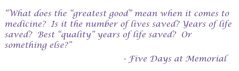
In Five Days at Memorial: Life and Death in a Storm Ravaged Hospital, Sheri Fink, an MD-turned-journalist, describes the fate of the patients and staff who faced the storm from within Memorial Hospital’s walls. As the decisions made during this unique, chaotic time cannot be evaluated from the place of our normal lives, she begins her expose by providing a historical and sociological context. When Katrina strikes, she provides a clear timeline of how the social framework and other mores governing our everyday lives start to break down, both inside and outside the hospital. We see how the uncoordinated, ineffective initial disaster response leads to further hopelessness.

Physicians and nurses start to make up their own triage methods, including giving last priority to any patient with a DNR status, irrespective of their current clinical condition. When the first patients are euthanized, Fink almost makes it seem like the only option. Although a reader might think that this situation was a complete anomaly born out of the unique events that occurred at one crazy hospital, Dr. Mary Mercer, the medical director of the San Francisco Base Hospital, adds that the “expectant death” category of triage was being used in many parts of New Orleans. Before Katrina, this category had never before been utilized in the United States; it had never been necessary. The second half of the book follows the legal battles that ensued, seemingly punishing those that had stayed behind to help.

An airboat helped evacuate patients and staff from Memorial Medical Center in New Orleans after HurricaneImage source:  Star Tribune

An airboat helped evacuate patients and staff from Memorial Medical Center in New Orleans after Hurricane

Image source:  Star Tribune

A few days after Katrina, Dr. John Brown and his DMAT team deployed to the Superdome. Because of safety concerns, they were moved to the airport, where many of Memorial’s evacuated patients and staff had already been waiting for days for food, water, and medical care. A few months later, Mike Marsh and other responders coordinated by the Department of Homeland Security met with Dr. Saussy, New Orleans’ EMS Medical Director, to complete an after action review, the formal evaluation of the strengths and weaknesses of a system and its disaster response. Marsh corroborates Fink’s account, and bolsters it with his EMS perspective. As with other medical infrastructure, the city’s 911 system had collapsed. The city emergency operations center flooded, ambulances flooded, people abandoned posts, the EMS base station was underwater, and a high percentage of the prehospital first responders were unaccounted for.

The federal after action report and media coverage led to several regulatory changes that have positively shaped disaster infrastructure in our country, including a national contract for ground ambulance support, coordination and integration of DMAT teams, the development of mutual aid for law enforcement, and contraflow evacuation methods. Other cities noted the importance of having an agreed-upon triage system in place before a disaster hits, and began to involve community members in the design process. 

Unfortunately,  these lessons haven’t reached everyone.  When reading the after action reports of subsequent disasters, common themes emerge.  More recently, when I was providing medical care at Standing Rock during a blizzard in December, I saw more parallels than I would have liked. We also had false information, communication breakdowns, a lack of unified chain of command, unclear triage methods, a patchwork of responders whose actions lacked coordination, and a lack of running water and functioning indoor plumbing. I wished that local leadership had a better understanding of the principles of disaster response, or that they had even read Five Days at Memorial.

After about an hour of conversing about various themes in the book, we noticed the omission of a major one: the mental health of patients, their families, and responders. During the recent Ghost Ship Fire in Oakland, the majority of the county’s response efforts revolved around emergency counseling and psychiatric support for the victims’ family and friends. Dr. Brown said that in all of his DMAT missions, several team members always quit, many never to be heard from again. Marsh told the story of a medic who responded to Katrina.  Although she witnessed countless human tragedies during her shifts without apparent difficulty, on her drive home, she would always break down because there, on the side of the road, was always lying the same dead baby.  At this point, Kelly Coleman, Alameda County’s Regional Disaster Medical Health Coordinator, mentioned the concept of responder guilt, a type of survivor guilt that first responders often experience. Although I had never before heard the term, it gave a name to an emotion that I knew I had also felt.   The pre-planning of disaster preparedness should include the mental health of first responders.

evicted.jpeg

All-too-soon, the clock struck eight, last sips of beer were finished, and plans were made for our next book: Evicted: Poverty and Profit in the American City, which we will be discussing in April.  Stay tuned for the scheduled date to join the discussion via twitter.


Do you have any books that you think we should read and discuss? If so, please share by emailing [email protected]

The content of this post is based on the book and group discussion, and not all statements have been independently verified.

The New 12-Lead: Prehospital Point of Care Ultrasound

by Brandon Bleess, MD

EMS MEd Editor: Maia Dorsett, MD PhD (@maiadorsett)

Case Scenario #1

image.001.jpeg

EMS is dispatched to scene of a witnessed cardiac arrest.  A 54 yo male was at a family gathering when he suddenly clutched his chest prior to collapsing and becoming unresponsive. First responders arrived within 4 minutes of initial call to find a bystander attempting CPR. On ALS arrival, first responders are performing compressions, have applied a monitor and shocked once for ventricular fibrillation.  Cardio Cerebral Resuscitation (CCR) is continued as patient deteriorates into asystole.  He has continuous CPR performed with a supraglottic airway placed as well as epinephrine given every 3-5 minutes and resuscitation is continued for 20 minutes.  The paramedic performs a cardiac Point of Care Ultrasound (POCUS) and finds the following:

Cardiac Standstill on Point-of-Care Cardiac Ultrasound

Case Scenario #2

EMS is dispatched to a vehicle motor vehicle collision (MVC).  Upon arrival EMS finds significant intrusion into the driver’s side of a vehicle that has been T-Boned by another vehicle.  The fire department is finishing extrication of the patient.  He is responsive to verbal stimuli and follows commands.  He has a heart rate of 122 and blood pressure of 120/56. Given the noise level on scene, the paramedic is unable to auscultate lung sounds in any fields.  The patient complains of abdominal pain and a seat belt sign is noted.  Local protocols state to transport to the closest facility, however the astute paramedic knows that the patient could be better served at a trauma center if surgery is needed.  Noting his vital signs and exam, he knows that intraperitoneal hemorrhage, tension pneumothorax, or pericardial effusion could be the cause of his presentation.  The paramedic performs a eFAST exam and finds the following:

Free Fluid in the Right Upper Quadrant on FAST exam

Normal Lung Slide

POCUS has improved the clinical practice of emergency medicine, begging the question of whether it should be incorporated into prehospital care.   Is POCUS practical for prehospital use?  How may it be used for triage and/or clinical management in the prehospital setting? 

Literature Review:

The use of ultrasound to improve clinical decision-making and management has ventured out of hospitals and into the prehospital realm.  In some clinical scenarios, including cardiac arrest and trauma triage, decreasing “time to ultrasound” may accelerate clinical decisions or lead to more appropriate utilization of healthcare resources. 

POCUS in Prehospital Management of Cardiac Arrest

Recent changes in management of Out-of-Hospital Cardiac Arrest (OHCA) from “load and go” to the “stay and play” method of cardiocerebral resuscitation (CCR) have shifted the burden of termination of resuscitation onto prehospital providers.  Multiple studies have addressed whether Point-of-Care Cardiac ultrasound would be useful in prehospital management of OHCA [1].

For example, a prospective study of 88 patients in cardiac arrest (PEA or asystole) conducted in Germany published in 2010 evaluated the prognostic value of cardiac ultrasound in OHCA.  Among patients with cardiac activity, 34% of patients survived to hospital admission compared to only 6% of those without cardiac activity on initial ultrasound [2].  They did not report survival to hospital discharge or neurologically-intact survival.

Another small prehospital study published in 2012 enrolled 42 patients in cardiac arrest with any rhythm [3].  Among 32 patients with no cardiac activity on initial field echocardiogram, only one survived to hospital admission.   In contrast, 4 of the 10 patients with cardiac activity survived to hospital admission. Only one of forty-two patients survived to hospital discharge (and did so with full neurology recovery).   He had cardiac activity on his prehospital ultrasound.

While these results were interesting, both studies were underpowered to detect the key outcome of neurologically-intact survival without cardiac activity on ultrasound due to the overall low incidence of survival from OHCA.

However, an adequately-powered multi-center Emergency Department study of 993 pre-hospital and ED patients with cardiac arrest in PEA or asystole was recently published [4].  Lack of cardiac activity portended an extremely poor likelihood of survival to hospital discharge (0.6%, neurologic status not reported).  In addition, POCUS was able to identify causes (Pulmonary embolism, cardiac tamponade) of cardiac arrest not amenable to traditional ACLS interventions. 

Given high utilization of resources with prolonged resuscitation and the potential to identify reversible causes of cardiac arrest, these results suggest that cardiac ultrasound may be beneficial in prehospital management of OHCA.

The FAST Exam and Trauma Triage

In emergency department patients with torso trauma, performing a FAST exam decreases time to operative care and the number of CT examinations of the torso [5].  FAST and eFAST (FAST + lung ultrasound) have since become key clinical decision making tools in the triage and management of trauma patients in the ED.  Extrapolating this to the field, could early identification of free fluid on abdominal exam better delineate which patients require one trauma center versus another?  Could lung ultrasound be used to help identify who needs needle decompression versus who does not, thus avoiding unnecessary intervention?

The prehospital FAST exam may allow for more appropriate transport destination decisions by providing valuable information to be obtained[6,7,8].  One prospective, multicenter study carried out in Germany study sought to compare the accuracy of physical exam and prehospital FAST exam to detect hemoperitoneum and to determine whether it changed clinical management [9].  They enrolled 230 patients with blunt trauma.  Among 202 patients who were fully scanned and were not lost to follow-up, 28 patients were found to have hemoperitoneum by ED ultrasound or CT imaging.  26 of these were identified prehospital, leading the authors to conclude that prehospital FAST has a sensitivity of 93 % (95 % CI 76 – 99 %) and specificity of 99 % (95% CI 97 -100 %).   However, as the study excluded patients lost to follow-up or in whom ultrasound was too technically difficult, the sensitivity of prehospital FAST for accurate detection of hemoperitoneum could be falsely inflated.   The study was interesting in that the there were several examples where prehospital detection of abdominal free fluid changed patient management, including minimizing prehospital interventions and alerting the receiving hospital to reduce time to surgical intervention.  As this was not a randomized trial, it was unclear whether this actually changed to the time to surgical intervention, but based on results of ED-based studies it is likely to have done so.

Several systematic reviews have examined the current evidence regarding the potential usefulness of prehospital ultrasound to change diagnosis or treatment of trauma patients  [10,11].  Their overwhelming conclusion?  The evidence is promising, although the quality of evidence very low and more studies are needed.

Practical for prehospital use?

Development of handheld, battery-powered, low-weight US machines has created the possibility of bringing US to the prehospital setting.  In addition, field ultrasound images can be transmitted en route to the emergency department (ED) similar to 12 lead EKGs [12,13,14].

A 2014 survey of medical directors using the NAEMSP mailing list demonstrated that 4.1% of EMS systems were already using ultrasound and that an additional 21.7% of systems were considering the implementation of pre-hospital ultrasound [15].  The vast majority cited equipment costs (89.4%), as well as training costs (73.7%), and challenges related to the training process (53.5%) as the major points of concern of why they the medical directors thought that it could not be implemented in their system. 

As a corollary to this, it is not surprising to see that medical directors would be willing to implement ultrasound into their system if there was decreased cost (69.7%), practice guidelines that included prehospital ultrasound (66.1%), a

nd studies demonstrating improvement in patient morbidity (73%) and mortality (71.8%).

Prehospital POCUS use has been more thoroughly investigated in Europe than in the United States [16,17].  This is somewhat of a confounding issue given that many EMS services and Europe use physicians in the field compared to the paramedic model in the United States.  So, is training paramedics to accurately perform prehospital POCUS feasible?  The current evidence suggests that it is.

Ultrasound education centers on two related but distinct skills:  Image acquisition and Image Interpretation. 

A 2015 study examined the ability of US EMS Providers’ (EMT, Paramedics, and Students) to interpret ultrasound images and identify pericardial effusion, pneumothorax, and cardiac standstill [18].  They were given a pre-test followed by an hour didactic session covering scanning techniques, normal anatomy, and image interpretation of both normal and pathologic videos.  After the didactic they were given an immediate post-test as well as a post-test one week later.

The study found that following a short educational intervention, paramedics could more accurately and confidently identify key ultrasound findings that would affect clinical management.  While this study only looked at image recognition and not image acquisition, it showed that the US EMS providers are able to identify pathologic conditions on ultrasound. 

Several studies have examined educational programs to train paramedics to both acquire and interpret prehospital ultrasound images.

A 2010 study looked at US paramedics and their ability to perform and interpret FAST exams and abdominal aortic (AA) exams [19].  Paramedics from two EMS agencies received a 6 hour training program with ongoing refresher education.  All ultrasound exams were then reviewed by a blinded physician overreader (PO).  A total of 104 patients were evaluated (84 FAST and 20 AA) using ultrasound, of which 76 FAST exams were adequate for evaluation and all 20 AA exams were adequate.  Of that, 6 FAST exams were deemed positive by the paramedics and the PO.  All 20 of the AA exams were deemed negative by the paramedics and the PO.  With these, there was a 100% proportion of agreement between the paramedics and the PO.  The study also looked at the amount of time that it would take paramedics to perform the exams as this could be a possible downside prior to transport.  The mean time for image acquisition for the FAST exam was 156 seconds (2.6 minutes) with the median being 138 seconds (range of 76-357 seconds).

Figure 3,  PAUSE study (Ref 20)

Figure 3,  PAUSE study (Ref 20)

A 2013 study looking at the viability of a Prehospital Assessment with Ultrasound for Emergencies (PAUSE) Protocol enrolled 20 firefighter/paramedics that did not have prior ultrasound training.  They underwent a 2 hour didactic session on the use of ultrasound on the lungs and heart to look for pneumothorax, pericardial effusion, and cardiac activity [20]. 

As noted in Figure 3 from the PAUSE study, 18 of the 20 subjects scored an 80% or higher and the mean score was 9.1 overall.

There was one image of cardiac standstill that 6 of the 20 paramedics answered incorrectly.  The authors note that the believed this to be due to the perceived cardiac movements as a result of the ultrasound probe being moved across the patient’s chest.

When evaluating image acquisition, 100% of the images for the evaluation of pneumothorax were noted to be satisfactory.  The Cardiac Ultrasound Structural Assessment Scale (CUSAS) was used to assess for adequate cardiac views for diagnosis [21].   The authors noted that for the purposes of determining cardiac standstill, being able to visualize any myocardium (CUSAS Score 3) should be adequate.  If this is true, there is a 100% success rate in the study.  They also believed that a significant pericardial effusion causing tamponade would likely be seen with CUSAS score of ≥4, as these images offer at least a partial view of the pericardium. Given these assumptions, in this study, 95% of the participants (19/20) were able to quickly acquire images that would likely be useful in assessing for both cardiac activity and a pericardial effusion.  In terms of time, views of the lung were acquired in less than 5 seconds. The views of the heart were acquired in less than 10 seconds for 16 paramedics. One paramedic took approximately 90 seconds, and the other three ranged between 10 and 25 seconds.

Take Home

In the hands of physicians and paramedics, POCUS is a promising technology to direct clinical care and utilization of prehospital resources.  However, the use of prehospital ultrasound must improve patient outcomes for it to become a reality and the standard of care.


Credits

Videos courtesy of Washington University in St. Louis Division of Emergency Medicine, Section of Emergency Ultrasound.

References

1.    Kellum MJ, Kennedy KW, Barney R, et al. (2008) Cardiocerebral resuscitation improves neurologically intact survival of patients with out-of-hospital cardiac arrest. Ann Emerg Med;52:244–52.
2.    Breitkreutz, R., Price, S., Steiger, H. V., Seeger, F. H., Ilper, H., Ackermann, H., Walcher, F. (2010). Focused echocardiographic evaluation in life support and peri-resuscitation of emergency patients: A prospective trial. Resuscitation, 81(11), 1527-1533.
3.    Aichinger, G., Zechner, P. M., Prause, G., Sacherer, F., Wildner, G., Anderson, C. L., Fox, J. C. (2012). Cardiac movement identified on prehospital echocardiography predicts outcome in cardiac arrest patients. Prehospital Emergency Care Prehosp Emerg Care, 16(2), 251-255.
4.    Gaspari, R., Weekes, A., Adhikari, S., Noble, V. E., Nomura, J. T., Theodoro, D., ... & Caffery, T. (2016). Emergency department point-of-care ultrasound in out-of-hospital and in-ED cardiac arrest. Resuscitation, 109, 33-39.
5.    Melniker, L. A., Leibner, E., McKenney, M. G., Lopez, P., Briggs, W. M., & Mancuso, C. A. (2006). Randomized controlled clinical trial of point-of-care, limited ultrasonography for trauma in the emergency department: the first sonography outcomes assessment program trial. Annals of emergency medicine, 48(3), 227-235.
6.    Chaudery, M., Clark, J., Wilson, M. H., Bew, D., Yang, G., & Darzi, A. (2015). Traumatic intra-abdominal hemorrhage control. Journal of Trauma and Acute Care Surgery, 78(1), 153-163.
7.    O'Dochartaigh, D., & Douma, M. (2015). Prehospital ultrasound of the abdomen and thorax changes trauma patient management: A systematic review. Injury, 46(11), 2093-2102.
8.    Ruesseler, M., Kirschning, T., Breitkreutz, R., Marzi, I., & Walcher, F. (2009). Prehospital and emergency department ultrasound in blunt abdominal trauma. Eur J Trauma Emerg Surg European Journal of Trauma and Emergency Surgery, 35(4), 341-346.
9.    Walcher F, Weinlich M, Conrad G, et al. Prehospital ultrasound imaging improves management of abdominal trauma. Br J Surg. 2006; 93:238–42.
10.    Jørgensen, H., Jensen, C. H., & Dirks, J. (2010). Does prehospital ultrasound improve treatment of the trauma patient? A systematic review. European Journal of Emergency Medicine, 17(5), 249-253.
11.    O’Dochartaigh, D., & Douma, M. (2015). Prehospital ultrasound of the abdomen and thorax changes trauma patient management: A systematic review. Injury, 46(11), 2093-2102.
12.    Sibert, K., Ricci, M. A., Caputo, M., Callas, P. W., Rogers, F. B., Charash, W., . . . Kocmoud, C. (2008). The feasibility of using ultrasound and video laryngoscopy in a mobile telemedicine consult. Telemedicine and E-Health, 14(3), 266-272.
13.    Strode, C. A. (2003). Satellite and mobile wireless transmission of focused assessment with sonography in trauma. Academic Emergency Medicine, 10(12), 1411-1414.
14.    Takeuchi, R., Harada, H., Masuda, K., Ota, G., Yokoi, M., Teramura, N., & Saito, T. (2008). Field testing of a remote controlled robotic tele-echo system in an ambulance using broadband mobile communication technology. J Med Syst Journal of Medical Systems, 32(3), 235-242.
15.    Taylor, J., McLaughlin, K., McRae, A., Lang, E., & Anton, A. (2014). Use of prehospital ultrasound in North America: a survey of emergency medical services medical directors. BMC Emergency Medicine, 14, 6
16.    Walcher F, Petrovic T, Heegaard W, et al.(2008) Prehospital ultrasound: perspectives from four countries. In: MAJ, MateerJ, BlaivasM, eds. Emergency Ultrasound. New York, NY: McGraw Hill.
17.    Nelson BP, Chason K. Use of ultrasound by emergency medical services: a review(2008). Int J Emerg Med. 1:253–9.
18.    Bhat SR, Johnson DA, Pierog JE, Zaia BE, Williams SR, Gharahbaghian L. (2015) Prehospital Evaluation of Effusion, Pneumothorax, and Standstill (PEEPS): Point-of-care Ultrasound in Emergency Medical Services. Western Journal of Emergency Medicine. 16(4):503-509.
19.    Heegaard, W., Hildebrandt, D., Spear, D., Chason, K., Nelson, B. and Ho, J. (2010), Prehospital Ultrasound by Paramedics: Results of Field Trial. Academic Emergency Medicine, 17: 624–630.
20.    Chin E, Chan C, Mortazavi R. (2013) A pilot study examining the viability of a Prehospital Assessment with UltraSound for Emergencies (PAUSE) protocol. J Emerg.44:142–149.
21.    Backlund, B., Bonnett, C., Faragher, J., Haukoos, J., and Kendall, J.  (2010) Pilot study to determine the feasibility of training Army National Guard medics to perform focused cardiac ultrasonography. Prehosp Emerg Care. 14: 118–123.

It Takes a Village...: Pediatric Out of Hospital Cardiac Arrest

by Melissa Puffenbarger MD

Expert review/editor Joelle Donofrio DO (@PEMEMS) & Hawnwan Moy MD (@Pecpodcast)

It’s eerily quiet in the Pediatric Emergency Department (ED) and everyone implicitly hopes that the peace will linger for the last hour of your overnight shift.  However, as an experienced Pediatric Emergency Medicine (PEM)/Emergency Medical Services (EMS) physician, you know that's probably not going to happen.  

Within minutes, your staff receives an emergent call from EMS. “We’re inbound with a 6-month-old male in cardiac arrest, compressions in progress, not intubated but being bagged, IO placed, 1 round of epi given, last rhythm check 2 minutes ago was PEA, and ETA 2 minutes.”  You can visibly see the anxiety build in the ED as everyone starts to shakes off their fatigue to get ready for this patient.

On arrival, EMS rushes the tiny patient into the resuscitation room.  As compressions are handed over to the Peds ED staff, the visibly shaken paramedic slowly drifts to the corner of the room looking on in concern.  As the resuscitation continues, you have a brief thought...with all the emphasis on adult prehospital cardiac arrest, what evidence do we have to provide the best care for pediatric out of hospital cardiac arrest (p-OHCA) patient?  

Literature Review:

When you hear about OHCA, the conversation will inevitably mention topics like pit crew CPR, the Cardiac Arrest Registry for Enhanced Survival (CARES) database, and the Resuscitation Outcomes Consortium (ROC). Yet, p-OHCA is often absent in these conversations, not because there is a lack of passion (there are a LOT of eager pediatric EM/EMS researchers out there), but because there are a lot of unanswered clinical questions concerning this topic.  Why is that?  First off, the number of p-OHCA is low. The incidence of p-OHCA is around 8 per 100,000 person-years with a dismal 6% survival to hospital discharge [7].  Additionally, only 13% of EMS runs are for pediatric patients [1].  As a result, not only do our EMS providers receive minimal pediatric clinical experience, but the low incidence makes p-OHCA research more difficult.

Nonetheless, to start to improve outcomes, we have to know where the baseline lies.  In a recent observational study utilizing data from the ROC, Fink et al. attempted to define how p-OHCA survival rates have changed in a 5-year time span from July 1, 2007, to June 30, 2012, by studying 1738 children with OHCA.  Unfortunately, the study showed that mortality rates and neurologic outcomes for pediatric out-of-hospital cardiac arrest have not improved [2]. Annual survival rates for p-OHCA were 6.7-10.2%, compared to a reported increase in survival rate of in-hospital cardiac arrest at 14-43% [2].  This large difference in survival between in-hospital and out-of-hospital arrests is likely related to multiple factors. These factors include time to compressions for an unwitnessed arrest, quality of bystander CPR, and a low frequency of initial shockable rhythms in pediatric patients.  

Although these findings are not a huge surprise, the real interesting data arises when this manuscript compares survival to discharge of the different regions of the ROC study.  For a brief refresher, the ROC is a collaboration of 10 regional sites in the United States and Canada.  Thus, when the authors compared regions to each other, ROSC rates of p-OHCA ranged from 2.5% to 34.7%.  Additionally, survival to discharge rates ranged from 2.6% to 14.7%.  We need to determine why ROSC and survival to discharge varied so widely across regions in order to replicate best practices in p-OHCA.  Fink et al. found that “...the regions with the greatest increases in survival over time exhibited increases in EMS-witnessed OHCA, increased the frequency of bystander CPR, and increased EMS-defibrillation compared to regions that did NOT see increases in survival over time [2].”    

What might be the first step in improving our p-OHCA ROSC and survival to discharge?  One place would be increasing provider knowledge and comfort when taking care of pediatric patients.   When EMS providers were asked to self-identify educational priorities, Paramedics, EMT-Basics, and first responders prioritized pediatric airway management, anxiety when working with children, and general pediatric skills as primary areas for targeted education [3]. Specifically, these providers identified a need for training regarding IV and IO access, when and how to perform an advanced airway, recognizing normal neonatal vital signs, and prevention of hypothermia [3]. Intuitively, targeting education to these areas and providing a foundation for continuously updating EMS skills and pediatric protocols can help bridge these knowledge gaps and perhaps help improve p-OHCA outcomes.  

The scant amount of literature available on p-OHCA supports the self-identified educational needs of our EMS providers. One study assessed pediatric airway management from a large database that included EMS encounters in 40 states and identified that EMS airway management should be a target for continuing skill development [4]. This study showed that endotracheal intubation (ETI) was the most commonly used advanced airway technique among EMS encounters.  There was a significantly lower success rate for out-of-hospital ETI vs. in-hospital (81.1% success rate for out-of-hospital in this series vs. reported 97-99% success rate among PEM physicians), and alarmingly low use of CO2-based placement confirmation [4]. The higher in-hospital success rate likely reflects access to adjunctive airway equipment as well as very different levels of experience with the pediatric airway. One series reported that paramedic students received only 6-10 intubation attempts in the OR during training, and most of these were adults [5]. Pediatric patients in full arrest are unique in that they most commonly have a primary respiratory issue, and focusing on providing adequate ventilation and oxygenation is the key to their resuscitation. While improving the EMS provider’s advanced airway skills may help patients in more extreme situations, the biggest impact will likely be seen in striving for perfection with basic airway management: positioning to open the airway, providing a good seal during BVM, and ventilating at an appropriate rate and volume.  Currently, there is no good data supporting prehospital pediatric intubation.    

In addition to skills in pediatric airway management, EMS CPR quality has also been shown to require improvement. A large prospective observational study demonstrated that prehospital CPR only met AHA guidelines during p-OHCA resuscitations 16% of the time and less than 25% of events met both rate and CPR fraction target [6]. While we know that many, many factors affect p-OHCA survival, this study identifies that consistently performing high-quality CPR is critical. The goals of high-quality CPR are the same for both pediatric and adult patients with a focus on providing adequate depth and rate of compressions, minimizing interruptions to compressions, and providing effective oxygenation and ventilation. Processes that may help maintain high-quality CPR in the field include asking EMS partners to coach, praise and correct each other as needed when performing CPR, periodic skill sessions, and staying up to date on any AHA guideline changes.

Remaining up-to-date on the most recent practice guidelines as well as maintaining proficiency of certain skills should be approached as a team effort. EMS physicians should provide scheduled educational sessions that meet the expressed needs of EMS providers and periodically review how to care for special patient populations such as the arresting child.  EMS providers should continue to improve pre-hospital care in their communities by evaluating themselves and each other, and remain involved in community outreach projects focused on prevention of injuries and improved bystander CPR.  As an example, a bill in California was passed that mandates high schools with a health requirement to graduate to require CPR training [8].  To take this one step further, as part of the San Diego EMS County cardiac arrest task force’s agenda, fire and EMS  are even teaching middle schoolers the art of bystander CPR.  It's actions like these that can really help our sick pediatric patients and EMS providers.  Finally, a culture of open dialogue with direct and timely feedback between ED personnel and EMS providers after transporting a critically ill patient will create an environment where all parties involved help improve the pre-hospital care of the pediatric patient.   

Take Home Points:

Although a rare event, in the case of pediatric out of hospital cardiac arrest, factors that have been shown to increase ROSC and survival to discharge include EMS-witnessed OHCA, increased frequency of bystander CPR, and increased EMS-defibrillation.  Additionally solid CPR mechanics, BASIC airway management, solid CPR education of the youth in our community and consistent, great pediatric education of our EMS providers allows us to provide the best care for the children in our communities.  As the old proverb goes, “It takes a village to raise a child.”  So too does it take a village- from our EMS providers, our community, our pediatric EMS researchers, to our medical directors- to save a child.


References

1. Shan, MN. et al. The epidemiology of emergency medical services use by children: an analysis of the National Hospital Ambulatory Medical Care Survey. Prehosp Emerg Care. 2008 Jul;12(3):269-76

2. Fink, E.L., Prince, D.K., et al. Unchanged pediatric out-of-hospital cardiac arrest incidence and survival rates with regional variation in North America. Resuscitation. 2016;107:121-128

3. Hansen, M., Meckler, G., et al. Children’s safety initiative: A national assessment of pediatric educational needs among emergency medical services providers. Prehosp Emerg Care. 2015; 19(2):287-291

4. Hansen, M., Lambert, W., et al.Out-of-hospital pediatric airway management in the United States. Resuscitation. 2016;90:104-110

5. Johnson, B.D., Seitz S.R., et al. Limited opportunities for paramedic student endotracheal intubation training in the operating room. Acad Emerg Med. 2006;13:1051-5

6. Sutton, R., Case, E., et al. A quantitative analysis of out-of-hospital pediatric and adolescent resuscitation quality – A report from the ROC epistry-cardiac arrest. Resuscitation. 2015;93:150-157

7. Atkins DL, Everson-Stewart S, Sears GK, et al.  Epidemiology and outcomes from out-of-hospital cardiac arrest in children: the Resuscitation Outcomes Consortium Epistry-Cardiac Arrest.  Circulation 2009;119:1282-91.  

8.  "Text." Bill Text - AB-1719 Pupil Instruction: Cardiopulmonary Resuscitation. Web. 10 Jan. 2017.

Images from:

1. http://www.ukprogressive.co.uk/wp-content/uploads/2016/12/hospital-emergency-room-1.jpg

2. http://www.christianitytoday.com/images/46719.png

3. http://cosmouk.cdnds.net/15/31/1600x800/landscape-1438173668-cute-success-kid.jpg

4. http://www.thedebutanteball.com/wp-content/uploads/2015/11/117dfdafbd688ea2f7745cb21b742895.1000x998x1.jpg


Time to Stop Beating a Dead Horse: Termination of Resuscitation in the Field

by Al Lulla, MD (@al_lulla) and Bridgette Svancarek, MD

Expert/Peer Reviewed by J. Brent Myers, MD, MPH    (@bmyersmd)

You arrive on scene to a cardiac arrest.  Your patient is a 64 yo male found pulseless by his wife when she returned from walking the family dog.  The patient’s initial rhythm was a narrow complex PEA.  Twenty-five minutes and 12 cycles of CPR later the rhythm remains unchanged.  The man’s wife is distraught and tearful.  Your clinical experience tells you that the likelihood of a good outcome is poor.

Termination of resuscitation (TOR) is commonplace in the ED and ICU setting, but there is a role for TOR in the pre-hospital setting as well. When is it appropriate to do so?  What are the risks and benefits of such this decision?

Literature Review

Nontraumatic out of hospital cardiopulmonary arrest (OHCA) is considered to be a catastrophic event that is associated with a poor prognosis. According to a report published by the American Heart Association, it is estimated that there is an annual incidence of 326,000 OHCAs occurring in the United States [1]. Several studies have reported that for the majority of patients, only those who regain pulses in the field may end up surviving to hospital discharge. For patients who do not regain pulses, outcomes are generally poor. Bonnin, et al. reported a 0.6% survival rate in patients who did not achieve return of spontaneous circulation (ROSC) within 25 minutes of paramedic arrival [2]. Another study performed in Japan demonstrated that death was 25.8 times more likely in patients without prehospital ROSC [3]. Despite these dismal statistics, in attempt to maximize chance for survival, patients who have suffered an OHCA are often continuously resuscitated by EMS and transported to the ED.

In 2011, the National Association of EMS Physicians (NAEMSP) released a position statement regarding termination of resuscitation in patients with non-traumatic cardiopulmonary arrest. In the statement, the NAEMSP affirmed the following: 

… As there are patients who will not be successfully resuscitated, an evidence-based methodology to determine those patients with out-of-hospital non traumatic cardiopulmonary arrest that will not result in a favorable outcome would contribute to the public health by conserving valuable health care resources and decreasing the number of emergency vehicles in transit with warning lights and sirens.

In addition to the poor outcomes associated with OHCA, the NAEMSP position statement touches upon several points that favor TOR versus transporting patients [4]:

·      Lights and sirens can be dangerous: According to the National Highway Traffic Safety Administration (NHTSA) Fatality Analysis System, approximately 59.6% of ambulance crashes occur during emergency use. Another study identified 45.9 ambulance crashes per 100,000 patients with lights and sirens versus 27.0 ambulance crashes per 100,000 patients without lights and sirens [5].

·      High quality CPR: Guidelines put forth by the American Heart Association highlight the importance of high quality CPR. There is some evidence to suggest that on scene chest compressions are higher quality in comparison to compressions done en route. Russi, et al. evaluated quality of chest compressions on scene vs transport in 140 patients. The study reported significantly lower quality compressions (i.e. decreased depth) during transport versus on scene. [6]

·      Financial cost and resource depletion: The cost of an unsuccessful resuscitation is significant, especially given scarce EMS resources. In addition, there is a cost to the general public when an EMS crew is out of service transporting a patient. For EMS physicians and providers in the field, it’s important to ask: is continuing resuscitation in the best interest of the patient? 

The points highlighted by the NAEMSP position paper as well as pre-hospital research which confirms poor outcomes for OHCA have served as an impetus for the implementation of TOR criteria in EMS systems. While multiple different sets of criteria have been studied and subsequently validated, by far the most widely accepted and researched were the criteria put forth by the Ontario Prehospital Advanced Life Support (OPALS) group.

From their registry of cardiac arrest patients, the OPALS investigators derived two sets of TOR rules, one for basic life support (BLS) providers and one for advanced life support (ALS) providers. These rules were retrospectively developed based on the goal of identifying all non-survivors. The BLS rule included three criteria of which all need to be fulfilled in order to terminate the resuscitation: unwitnessed by EMS, no AED or shock delivered, and no ROSC. The ALS criteria included the BLS rules and two additional criteria: the arrest had to be unwitnessed by a bystander, and no bystander CPR was performed.

The data from the OPALS cardiac arrest registry showed that patients who fulfill all of the TOR criteria do not have good outcomes. For the BLS TOR protocol, Verbeek et al reported the rule to be 100% sensitive in identifying survivors and had a negative predictive value of 100% in identifying non-survivors in patients with OHCA [7]. These findings were validated in subsequent studies. Sasson, et al. demonstrated in a retrospective cohort study that in 2592 patients who suffered from OHCA and met BLS TOR criteria, only 0.2% survived to hospital discharge (98.7% specificity, CI 97.0-99.6). In the 1192 patients who met ALS TOR criteria, 0 survived to hospital discharge (100% specificity, CI 99.1-100). In essence both rules have close to 100% positive predictive value for predicting death in patients with OHCA [8].

Morrison, et al. had similar findings in their study, which just looked at BLS termination criteria regardless if there were BLS or ALS providers on scene. They found that in 776 patients who fulfilled BLS TOR criteria, only 4 patients (0.5%) survived, with a positive predictive value of 99.5% for predicting death. Of the 4 survivors, 3 were characterized as having good cerebral performance, with 1 patient having severe neurological disability. The study showed that implementation of the BLS TOR criteria would theoretically reduce rate of transport by 62.6%!  How to reconcile this benefit with three neurologically-intact survivors who met BLS TOR criteria was not explicitly addressed. [9]

No matter which way you look at it, the research on pre-hospital TOR is clear: OHCA patients who fulfill BLS or ALS TOR criteria most often do not survive to hospital discharge. Studies consistently show a survival of less than 0.5% if BLS TOR criteria are followed and 0% if ALS criteria are followed. Studies also show transportation rates of 40-60% if BLS criteria are followed and around 80% if ALS criteria are followed.  In spite of this, research suggests that there are barriers to the implementation of TOR criteria by EMS providers. The Termination of Resuscitation Implementation Trial (TORIT) was a multi-center prospective trial that evaluated the implementation of TOR rules in patients with OHCA. The investigators showed that in 953 patients who were BLS TOR eligible, EMS providers correctly applied the rule in 755 patients (79%) and did not apply the rule in 198 patients (21%). All of the 198 patients in whom the rule was not applied (i.e. they were transported to the hospital despite meeting BLS TOR criteria) did not survive. For these patients, providers were surveyed regarding their decision to transport. Family distress was the most commonly cited reason for continuing resuscitation and transporting patients [10]. 

Is there a magic number?

Studies have shown that pre-hospital ROSC is the most important predictor of survival for patients with OHCA. An important question often arises: how long should EMS providers work these patients in the field? The prospective 1993 study by Bonnin, et al. showed that in 1471 patients with OHCA, only 370 patients achieved ROSC on scene. Of these 370 patients, all patients achieved ROSC within 25 minutes of paramedic arrival. Newer research coming out of Wake County EMS has shown that working patients longer may be of benefit. In 2905 adult OHCA patients that were examined retrospectively, 363 survived (12.5%). Of the survivors, 300 patients (83%) were neurologically intact. The investigators found that duration of prehospital resuscitation was less than 40 minutes in 90% of neurologically intact survivors [11]. Other studies have shown neurologically intact survival with duration of resuscitation times ranging between 35 to 60 minutes. This broad range may likely be attributed to variation in EMS practice and available resources (i.e. therapeutic hypothermia) as it pertains to agency specific protocols for OHCA.

And what about traumatic cardiopulmonary arrest?

Prehospital TOR may also play a significant role in patients suffering from traumatic arrest as well. While there are sets of robust decision rules validated for TOR and nontraumatic cardiopulmonary arrest, the research on TOR in traumatic arrest patients is scarce.

In 2012, the NAEMSP in conjunction with the American College of Surgeons Committee on Trauma (ACSCOT) released a joint position statement addressing this issue. Based on a review of the literature, the NAEMSP-ACSCOT paper estimated that survival rates for traumatic cardiopulmonary arrest is approximately 2%. This position paper differentiates between withholding resuscitation in cardiopulmonary arrest and TOR.

The guidelines recommend withholding resuscitation in the following patients with traumatic arrest [12]:

·      Whom death is the predictable outcome

·      Injuries incompatible with life (i.e. decapitation, hemicorporectomy)

·      Blunt or penetrating trauma with evidence of prolonged cardiac arrest (i.e. rigor mortis)

·      Blunt trauma patients who are apneic, pulseless, without organized EKG activity

·      Penetrating trauma patients who are apneic without signs of life (i.e. no spontaneous movement, EKG activity, pupillary response)


The paper makes the following comments regarding TOR in traumatic arrest:

·      The primary focus should be evacuation to an appropriate facility for definitive care

·      EMS systems should implement protocols that allow for TOR in cases of traumatic arrest

·      TOR should be considered in patients without signs of life or without ROSC

·      Protocols should be in place that require for a specific interval of CPR (for example, up to 15 minutes prior to termination)

·      TOR protocols should be accompanied by procedures to ensure appropriate management of the deceased patient and support services for family members

·      Physician oversight is a mandatory component of TOR protocols

·      TOR protocols should include locally specific clinical, environmental or population based situations for which the protocol may not be applicable

·      Further research is required to determine optimal duration of CPR before TOR

A criticism of the NAEMSP-ACSCOT guidelines is their degree of detail and likely inability to be implemented in the field. A recent paper published in 2016 from Taiwan looked at a simplified decision rule for TOR in patients with traumatic cardiopulmonary arrest modified from the NAEMSP-ACSCOT guidelines. The simplified decision rule included two criteria: 1) blunt trauma injury AND 2) presence of asystole. The study found that this TOR rule could accurately predict 100% of non-survivors and had the potential to decrease ambulance transports for traumatic cardiopulmonary arrest between 16.4-29.0% [13].

Take home points

·      The majority of patients with nontraumatic OHCA who do not achieve ROSC in the field have a very poor prognosis

·      Identifying patients with OHCA who will most likely have an unfavorable outcome and not benefit from transport with lights and sirens is important with regards to EMS safety and utilization. 

·      The BLS and ALS TOR criteria derived from the OPALS cardiac arrest registry have been validated and are good predictors for which patients can be pronounced dead in the field

·      TOR may have an important role in patients suffering from traumatic arrest, however further research is still needed

·      There is still no widely accepted guidelines in terms of duration of prehospital resuscitation for OHCA, however several studies have shown successful outcomes ranging from 25 to 60 minutes

·      There are still barriers to implementation of TOR criteria which may explain underutilization by EMS agencies.  As family distress is the most commonly cited region for falling out of protocol, additional training of EMS personnel in communication skills, as well as public education will be important to successful implementation.

EMS MEd Editors: Maia Dorsett (@maiadorsett) & Hawnwan P. Moy (@PECpodcast)

References:

1.                Mozaffarian D, Benjamin EJ, Go AS, et al. Heart disease and stroke statistics--2015 update: a report from the American Heart Association. Circulation. 2015;131(4):e29-322.

2.                Bonnin MJ, Pepe PE, Kimball KT, Clark PS. Distinct criteria for termination of resuscitation in the out-of-hospital setting. JAMA. 1993;270(12):1457-62.

3.                Goto Y, Maeda T, Goto YN. Termination-of-resuscitation rule for emergency department physicians treating out-of-hospital cardiac arrest patients: an observational cohort study. Crit Care. 2013;17(5):R235.

4.               Millin MG, Khandker SR, Malki A. Termination of resuscitation of nontraumatic cardiopulmonary arrest: resource document for the National Association of EMS Physicians position statement. Prehosp Emerg Care. 2011;15(4):547-54.

5.               Saunders CE, Heye CJ. Ambulance collisions in an urban environment. Prehosp Disaster Med. 1994;9(2):118-24.

6.     Russi CS, Myers LA, Kolb LJ, Lohse CM, Hess EP, White RD. A Comparison of Chest Compression Quality Delivered During On-Scene and Ground Transport Cardiopulmonary Resuscitation. West J Emerg Med. 2016;17(5):634-9.

7.                Verbeek PR, Vermeulen MJ, Ali FH, Messenger DW, Summers J, Morrison LJ. Derivation of a termination-of-resuscitation guideline for emergency medical technicians using automated external defibrillators. Acad Emerg Med. 2002;9(7):671-8.

8.                Sasson C, Hegg AJ, Macy M, et al. Prehospital termination of resuscitation in cases of refractory out-of-hospital cardiac arrest. JAMA. 2008;300(12):1432-8.

9.                Morrison LJ, Visentin LM, Kiss A, et al. Validation of a rule for termination of resuscitation in out-of-hospital cardiac arrest. N Engl J Med. 2006;355(5):478-87.

10.            Morrison LJ, Eby D, Veigas PV, et al. Implementation trial of the basic life support termination of resuscitation rule: reducing the transport of futile out-of-hospital cardiac arrests. Resuscitation. 2014;85(4):486-91.

11.          Bachman MW, Williams JG, Myers JB, et al. Duration of prehospital resuscitation for adult out-of-hospital cardiac arrest: Neurologically intact survival approaches overall survival despite extended efforts. Prehosp Emerg Care. 2014;18(1):134–135.

12.          Hopson LR, Hirsh E, Delgado J, et al. Guidelines for withholding or termination of resuscitation in prehospital traumatic cardiopulmonary arrest: a joint position paper from the National Association of EMS Physicians Standards and Clinical Practice Committee and the American College of Surgeons Committee on Trauma. Prehosp Emerg Care. 2003;7(1):141-6.

13.          Chiang WC, Huang YS, Hsu SH, et al. Performance of a simplified termination of resuscitation rule for adult traumatic cardiopulmonary arrest in the prehospital setting. Emerg Med J. 2016;

Should EMS providers administer Ondansetron for pregnancy-related vomiting?

Summary and analysis by Maia Dorsett, MD, PhD     @maiadorsett

Framing the Problem

Nausea and vomiting in pregnancy is not rare, occurring in about 50% of all pregnant women. [1]  Symptom onset typically occurs within four weeks of the last menstrual period and peaks at nine weeks of gestation, with the vast majority of cases resolving by 20 weeks. The most severe form, hyperemesis gravidarum, is characterized by persistent vomiting, weight loss of more than 5%, ketonuria, electrolyte abnormalities and dehydration. [1]  This occurs in a minority of patients (0.3 to 1.0%), although patients with a less severe form may also access the emergency department by utilization of ambulance services. [1]  The frequency of EMS utilization for patients with hyperemesis gravidarum is unclear.

With regard to the most effective treatment of hyperemesis, the proverbial wind has blown in several directions.  Lessons learned from the tragedy of thalidomide, used in the late 1950’s as a treatment for nausea in pregnant women before being banned in 1961 due to severe teratogenic effects, have raised the bar of safety considerations for the administration of medications to pregnant women. [2] Partially in response to the thalidomide tragedy, in 1979 the FDA established five letter risk categories - A, B, C, D or X - to help clinicians evaluate the risk versus benefit of medication use in pregnancy (see Figure, [3]).  However, the pregnancy letter categories were often falsely interpreted to be an interpretation of teratogenic risk, leading to misunderstanding among clinicians and patients.   Recently, the FDA has recommended eliminating the letter categories in favor of the Pregnancy and Lactation Labeling Final Rule (PLLR) that went into effect on June 30, 2015 .  The PLLR requires a description of the “risk summary” and associated data regarding safety or harm in pregnancy and lactation. 

The anti-emetic ondansetron is approved by the FDA for the treatment of nausea and vomiting associated with chemotherapy or surgery.  However, its effectiveness at treating nausea and vomiting with little sedating side effects, as well as its classification as pregnancy category “B”, has now made it the most commonly used prescription oral antiemetic in pregnancy (at least in 2008). [4,5]  More than half of women treated in emergency departments will receive intravenous ondansetron. [4,6]  Retrospective cohort studies published in 2013 and 2014 came to conflicting conclusions regarding the safety of ondansetron in pregnancy, with one article finding a slight increase in the risk of cardiovascular defects. [see below for more in-depth review; 7,8]  In September 2015, the American Society of Obstetricians and Gynecologists published a Practice Bulletin on Nausea and Vomiting in pregnancy.  This practice bulletin recommended doxylamine and pyridoxine as first line treatment, and wrote that:

There are insufficient data on fetal safety with ondansetron use and further studies are warranted…. Thus, although some studies have shown an increased risk of birth defects with early ondansetron use, other studies have not and the absolute risk to any fetus is low. As with all medications, the potential risks and benefits should be weighed in each case.” [5]

From the perspective of the EMS provider, should we giving ondansetron to pregnant women in their first trimester with nausea and vomiting?  Our discussion forum reviewed one such case.


Case and Discussion

The Case:

EMS is called to the house of a 24 yo female who is 8 weeks pregnant.  She has had countless episodes of nausea and vomiting over the last 4 days.  She has been unable to take anything by mouth over this period and has not urinated in the last day.  Heart rate is 115, blood pressure 101/60.  The crew starts an IV and begins administration of IV fluids.  The patient asks if they can give her anything for the nausea on the way to the hospital.  The current protocol for nausea and vomiting recommends administration of 4 mg of IV ondansetron.  Should the paramedics give it?

Multiple perspectives/comments regarding the case were shared (thanks to all who contributed!).  These comments can be separated into a number of points or themes:

Theme #1Outpatient treatment of nausea and vomiting is a clinically distinct situation from the acute management of intractable vomiting.

“I'd just be careful with that guideline. I generally do use B6 & pyridoxine but that is after they stop vomiting. I think people with some nausea & vomiting who can manage their symptoms at home are different than those that are coming to the ED as they can't stop vomiting and need a SL or IV medication.” – E. Schwarz


“I agree with Evan that pyridoxine (vitamin B6) is helpful in reducing the tendency toward nausea & vomiting in pregnancy, but it is not an immediately effective treatment for HG even when given IV (which I sometimes do since the woman may have continued difficulty with oral intake and absorption).” – M. Mullins

This is a really important point.  We often inappropriately extrapolate general recommendations from one clinical situation and apply them to another.  The ACOG guidelines refer to treatment of nausea and vomiting in pregnancy, the majority of which is treated at home.  The patient population using EMS for refractory vomiting and/or have hyperemesis gravidarum is different from those who would tolerate oral doxylamine and pyridoxine.  Even when it comes to oral treatment, one small randomized trial involving 36 women compared 5 days of treatment with ondansetron versus doxylamine/pyridoxine. [10] This study found that ondansetron was superior in reducing nausea/vomiting in pregnancy.   The ACOG guidelines write that “the potential risks and benefits should be weighed in each case”,  leaving room to use alternatives to doxylamine and pyridoxine in a patient who is not tolerating oral intake or with clinical signs of dehydration. 

Theme #2:  The data regarding ondansetron and birth defects is not particularly convincing and most EMS protocols do not distinguish between pregnant and non-pregnant patients when it comes to the treatment of nausea/vomiting.

“In addition, many are still not convinced that ondansetron is truly problematic in pregnancy.” – E. Schwarz


“no limitations to choice of anti-emetic at my agency.” – S. Pearson


“I remain unconvinced the Ondansetron is a cause of birth defects rather than being associated with problem pregnancies with more nausea. Although it may or may not be effective in HG, it is Category B and remains the first line anti-emetic for paramedics in the field.” – M. Mullins


“Our (statewide) protocols use ondansetron as first-line treatment for nausea/vomiting, and do not exclude or distinguish between pregnant and non-pregnant patients. The most recent data on the subject (Ondansetron in pregnancy and risk of adverse fetal outcomes in the United States. Reprod Toxicol. 2016 Jul;62:87-91.) is reassuring and throws into question the previously reported association with cardiac defects.” – M. Holtz


The overall prevalence of cardiac or major congenital anomalies is low, and therefore one should have larger sample sizes to detect an increase in teratogenic risk.  To date, there are only three peer-reviewed and published studies with greater than 1000 pregnancies with ondansetron exposure that have evaluated for an association between ondansetron and birth defects.  Given the inability to perform a randomized-control trial to evaluate for risk of birth defects due to ethical concerns, all three of these studies are retrospective cohort studies.

                  1. Pasternak et. al. (2013) [7]: This was a cohort study of 441,511 pregnancies in Denmark utilizing data from the National Patient Register and medical Birth Registry from 2004-2011.  The authors studied women with ondansetron exposure prior to the 12th week of pregnancy (1233 pregnancies) and matched these to unexposed women (4,932 pregnancies).  After adjusting for possible confounding variables (including hospitalization for hyperemesis and maternal comorbidities including diabetes), the authors did not find any significant association between ondansetron-exposure and the incidence of major birth defects (OR 1.12, 9%% 0.69 – 1.82) or cardiovascular defect (OR 1.04, 95% CI 0.52 – 1.95). 

                  2. Danielsson et. al. (2014) [8]: This retrospective cohort study in Sweden identified women through midwife interviews at the first antenatal visit and through the Swedish Prescription Drug registry. The study included approximately 1.5 million births, with 1349 ondansetron-exposed infants. They did not find a significant difference in risk of “major malformation” (OR 1.11, 95% CI 0.81-1.53).  However, they did find a slightly increased risk of cardiovascular defects in the ondansetron-exposed group (OR 1. 62, 95% CI 1.04-2.14).

                  3. Fejzo et. al. (2016) [11]: This was a retrospective cohort study of women in the United States recruited through the Hyperemesis Education and Research Foundation between 2007 and 2014.  Normal controls were recruited by study participants.  The study recruited women in three groups: (1) Hyperemesis with ondansetron (1070 pregnancies), (2) Hyperemesis without ondansetron (771 pregnancies), and (3) No hyperemesis without ondansetron (1555 pregnancies).  Participants filled out online surveys on fetal outcome following their due date.  They found that the rate of birth defects was equally reported among the HG groups (3.47% in HG/ondansetron group vs. 3.40% in HG/no ondansetron group, p = 1.0), and increased in comparison to the control group (1.87%), suggesting that birth defects may be associated with problem pregnancies with more nausea. They also found that women with a history of HG who took ondansetron were significantly less likely to report miscarriage or termination of their pregnancy due to hyperemesis gravidarum.  This study was limited by potential recall bias, as these were all self-reported outcomes.

The combination of dicyclomine and pyridoxine was transiently taken off the market in 1983 because of allegations of teratogenicity only to be reinstated later as first line therapy and the first FDA-approved treatment for hyperemesis gravidarum [1].  We will see what the future holds for ondansetron, but the above evidence is far from convincing of direct teratogenic effects of ondansetron-exposure rather than confounding effects.


Theme#3:  What about alternatives?

“Actually, if you look at a drug's mechanism of action, metoclopramide makes a lot more sense than ondansetron. Metoclopramide lowers GI esophageal sphincter tone and speeds gastric emptying, precisely the opposite of what progesterone does to the pregnant woman's GI system. It puzzles me that we target antibiotics mechanistically to treat microbes, yet we don't target anti-emetics to treat nausea based on the presumed cause. I'm just sayin.... I have treated several ondansetron-refractory ladies with metoclopramide with excellent results (And by the way, if you are worried that Metoclopramide may not be as "safe" as ondansetron, perish the thought. Metoclopramide, just like ondansetron, is "Class B")” – G. Gaddis


Within the scope of retrospective data, Metoclopramide appears to be safe in pregnancy.  A retrospective cohort study involving 113, 612 pregnancies with 3458 exposures to metoclopramide in the first trimester of pregnancy found no increased risk of adverse outcomes. [12] In a randomized-control trial of ondansetron and metoclopramide for hyperemesis gravidarum, the two drugs performed comparably in nausea-reduction, but there was more reported side-effects of drowsiness and dry mouth in the metoclopramide-treated group. [13]


Take Home Points

The evidence that ondansetron causes harm in pregnancy is far from conclusive.  As EMS takes care of women with more severe symptoms, intravenous ondansetron can be considered as a therapy in pregnant women who present with intractable nausea and vomiting.


References:

1. Niebyl, J. R. (2010). Nausea and vomiting in pregnancy. New England Journal of Medicine, 363(16), 1544-1550.

2. Kim, J. H., & Scialli, A. R. (2011). Thalidomide: the tragedy of birth defects and the effective treatment of disease. Toxicological Sciences, 122(1), 1-6.

3. Mospan C. New Prescription Labeling Requirements for the Use of Medications in Pregnancy and Lactation. CE for Pharmacists. Alaska Pharmacists Association. April 15, 2016. Accessed July 25, 2016 at http://www.alaskapharmacy.org/files/CE_Activities/0416_State_CE_Lesson.pdf

4. Siminerio, L. L., Bodnar, L. M., Venkataramanan, R., & Caritis, S. N. (2016). Ondansetron use in pregnancy. Obstetrics & Gynecology, 127(5), 873-877.

5. Mitchell, A. A., Gilboa, S. M., Werler, M. M., Kelley, K. E., Louik, C., Hernández-Díaz, S., & Study, N. B. D. P. (2011). Medication use during pregnancy, with particular focus on prescription drugs: 1976-2008. American journal of obstetrics and gynecology, 205(1), 51-e1.

6. Mayhall, E. A., Gray, R., Lopes, V., & Matteson, K. A. (2015). Comparison of antiemetics for nausea and vomiting of pregnancy in an emergency department setting. The American journal of emergency medicine, 33(7), 882-886.

7. Pasternak, B., Svanström, H., & Hviid, A. (2013). Ondansetron in pregnancy and risk of adverse fetal outcomes. New England Journal of Medicine, 368(9), 814-823.

8. Danielsson, B., Wikner, B. N., & Källén, B. (2014). Use of ondansetron during pregnancy and congenital malformations in the infant. Reproductive Toxicology, 50, 134-137.

9. Goodwin, T. M., & Ramin, S. M. (2015). Practice Bulletin Summary No. 153: Nausea and Vomiting of Pregnancy. OBSTETRICS AND GYNECOLOGY, 126(3), 687-688.

10. Oliveira, L. G., Capp, S. M., You, W. B., Riffenburgh, R. H., & Carstairs, S. D. (2014). Ondansetron compared with doxylamine and pyridoxine for treatment of nausea in pregnancy: a randomized controlled trial. Obstetrics & Gynecology, 124(4), 735-742.

11. Fejzo, M. S., MacGibbon, K. W., & Mullin, P. M. (2016). Ondansetron in pregnancy and risk of adverse fetal outcomes in the United States. Reproductive Toxicology, 62, 87-91.

12. Matok, I., Gorodischer, R., Koren, G., Sheiner, E., Wiznitzer, A., & Levy, A. (2009). The safety of metoclopramide use in the first trimester of pregnancy. New England Journal of Medicine, 360(24), 2528-2535.

13. Abas, M. N., Tan, P. C., Azmi, N., & Omar, S. Z. (2014). Ondansetron compared with metoclopramide for hyperemesis gravidarum: a randomized controlled trial. Obstetrics & Gynecology, 123(6), 1272-1279.


Prehospital Ketamine: The sweet ain't as sweet without the sour

By: Hawnwan Philip Moy MD @pecpodcast

Expert Reviewed by: Minh Le Cong MD @ketaminh

Case Scenario

It’s an unusually warm Halloween night and you, the medical director, are riding along with the EMS supervisor when you hear Medic 2 urgently request assistance on scene with a “bizarre behavior” patient.  Your supervisor gives you a side long glance and says, “...And we were having such a good night!”  He quickly puts his truck in gear and you both speed off to the scene.

On arrival...you find a large, athletic male patient in his 30s, who is only wearing purple, tattered pants, painted green, screaming, “Hulk Smash!!!” as he proceeds to...well...SMASH EVERYTHING!!!


You get a quick report from your exasperated paramedic: “This is a 30 year old male with no past medical history (we think) who took A LOT of...SOMETHING.  We (2 fireman, 2 policeman, and his partner) pinned him down and tried to de-escalate him verbally...but he’s still Hulking out!  What do we do?" 

As the patient turns to you (UH OH!) and you ask yourself...is it time to use our Ketamine protocol?  You remember reading that Ketamine can help sedate these patients...but is Ketamine safe to use in the prehospital environment?  What are its pros and cons?!?!?

Fortunately, with your adrenaline-filled, razor sharp mind is firing on all cylinders... time slows down and you quickly recall the your most up to date research on Ketamine.

Background

Excited Delirium (ExDS) is a syndrome described by the American College of Emergency Physicians (ACEP) as those patients with altered mental status who demonstrate severe agitation with combative and/or assaultive behavior.  Also known as Agitated Delirium, Excited Derlium, or Sudden Death in Custody Syndrome, ExDS is characterized by the following [1, 2]:

  • Hyper-aggressive or Bizarre behavior

  • Lack of Sensitivity to Pain

  • Hyperthermia

  • Diaphoresis

  • Attraction to light or shiny objects

The scariest thing about ExDS is not just the harm it can do to our prehospital providers, but the harm it could cause to the patient himself.  These patients can die with mortality rates of up to 10% with causes that are relatively unknown (click here to see ExDS from the beginning to the death of the patient) [1, 2].  Many prehospital providers are called to utilize a variety of medications (see below) to try to sedate the patient before harm to providers or harm to the patient himself can occur [3]. 

3. Vilke, G. M., Bozeman, W. P., Dawes, D. M., DeMers, G., & Wilson, M. P. (2012). Excited delirium syndrome (ExDS): treatment options and considerations. Journal of forensic and legal medicine, 19(3), 117-121.

3. Vilke, G. M., Bozeman, W. P., Dawes, D. M., DeMers, G., & Wilson, M. P. (2012). Excited delirium syndrome (ExDS): treatment options and considerations. Journal of forensic and legal medicine19(3), 117-121.

At present, the most popular medications for chemical restraint are Benzodiazepines with Antipsychotics.  Unfortunately, Benzodiazepines have a relatively long onset (18 minutes) as do antipsychotics like Haldol (17 min)[4-7].  These prolonged times place prehospital providers at a higher risk of physical harm.

To attempt to ameliorate this inefficiency, researchers have decided to visit our old friend Ketamine as a potential treatment option.

Literature Review

Ketamine

the good, the bad, and the ugly

In 2014, Scheppke et al. retrospectively studied 52 agitated patients who were given 4mg/kg IM of Ketamine8.  The average time to sedation was 2 minutes (yeah!).

However, 3/52 patients had significant respiratory depression with 2 of those 3 patients requiring intubation in the Emergency Department (ED).  Should we be concerned?  Maybe.  On one hand, intubating is a high risk procedure and should make every medical director a little squeamish.  However,  those three respiratory depressed patients also received IV Midazolam to prevent an emergence reaction.  As a result Scheppke et al. concluded that “Ketamine may be safely and effectively used by trained paramedics following a specific protocol.” A major limitation to this study is that the authors did not evaluate outcomes of these patients in the ED.  However, so far so good for Ketamine in that 1) providers can provide it safetly and 2) it works pretty darn fast.

Earlier this year (2016), Cole et al. performed a prospective study in their urban/suburban midwest community that services approximately 1,000,000 citizens while transporting 70,000 patients a year [9].  To minimize bias from seasonal changes, this service provided 10 mg of Haldol intramuscularly (IM) for severely agitated patients (defined as an altered mental status score of 2 or 3) for the first 3 months of the year.  Subsequently,  the authors changed the sedation medication to Ketamine 5mg/kg IM for the next 6 months.  Afterwards, in the final three months of the year, they switched the sedative medications back to Haldol.  So what did they observe?  A total of 146 patients were treated with a median time for sedation of 5 minutes compared to Haldol’s 17 minutes to sedation.  It appears that time to sedation using Ketamine was much faster in making the scene safe for our providers...but at what cost?!?!

 It turns out that the Ketamine cohort had more side effects with more patients vomiting, more patients suffering laryngospasm (5% compared to 0.3%), and more patients being intubated (an intubation rate of 39% in the Ketamine cohort compared to 4% in the Haldol cohort).  

Now, before we jump to any drastic conclusions about airway compromise, let’s take a little deeper dive into these intubated patients of this study.  First, there was no association with the dosage of Ketamine and intubation rates.  Next, the reasons for intubating these patients were documented as “Not Protecting Airway NOS.”  Cole et al hypothesized that perhaps receiving physicians may be uncomfortable receiving patients in this dissociated state or may have, “misapplied the axiom ‘intubation for a GCS of 8.’”  Certainly, those of us who are Emergency Physicians (EP),  have had drunk patients arrive in the ED with a Glasgow Coma Scale (GCS) less than 8 and let them sleep it off without even a nasal cannula.  Perhaps, when Ketamine is involved, EPs can also take this into account.  However, is it because the EP wasn’t used to dealing with patients in the K-hole or was it truly an airway issue where the EP had to secure a compromised airway?  Honestly, we can’t say for sure, but it is something to think upon when considering this manuscript for Ketamine in your system.

Finally, Olives et al. recently published their findings on the use of Ketamine (5mg/kg IM) for severely agitated patients in the prehospital environment [10].  In this 2 year retrospective study, they studied a total of 135 patients who displayed “...active physical violence to himself/herself or others and usual chemical or physical restraints may not be appropriate or safely used.”  Prehospital providers reported an initial improvement in agitation in 91.8% of ketamine treated agitated patients.  Awesome news! 

So it appears that Ketamine is safe for our providers and easy to provide.  But...wait for it...endotracheal intubation was performed on 85 patients (63%) in which 4 patients (2.96%) were intubated by prehospital providers.  Of note, laryngospasm, hypersalivation, and pulmonary edema were not listed as any complications and there was no difference (p=0.68) in the dosage of ketamine for those intubated (5.25 mg/kg IM) and those not intubated (5.14 mg/kg IM).  :(

Again, before jumping to any conclusions, let’s look at Olives et al’s analysis of those intubated patients.  Among the four patients in whom prehospital intubation was undertaken, one experienced post ketamine vomiting and jaw clenching resulting in intubation and another suffered severe hypoxemia.  Fair enough.  The other two patients were combative, altered on scene, and required both physical and chemical restraints.  After sedation both patients went into cardiac arrest and died.  The medical examiner determined that cause of death was Citalopram and Amphetamine toxicity for one patient and seizure disorder, sub therapeutic dose of valproic acid, hypertension and history of substance abuse for the other patient.  Essentially, Ketamine nor hypoxia was not deemed to be the cause of the patient’s death.  However...did Ketamine exacerbate the situation or help?  Again hard to say.  Now for the other 81 patients intubated in the ED.  Olives et al. found that intubation had a higher association if they were male and presented to the ED overnight.  More interestingly, among the 31 staffed EPs at that facility, two providers accounted for 50.9% (28/55) of overnight encounters, but 65.9% (27/41) of overnight intubations.  The same two providers comprised just 7.5% (6/80) of daytime encounters, but 11.4% (5/44) of all daytime intubations.  So could this be physician practice, resource availability associated with time of day, or staffing that justified intubation?  Again, it is hard to say, but certainly something to consider when looking at these numbers.  Of note, arterial PH of those intubated versus those not intubated were similar (7.33 v. 7.32), lactate was slightly higher in the non intubated group (5.6 v. 7.05), and ethanol levels were similar as well (0.18 g/dL v. 0.19 g/dL).  At least physiologically, intubated versus nonintubated groups appeared to be very similar, suggesting that intubation was more of a clinical decision.  Again this is hard to tell as this is sometimes difficult to articulate in medical charts.   

Bottom Line:

Overall, ExDS is dangerous for both our patients and our providers in the field.  Ketamine is one shield that we can use to help sedate our patients and keep our providers safe.

It has a great treatment profile, especially for prehospital care in that it sedates quickly, can be administered in a safe manner, and has a relatively short duration of action.  However, the sweet ain’t as sweet without the sour. There are those side effects that you have to watch out for...mainly airway issues that are up for debate, but should be considered.  That being said, there is always a risk-benefit assessment in everything we do in medicine. In a prehospital environment, the benefits of Ketamine administration for quick and safe sedation in a truly chaotic, dangerous environment does appear to outweigh the risk to your providers.  More importantly, Ketamine's quick sedation profile protects your patients from themselves and the harms of physical restraint. Thus, if you decide to use Ketamine in your system, ensure that you have adequate safeguards within your protocols to monitor airways (i.e. ETCO2 & paramedic training) as well as assuage receiving facility's discomfort by informing them that Ketamine may be provided in the prehospital environment, careful monitoring of these patients is important, and GCS <8 may not always indicate intubation (i.e. we do not intubate every inebriated patient in the ED).  

Bonus:

For your entertainment, here is a video of when I had to be procedurally sedated with Ketamine and Propfol.  I suffered a nasty open left subtalar ankle dislocation (there are pics, ask me about them via email or at the NAEMSP Conference and I'll show them to you!).  I have no recollection of this at all...but man...what a video! We swear there no editing was done :)!  Enjoy and Happy Holidays!

Screen Shot 2016-12-11 at 2.30.34 PM.png

References:

1. Vilke, G. M., DeBard, M. L., Chan, T. C., Ho, J. D., Dawes, D. M., Hall, C., ... & Bozeman, W. P. (2012). Excited delirium syndrome (ExDS): defining based on a review of the literature. The Journal of emergency medicine, 43(5), 897-905.

2. Vilke, G. M., Payne-James, J., & Karch, S. B. (2012). Excited delirium syndrome (ExDS): redefining an old diagnosis. Journal of forensic and legal medicine, 19(1), 7-11.

3. Vilke, G. M., Bozeman, W. P., Dawes, D. M., DeMers, G., & Wilson, M. P. (2012). Excited delirium syndrome (ExDS): treatment options and considerations. Journal of forensic and legal medicine, 19(3), 117-121.

4.  Nobay F, Simon BC, Levitt MA, Dresden GM.  A prospective, double-blind, randomized trial of midazolam versus haloperidol versus lorazepam in the chemical restraint of violent and severely agitated patients.  Acad Emerg Med.  2004; 11(7):744-749.

5. Spain D, Crilly J, Whyte I, Jenner L, Carr V, Baker A.  Safety and effectiveness of high-dose midazolam for severe behavioral disturbance in an emergency department with suspected psychostimulant-affected patients.  Emerg Med Australas.  2008; 20(2):112-120.

6. Isbister GK, Calver LA, Page CB, Stokes B, Bryant JL, Downes MA.  Randomized controlled trial of intramuscular droperidol versus midazolam for violence and acute behavioral disturbance: the DORM study.  Ann Emerg Med.   2010;56(4):392-401.c1.

7. Takeuchi A, Ahern TI, Henderson SO.  Excited Delirium.  West J Emerg Med.  2011;12(1):77-83.

8.  Scheppke, K. A., Braghiroli, J., Shalaby, M., & Chait, R. (2014). Prehospital use of IM ketamine for sedation of violent and agitated patients. Western Journal of Emergency Medicine, 15(7), 736.

9. Cole, J. B., Moore, J. C., Nystrom, P. C. et al. (2016). A Prospective study of ketamine versus haloperidol for severe prehospital agitation.  Clinical Toxicology, 54(7), 556-562.

10.  Olives, T. D., Nystrom, P. C., Cole, J. B., Dodd, K. W., Ho, J. D. (2016).  Intubation of Profoundly Agitated Patients Treated with Prehospital Ketamine. Prehospital and Disaster Medicine, 31(6), 1-10.  


Medicine in Public: Messaging is a Core Skill in EMS Education

Sabina Braithwaite, MD, MPH, NRP, FACEP

The current EMS Fellowship curriculum emphasizes clinical skills needed to be a competent EMS physician.  In addition to medical knowledge, prehospital patient care, and systems-based practice, there are milestones for team and patient centered communication. These milestones focus on communication between EMS physician and EMS providers, EMS physician and patient, but do not expressly focus on communication between EMS physician and the public in general.

But unlike many other subspecialities of medicine, EMS is largely performed in public venues, now under the ever-watchful (and recording) eye of social media.  When taking care of complex patients in a complex system, it is guaranteed that bad things will happen. When that day comes (and it will), EMS physicians must be prepared to be questioned privately, in public, on TV, in front of a county commission hearing, and who knows where else, about what their role in the EMS system is and why that bad thing happened. 

Knowing this, we must make sure to prepare postgraduate EMS physicians with  additional communication skills that are at best under-emphasized in the current curriculum.  These skills are absolutely central to success as an EMS physician specifically, but also more broadly in any administrative role taken on.

One of these key skills is “messaging.”  A message is a brief, value-based statement aimed at a targeted audience that captures a positive concept.  This means that even when addressing a negative subject, the message should be positive.  Without a firm grasp on messaging, it is pretty easy to get into trouble in any of a number of ways when bad things happen.

So how do you become effective at messaging?  Here are my Top 6 tips: 

1.      Proactively work to get a positive public image.  This isn’t too hard to do: EMS are the “good guys” that help people in need.  But get that positive message out in front of the public, the funders (county / state government), and whoever else needs to know BEFORE the bad thing happens.  Develop a positive relationship with your print / TV media so they know they can call on you for facts when “it” hits the fan.  Get a reputation as an accessible expert who is part of a large team of folks there to help the public.  Volunteer to do a piece on heat exhaustion in the summer when it gets hot, pool safety for Memorial Day weekend, etc.  Every TV station in the land does those same pieces every year, and they will love you for not making them hunt someone down.  That way when you almost inevitably end up in front of a microphone / camera on a bad day, at least you will have done it before under non-confrontational circumstances.


2.      Always force yourself to write down the two or three bullet points that are the major message you want to get across.  You have time to do this no matter what, and it will keep you focused so you don’t wander off down some rabbit hole.  Have a “hook” that you can go back and rephrase, reiterate and come back to during the interview to emphasize your point.  Remember the KISS principle (Keep It Simple, Stupid).  Have a couple strong facts to support your points.  Anticipate the negative questions you will likely be asked and have an answer ready.  And shut your mouth and stop making sounds when you have made your point.  Resist the urge to over-explain.

Example:  Your system plans to start field cease resuscitation for OOHCA.  The public’s biggest concern will be that patients aren’t getting as high a level of care as they would if they were in a hospital [negative].  So create a positive message:  We want the public to know that we are taking a new approach to cardiac arrest similar to other progressive EMS systems [of course your system is progressive, right?], and we are now bringing everything the patient would get in an emergency department right to your home, where a whole team of professionals will try to help your loved one survive neurologically intact, starting with the dispatchers who will help you start CPR, which is one of the most important factors in helping your loved one survive ….. [you get the picture, team effort, everyone is there to help you, we are bringing it all to you, your job is to do CPR when we tell you, etc. Then explain when you will transport patients to the hospital.]


3.      Tell a story that illustrates your point.  This works particularly well with legislators and government officials, because it makes it personal and helps explain the problem or issue in a way people can relate to.

Example:  when explaining change in approach to cardiac arrest to the county commission, do a demo of your new “pit crew” approach, bring a survivor along, and tell them how many more people are surviving and going back to work (not a nursing home in diapers!) because of the great teamwork approach your system is using.  


4.      Be energetic, authoritative, and human.  Don’t hide behind doctor-speak - use straightforward language.  You know your stuff – show everyone!  For in-person interviews, be very mindful of the nonverbal messages you send and be sure they are in sync with what your voice is saying.  If doing a phone interview, stand up and walk around, it gives your delivery more energy and inflection.  Be sure the interviewer knows your name (spelling), title, and role in whatever you are being asked to comment on.  Feel free to educate them on what a medical director is / does, and also feel free to offer them some questions they can ask you when you are doing a public relations-type piece, so it is more likely that your major points will get across. 


5. The microphone is always on.  Corollary:  there is no such thing as “off the record.”  And remember, the 10-20 seconds that ends up on the evening news can be any 20 seconds out of the 5-10 minutes you talked, so don’t stray off message and have comments that can be taken out of context.


6.  Most importantly, KNOW YOUR AUDIENCE.  Be sure you are tuned in to station WII-FM – “what’s in it for me?”.  If you can anticipate what your audience wants to know / is worried about / can connect with and incorporate that information into your message, you can be sure that the message you think you are sending out is also the message they actually receive.  For the public, they want to know they are getting the best care possible.  For legislators, they want to know that the public isn’t going to complain to them, and that they can truthfully tell their constituents that public funds are being expended wisely, and possibly even that they have supported some fabulous thing that you are doing in your system that has improved patient outcomes.


EMS is a uniquely public specialty of medicine.  It is therefore of paramount importance that EMS physicians – in –training are taught how to navigate medicine in the public eye.   It not only has the potential to save them a lot of heartache, but also prepare them to be more effective advocates for themselves – and most importantly – the patients we serve.


Interested in learning more?

If you ever get the opportunity to go to the Media Training offered by ACEP at Scientific Assembly or at the Leadership meeting, definitely do so, it will really open your eyes and give you valuable pointers.  There also is considerable in-depth information on this subject in the chapter in the NAEMSP textbook Vol2 Ch15:  EMS Physicians as public spokespersons.


EMS MEd Editor: Maia Dorsett

Transporting Stroke Patients in the Era of Endovascular therapy

by Richard T. Benson II, MD, Joseph Grover, MD, Jane Brice, MD, MPH

Clinical Scenario:

EMS is dispatched to a possible stroke patient whose last seen normal time was 1.5 hours prior to dispatch.  The patient lives 15 minutes from a primary stroke center and 45 minutes from a comprehensive stroke center.  The patient has complete hemiparesis on the right, aphasia, with facial droop.  Which facility should the patient be transported to via EMS?

Literature Review:

Stroke is the fifth leading cause of death in the United States and is a major cause of adult disability [1].  About 800,000 people in the United States have a stroke each year; and on average, one American dies from a stroke every 4 minutes [1,2]. Most strokes (85%) are ischemic, meaning an artery that supplies oxygen-rich blood to the brain becomes blocked. There are also hemorrhagic strokes (15%) where an artery in the brain leaks out blood or completely ruptures. An acute stroke represents a neurologic emergency that requires time-dependent treatments to mitigate the insult. Over the past 20 years, EMS has played a vital role in the triage and management of stroke.  In 1995 an article published in the New England Journal of Medicine (NEJM) revolutionized the care of stroke patients when it demonstrated the benefit of tissue plasminogen activator (tPA) in the treatment of acute ischemic stroke [3].  Nationally, this led to the creation of Primary stroke centers – facilities capable of administering systemic tPA. Because of this, many EMS systems created specific destination plans, with the goal of getting possible stroke patients to a Primary stroke center as fast as possible. In addition, stroke scales were developed to aid providers in the early recognition of stroke. Ultimately, providers were able to appropriately triage possible stroke patients, transporting them to capable facilities, and provide pre-notification to those facilities or “Code strokes.” This decreased in-hospital delays and led to quicker dispositions and treatments. Ekundayo et al demonstrated that EMS play a major role in stroke management, transporting the majority of stroke patients (63.7%), as well as, those stroke patients with the higher stroke severity scores [4].  In 2015, almost 20 years after the FDA approved tPA for the management of stroke, the NEJM published five studies which demonstrated the superiority of endovascular treatment for Large Vessel Occlusion (LVO) strokes over the standard treatment of tPA alone. 5-9 It should be noted, that approximately 70% of the patients in the mentioned trials received intravenous (IV) tPA, in addition to endovascular treatment. This led to the creation of Comprehensive stroke centers – facilities capable to administering IV tPA and administering intra-arterial thrombolysis. These studies provide compelling evidence to support transporting LVO stroke patients to a comprehensive stroke center over primary stroke center, if available.  With EMS transporting the majority of stroke patients and those patients with the highest stroke severity scores, this fundamental change in the management of stroke patients with LVOs should have a significant impact on how EMS systems triage and transport stroke patients. 

The greatest difficulty for current EMS systems is differentiating LVO stroke patients from the standard stroke patient in the hopes of getting them to the most appropriate stroke center.  Many EMS systems utilize either the Cincinnati Prehospital Stroke Scale (CPSS) or the Los Angeles Prehospital Stroke Scale (LAPSS) for their stroke recognition.  Multiple studies have been done looking at these scales with the results showing varying sensitivities with relatively low specificities [10-11].  None of these current scales differentiate a LVO from a stroke and therefore could not aid providers in making a decision to bypass a primary stroke center for a comprehensive stroke center.  To address this need newer scales have been developed to aid providers in this regard.  The simplest scale utilizes severe hemiparesis as their sole criteria, and found 26.7% had an LVO that was treated with thrombectomy [12]. Other scales have been developed including the Cincinnati Prehospital Stroke Severity Scale (CPSSS), RACE scale, and the FAST-ED scale which show promising results in differentiating LVO from a standard stroke [13,14,15].  The associated time delays of transferring a patient from one facility to the other would likely prevent patients from receiving endovascular therapy and therefore every effort must be made to accurately triage these patients to the most appropriate facility first [16].

Take Home Points:

Over the past 20 years, the early recognition and treatment of strokes have come a long way, with the help of stroke scales and tPA administration. EMS has always been a key component in effective and timely management of these patients. Emerging evidence demonstrates the superiority of combined systemic IV tPA with endovascular treatment in patients with LVO over systemic tPA alone, challenging EMS systems to develop sensitive screening tools for LVO and updated destination plans.

References

1. Kochanek KD, Xu JQ, Murphy SL, Arias E. Mortality in the United States, 2013. NCHS Data Brief, No. 178. Hyattsville, MD: National Center for Health Statistics, Centers for Disease Control and Prevention, US Dept. of Health and Human Services; 2014.

2. Mozzafarian D, Benjamin EJ, Go AS, et al. Heart disease and stroke statistics—2015 update: a report from the American Heart Association. Circulation. 2015:e29–322.

3. Tissue plasminogen activator for acute ischemic stroke. the national institute of neurological disorders and stroke rt-PA stroke study group. N Engl J Med. 1995;333(24):1581-1587.

4. Ekundayo OJ, Saver JL, Fonarow GC, et al. Patterns of emergency medical services use and its association with timely stroke treatment: Findings from get with the guidelines-stroke. Circ Cardiovasc Qual Outcomes. 2013;6(3):262-269

5. Berkhemer OA, Fransen PS, Beumer D, et al. A randomized trial of intraarterial treatment for acute ischemic stroke. N Engl J Med. 2015;372(1):11-20.

6. Campbell BC, Mitchell PJ, Kleinig TJ, et al. Endovascular therapy for ischemic stroke with perfusion-imaging selection. N Engl J Med. 2015;372(11):1009-1018.

7. Goyal M, Demchuk AM, Menon BK, et al. Randomized assessment of rapid endovascular treatment of ischemic stroke. N Engl J Med. 2015;372(11):1019-1030.

8. Jovin TG, Chamorro A, Cobo E, et al. Thrombectomy within 8 hours after symptom onset in ischemic stroke. N Engl J Med. 2015;372(24):2296-2306.

9. Saver JL, Goyal M, Bonafe A, et al. Stent-retriever thrombectomy after intravenous t-PA vs. t-PA alone in stroke. N Engl J Med. 2015;372(24):2285-2295.

10. Asimos AW, Ward S, Brice JH, Rosamond WD, Goldstein LB, Studnek J. Out-of-hospital stroke screen accuracy in a state with an emergency medical services protocol for routing patients to acute stroke centers. Ann Emerg Med. 2014;64(5):509-515.

11. Oostema JA, Konen J, Chassee T, Nasiri M, Reeves MJ. Clinical predictors of accurate prehospital stroke recognition. Stroke. 2015;46(6):1513-1517.

12. Gupta R, Manuel M, Owada K, et al. Severe hemiparesis as a prehospital tool to triage stroke severity: A pilot study to assess diagnostic accuracy and treatment times. J Neurointerv Surg. 2015.

13. Katz BS, McMullan JT, Sucharew H, Adeoye O, Broderick JP. Design and validation of a prehospital scale to predict stroke severity: Cincinnati prehospital stroke severity scale. Stroke. 2015;46(6):1508-1512.

14. Perez de la Ossa N, Carrera D, Gorchs M, et al. Design and validation of a prehospital stroke scale to predict large arterial occlusion: The rapid arterial occlusion evaluation scale. Stroke. 2014;45(1):87-91.

15. Prabhakaran S, Ward E, John S, et al. Transfer delay is a major factor limiting the use of intra-arterial treatment in acute ischemic stroke. Stroke. 2011;42(6):1626-1630.

16. Lima FO, Silva GS, Furie KL, et al. Field Assessment Stroke Triage for Emergency Destination: A Simple and Accurate Prehospital Scale to Detect Large Vessel Occlusion Strokes. Stroke. 2016 Jun 30


EMS MEd Editor: Maia Dorsett

How the books we read inspire change in our EMS world -- A Fresh Perspective

Melody Glenn, MD

One by one, people start to arrive at Greasebox, a local diner in Oakland, CA, for our inaugural multidisciplinary book club session.  Only a few of the faces are familiar to me -- my colleagues in emergency medicine and EMS, but the rest introduce themselves as students and practitioners working in sociology and public health. I am not sure if they were attracted by our objective -- to discuss issues loosely related to EMS from a multifaceted approach -- or that the author, Seth Holmes MD PhD, of our first book, Fresh Fruit, Broken Bodies, was here to provide his own personal insights to our bookclub.  

In his book, Dr. Holmes delves into a time between his medical school and residency, when he lived and worked with a group of migrant farmworkers, beginning with their dangerous journey across the US-Mexico border. Although he is detained in an Arizona Jail, he continues forward. He follows his adoptive family to various farms along the West Coast, living in substandard living conditions and developing chronic pains from hours in the fields. Why would a young physician want to subject himself to living a life in destitution, danger, and hard-labor? For his anthropology fieldwork, Dr. Holmes’ was committed to observing and recording the health and socioeconomic issues related to a population that is largely undocumented and thus uninsured.  As a result, almost all migrant workers find it difficult to access many American medical services outside of primary care clinics, emergency departments, and Emergency Medical Services (EMS).    

Their jobs and living conditions expose them to a high burden of illness and trauma. Agricultural workers have a fatality rate five times that of all workers, and increased rates of nonfatal injuries, musculoskeletal pain, heart disease, cancer, stillbirth, and congenital birth defects. Their children have high rates of malnutrition, vision problems, dental problems, anemia, and lead poisoning.  Specifically,  Holmes uses three case studies -- Albelino’s knee, Crescencio’s headaches, and Bernado’s stomach pains -- to clearly demonstrate how our medical system fails this population because we don’t recognize the environment they live in or the root causes of their pathology. The medical system, and their physicians, failed them.

However, as Holmes was not yet a practicing physician at the time of writing his book, his analysis sometimes feels too disconnected from the realities of clinical constraints and modern healthcare. Even if we identify the structural factors causing our patients’ illnesses, we, EMS providers and EM physicians, do not have the tools to solve them.  In my 15-minute emergency department visit, I can’t arrange for fair working conditions or safe housing.  It is even more constrained in the prehospital world.  As emergency providers, what can we really do?

In response, Dr. Holmes refers us to a paper that he wrote with Phillippe Bourgois titled, Structural Vulnerability: Operationalizing the Concept to Address Health Disparities in Clinical Care,” which included a vulnerability assessment tool that can be utilized in our medical assessments.  He also recommended including our patients’ barriers to care in our patient’s H&P, even if we cannot directly fix them, so that they become a formalized part of the patient narrative for other providers to see and incorporate. Lastly, if we see certain issues come up over and over again in multiple patients’ assessments, we can work with local advocacy and policy groups to affect large-scale change.  

The next week, we tried to put these words into action; an EM resident from the book club used the vulnerability assessment tool during a clinical shift. He noticed that although it took only 35 seconds to read out the questions, the answers took significantly more time. He also did not feel qualified to address their needs.  Instead of having the physician ask the questions on the assessment tool, perhaps there could be a health coach on-staff in order to screen high-risk patients and integrate local social service organizations into the ED milieu.

And that’s when it hit me...maybe there is a role for EMS in this whole complicated issue.  We know that  paramedics actually meet patients where they are -- whether that is the street or their home -- and have the unique ability to see first hand some of the structural barriers to effective medical treatment that patients might face, such as homelessness, polysubstance abuse, lack of insurance, and lack of a social support network.   As a result, I’m working with local partners to incorporate aspects of the structural vulnerability assessment tool into the ePCR so that paramedics have a way to catch patients that they deem high risk of failing medical treatment because of these barriers. For example, when you make your 5th call on that patient living in a dirty Single Room Occupancy (SRO), who is lying in a urine-soaked mattress with a half-drank bottle of vodka at his bedside -- are you surprised that he is again suffering from a CHF exacerbation? If a patient scores high on the prehospital screening tool, they can be referred to a community paramedicine initiative.

Overall, Dr. Holmes’ book Fresh Fruit, Broken Bodies is a solid narrative that takes you through a personal journey of the hardships experienced by our migrant workers, illuminating many of the structural issues that get in the way of their health. As the book’s success weighs heavy on its thoughtful content and sociologic theory, don’t expect it read like a page-turning novel. I believe that chapters 1, 4, and 5 have the most relevance to healthcare providers, and recommend you read them in order to deepen your understanding of the barriers that this population faces. Even if you do not work directly with undocumented people or farmworkers, you will come away with a new perspective that can improve your patient care.


Next book: Five Days at Memorial by Sheri Fink

Five Days at Memorial is the Pulitzer Prize winning work by Sheri Fink, New York Times reporter and physician, about a hospital that faced difficult life-and-death decisions during Hurricane Katrina.

When: January 5, 2016, 6-8 pm Pacific

How to participate: Share thoughts and questions below, or tweet them live to @MGlennEM during the event!

If you want to listen to a great podcast to get you excited, check out the radiolab episode!

Now Is the Age of EMS: It is Time to Revolutionize Our Practice

David K. Tan, M.D.

            Growing up, one of my favorite TV shows was “Emergency!”  I suspect many of our readers with a few grey hairs share my fond memories of watching Johnny and Roy gallantly saving lives every week while receiving sage advice from Dr. Brackett and Dr. Early when they brought the patients to Rampart ER.  It is fascinating to watch the portrayal of the early paramedics and what was necessary to become the first physician surrogates during the birth of EMS.  Training was a few weeks long, online medical control was required to defibrillate VF patients, every IV started in the field required permission as well as a 24-hour follow-up report, and an EOA was considered their advanced airway. 

            Fast forward to today, and look at how times have changed!  We now expect paramedics to interpret Sgarbossa’s criteria on a 12-lead EKG and, in some locales, initiate thrombolytic therapy for it, perform surgical crics, calculate cardioactive pharmaceutical drip rates, and engage in community paramedicine as part of the continuum of medical care.  EMS no longer merely brings patients to health care.  EMS is health care.    We continually pile more and more expectations onto EMTs as well as paramedics, yet we still treat the profession as a vocational tech skill as opposed to a practice of Medicine.   Paramedicine is not skilled labor.  It is a medical practice deserving of the requisite training and education worthy of the trust that citizens place in our abilities and, more importantly, our aptitude for decision-making and critical thinking.  Such capabilities require an entirely new paradigm in EMS education, yet if anyone dares mention that paramedic licensure should be via a college degree pathway instead of a “certificate,” prepare for an onslaught of naysayers! 

            Something else that struck me about Johnny and Roy is that they had a personal relationship with their medical control physicians.  Dr. Brackett did not abrogate his responsibility to teach and mentor his medics to a staff member.  He spent time with them, taught them his approach to patient assessment, gave feedback to the crew, and imparted his sense of the sacred trust between a caregiver and the patient.  Merely by the tone of their voice over the radio, either party instinctively knew if something was awry or if a patient was going to be particularly ill or a situation particularly difficult.  EMS Physicians must find time to break away from the desk and jump on the box or the apparatus, respond unannounced on scenes as a resource and teacher, and spend time in the classroom imparting knowledge impossible to glean from books. 

            As such EMS agencies must recognize that EMS, as a practice of Medicine, requires a compensated physician, ideally board-certified in EMS Medicine, with the requisite time and infrastructure to realize the new reality of this unique practice of Medicine.  Government, from local to state, must support medical oversight including having a functional state EMS office and a state EMS Medical Director. 

            Now is the most exciting time to be an EMS provider in the age of Mobile Integrated Healthcare, recognition by the proverbial House of Medicine as a bona fide subspecialty, and the growth of evidence-based protocols and practice where EMS can actually influence hospital policy.  It is a quantum leap from where Johnny and Roy started, and we now must begin treating this new frontier with a new vision by demanding more from ourselves and each other. 

Good Care Starts Early: Pre-Hospital Lung Protective Ventilation

Daniel Kolinsky MD, Nicholas M Mohr, MD MS & Brian M Fuller, MD, MSCI

Case Scenario

‘Not again,’ you think to yourself as you listen to the dispatch report. “Call for inter-hospital transport. The patient is a 58 year-old male with a recent diagnosis of pneumonia, in the ED with acute respiratory failure, and is now intubated. Needs transport to the ICU.” This presentation is all too familiar. You remember transporting a similar patient two hours ago. How could you forget? He was hypoxic in the 80’s from his pneumonia.

On arrival to the ED, you get report from the nurses. During sign out you notice that the patient’s ventilator settings are different, specifically the tidal volume is substantially higher than the previous patient’s. You remember that lung-protective ventilation improves outcome in patients with ARDS. You wonder if the same lung-protective strategy should be used in patients at risk for ARDS?


Clinical Question

Does the early use of lung-protective ventilation reduce the incidence of ARDS?

Literature Review

Pre-hospital care of the critically ill and injured patient often requires airway management and subsequent mechanical ventilation. Modern transport ventilators can support critically ill patients across the spectrum of illness severity, and also provide more reliable tidal volume and respiratory rates than manual bag-valve positive pressure ventilation. [1] Furthermore, they also free up the advanced care medic to perform other necessary patient care activities. [2]

Although portable mechanical ventilators have advanced critical care transport capabilities, they are not without risk. Ventilator associated lung injury (VALI) is a general term that refers to how a ventilator can propagate injury in already damaged lungs, or initiate injury in at-risk lungs. [3] Lung-protective ventilation aims to mitigate VALI by reducing the mechanical power applied to the lungs. [4] In patients with established ARDS, lung-protective ventilation with low tidal volume and effective PEEP is standard of care. [5-6] There is also a growing body of evidence from critically ill patients in the ICU and operating room demonstrating that low tidal volume ventilation [6-8 mL/kg predicted ideal body weight (PBW)] is associated with improved outcomes in mechanically ventilated patients without ARDS. [7-12] Although the data are not definitive, the current body of evidence suggests that using lung-protective ventilation strategies can mitigate VALI and prevent progression to ARDS.

Pre-hospital transport and the emergency department (ED) are the common entry points into the hospital for critically ill patients, yet only recently has research been devoted to mechanical ventilation in these arenas. Low tidal volume ventilation initiated in the ED is more likely to be continued in the ICU. [13-14] Additionally, it has been demonstrated that the mechanical ventilation strategy started in the pre-hospital setting is often continued in the ED and in the ICU.15 Together, these studies demonstrate that “ventilator inertia” is real and reinforce the importance of initiating lung protective ventilator strategies from the outset. Unfortunately, compliance with lung protective ventilation strategies in the pre-hospital setting (13%) and ED (range 27.1%-55.7%) leaves much room for improvement. [13-15]

As more studies show that earlier diagnoses with commensurate time-sensitive interventions for the critically ill improves outcomes, pre-hospital personnel will be expected to implement these new standards into practice. [16] Among these interventions, ventilator management is paramount as mechanical ventilation is one of the most common indications for intensive care. [17-18] Providers transporting these patients in the post-intubation period must think about the potential for VALI, as ARDS develops early in the course of critical illness. [12]

Setting the Ventilator

In order to determine the appropriate tidal volume for lung protective ventilation, one needs to know the patient’s gender and height in order to calculate the PBW. PBW can then be derived from a table for low tidal volume ventilation.

Other ventilator parameters to monitor when using lung protective ventilation are positive end-expiratory pressure (PEEP) and the plateau pressure. PEEP can be used to keep diseased alveoli open and limit physiologic shunting thus reducing hypoxemia. Setting the PEEP to 5 cm H2O and titrating PEEP and fraction of inspired oxygen (FiO2) combinations using a PEEP table is a simple way to maintain alveolar recruitment and limit derecruitment injury (i.e. atelectrauma). Targeting oxygen saturations of 88% or greater can limit the dangers of hyperoxia as well.[19] Additionally, maintaining plateau pressures less than 30 cm H2O helps to limit alveolar stretch.

Take Home Points

Acknowledgement that the pre-hospital period is part of the continuum of critical care has led to a focus on implementing best care practices early. In the intubated patient, this includes ventilator management and institution of lung-protective ventilation. Currently, there is a growing body of evidence for using lung-protective ventilation to reduce VALI and to prevent to ARDS. Several large studies testing prophylactic lung protective ventilation are underway. [20-21] Their results will provide further insight into the use of early lung-protective ventilation to improve outcomes.

References

1.     Gervais HW, Eberle B, Konietzke D, Hennes HJ, Dick W, “Comparison of blood gases of ventilated patients during transport”. Critical Care Medicine 1987;15:761-763.

2.     Weiss, Steven J., et al. "Automatic Transport Ventilator Versus Bag Valve In The EMS Setting: A Prospective, Randomized Trial." Southern Medical Journal 98.10 (2005): 970-976.

3.     Slutsky AS, Ranieri VM. Ventilator-induced lung injury. N Engl J Med. 2013 Nov 28;369(22):2126-36.

4.     Gattinoni L, Tonetti T, Cressoni M, Cadringher P, Herrmann P, Moerer O, Protti A, Gotti M, Chiurazzi C, Carlesso E, Chiumello D, Quintel M. Ventilator-related causes of lung injury: the mechanical power. Intensive Care Med. 2016 Oct;42(10):1567-75.

5.     The Acute Respiratory Distress Syndrome Network: Ventilation with lower tidal volumes as compared with traditional tidal volumes for acute lung injury and the acute respiratory distress syndrome. N Engl J Med 2000, 342:1301-1308.

6.     Putensen C, Theuerkauf N, Zinserling J, Wrigge H, Pelosi P. Meta-analysis: ventilation strategies and outcomes of the acute respiratory distress syndrome and acute lung injury. Ann Intern Med. 2009;151:566–76.

7.     Determann RM, Royakkers A, Wolthuis EK, Vlaar AP, Choi G, Paulus F, et al. Ventilation with lower tidal volumes as compared with conventional tidal volumes for patients without acute lung injury: a preventive randomized controlled trial. Crit Care 2010;14:R1.

8.     Mascia L, Pasero D, Slutsky AS, Arguis MJ, Berardino M, Grasso S, Munari M, Boifava S, Cornara G, Della Corte F, Vivaldi N, Malacarne P, Del Gaudio P, Livigni S, Zavala E, Filippini C, Martin EL, Donadio PP, Mastromauro I, Ranieri VM. Effect of a lung protective strategy for organ donors on eligibility and availability of lungs for transplantation: a randomized controlled trial. JAMA. 2010 Dec 15;304(23):2620-7.

9.     Futier E, Constantin JM, Paugam-Burtz C, Pascal J, Eurin M, Neuschwander A, et al. A trial of intraoperative low-tidal-volume ventilation in abdominal surgery. N Engl J Med 2013;369:428–37.

10. Serpa Neto A, Cardoso SO, Manetta JA, et al. Association between use of lung- protective ventilation with lower tidal volumes and clinical outcomes among patients without acute respiratory dis- tress syndrome: a meta-analysis. JAMA. 2012;308(16):1651-1659.

11. Serpa Neto A, Simonis FD, Barbas CS,et al. Association between tidal volume size, duration of ventilation, and sedation needs in patients without acute respi- ratory distress syndrome: an individual patient data meta-analysis. Intensive Care Med. 2014;40(7):950-970.

12. Fuller BM, Mohr NM, Drewry AM, Carpenter CR. Lower tidal volume at initiation of mechanical ventilation may reduce progression to acute respiratory distress syndrome: a systematic review. Crit Care. 2013;17(1):R11.

13. Fuller BM, Mohr NM, Miller CN, Deitchman AR, Levine BJ, Castagno N, Hassebroek EC, Dhedhi A, Scott-Wittenborn N, Grace E, Lehew C, Kollef MH. Mechanical Ventilation and ARDS in the ED: A Multicenter, Observational, Prospective, Cross-sectional Study. Chest. 2015 Aug;148(2):365-74.

14. Fuller BM, Mohr NM, Dettmer M, Kennedy S, Cullison K, Bavolek R, Rathert N, McCammon C. Mechanical ventilation and acute lung injury in emergency department patients with severe sepsis and septic shock: an observational study. Acad Emerg Med. 2013 Jul;20(7):659-69.

15. Stoltze AJ, Wong TS, Harland KK, Ahmed A, Fuller BM, Mohr NM. Prehospital tidal volume influences hospital tidal volume: A cohort study.J Crit Care. 2015 Jun;30(3):495-501.

16. Seymour CW, Rea TD, Kahn JM, Walkey AJ, Yealy DM, Angus DC. Severe sepsis in pre-hospital emergency care: analysis of incidence, care, and outcome. Am J Respir Crit Care Med 2012;186:1264–71.

17. Esteban A, Anzueto A, Frutos F, et al; Mechanical Ventilation International Study Group. Characteristics and outcomes in adult patients receiving mechanical ventilation: a 28-day interna- tional study. JAMA. 2002;287(3):345-355.

18. Needham DM, Bronskill SE, Calinawan JR, Sibbald WJ, Pronovost PJ, Laupacis A. Projected incidence of mechanical ventilation in Ontario to 2026: preparing for the aging baby boomers. Crit Care Med. 2005;33(3):574-579.

19. Girardis M, Busani S, Damiani E, Donati A, Rinaldi L, Marudi A, Morelli A, Antonelli M, Singer M. Effect of Conservative vs Conventional Oxygen Therapy on Mortality Among Patients in an Intensive Care Unit: The Oxygen-ICU Randomized Clinical Trial. JAMA. 2016 Oct 5.

20. Fuller BM, Ferguson I, Mohr NM, Stephens RJ, Briscoe CC, Kolomiets AA, Hotchkiss RS, Kollef MH. Lung-protective ventilation initiated in the emergency department (LOV-ED): a study protocol for a quasi-experimental, before-after trial aimed at reducing pulmonary complications. BMJ Open. 2016 Apr 11;6(4):e010991.

21. Simonis FD, Binnekade JM, Braber A, Gelissen HP, Heidt J, Horn J, Innemee G, de Jonge E, Juffermans NP, Spronk PE, Steuten LM, Tuinman PR, Vriends M, de Vreede G, de Wilde RB, Serpa Neto A, Gama de Abreu M, Pelosi P, Schultz MJ. PReVENT--protective ventilation in patients without ARDS at start of ventilation: study protocol for a randomized controlled trial. Trials. 2015 May 24;16:226.